
ABSTRACT
‘Smaller is stronger’ has been a well-established tenet for the mechanical strength of small-volume single crystals. However, emerging evidence suggests that this trend does not always hold in the deep sub-micrometre regime (<∼102 nm) when surface-mediated displacive and diffusive processes dominate. Here we present a perspective to highlight typical scenarios in which the single-crystal strength is influenced by surface stresses, diffusion-mediated surface creep, or dislocation-starvation-engendered high-stresses in the crystal interior, leading to ‘smaller is weaker’ or ‘plateau limited by ideal strength’. Such an unusual size dependence/independence of strength has implications for the reliability and stability of nano-structured devices.
GRAPHICAL ABSTRACT
IMPACT STATEMENT
Due to significant surface effects, single crystals in the deep sub-micron regime (< ∼102 nm) can be ‘smaller is weaker’ or reach a ‘saturated strength plateau’, defying the well-known ‘smaller is stronger’ trend.
Introduction
In recent years, ‘smaller is stronger’ has been a popular tenet when it comes to describing the sample size dependence of the mechanical strength of micro- and nano- sized metal crystals. This trend can be interpreted as due to the interaction of dislocations with obstacles that impede dislocation line length growth. The magnitude of this Orowan-type strengthening depends on a characteristic distance L between pinning points, which defines a critical curvature through which the dislocations have to squeeze by to propagate. For single-crystalline metals with very small volume, the moving dislocations sense and interact with the sample surfaces. For example, according to the dislocation source truncation hardening [Citation1] interpretation of ‘smaller is stronger’, the flow stress scales inversely with the mobile free-arm length between an internal pinning point and the sample surface. In other words, now the limiting factor lies in overcoming the critical curvature for dislocation bow-out, increasing the flow stress needed with decreasing sample size.
The purpose of this perspective is to draw the attention of the community to a broader perspective of how crystal dimensions influence mechanical strength. In particular, we focus on the observation that ‘smaller is stronger’ is not the whole story in the size regime where sample dimension matters. For metal samples having very small sizes in the nanomaterials regime, scenarios arise that defy the ‘smaller is stronger’ norm. Since there have already been many papers and reviews about ‘smaller is stronger’ [Citation2,Citation3], it is important to outline when this tenet no longer holds, so as to paint a more complete picture of the sample size dependence of mechanical strength. Specifically, we will present three examples to make the case that the strength is not necessarily an increasing function of decreasing sample size, but can reach a plateau akin to hitting a ceiling, or even enter a regime where ‘smaller is much weaker’. The latter scenario is akin to inverse Hall-Petch relationship postulated for nanocrystalline metals (polycrystalline metals when the size of the internal grains are on the nanoscale) [Citation4], which is yet another interesting topic that deserves a separate discussion elsewhere.
Strength controlled by dislocation nucleation on surfaces: a myriad of possibilities
First, in nano-sized crystals we enter the ‘dislocation starvation’ regime [Citation5–7] or the dislocation-free regime [Citation8–11], and the displacive plasticity of the pristine metal would rely on surface dislocation sources [Citation12–16]. Particularly, the yielding has to come from the thermally activated nucleation of dislocations on surfaces. In this case of dislocation-nucleation-controlled plasticity, it is generally believed that dislocation nucleation from surface sites would be only weakly size-dependent [Citation12]. Here we point out that this does not necessarily hold. For metals with medium to high surface energy, when the size is below ∼50 nm the surface stress induced internal stress will significantly depress the dislocation nucleation barrier upon external loading (with the same sign as the pre-existing internal stress) [Citation16]. In other words, intrinsically there can be a ‘smaller is obviously weaker’ regime, for strength controlled by surface dislocation nucleation. Other surface defects, such as surface steps [Citation17,Citation18], can also facilitate dislocation nucleation and reduce strength—those are additional extrinsic effects that break the ‘smaller is stronger’ norm and they come with large data scatter depending on sample preparation and surface conditions.
Specifically, a ‘smaller is weaker’ trend in strength controlled by surface dislocation nucleation has been recently demonstrated by Li at al. using atomistic simulations [Citation16]. By using a modified activation-relaxation technique nouveau [Citation19] to efficiently search the saddle configurations, the authors calculated the zero-T activation enthalpy () of surface dislocation nucleation for a series of [100] oriented square copper nanowires with different sizes (from 2 to 50 nm in diameter). As shown in Figure A, under uniaxial compressive loading,
shows strong sample size effects, i.e. at a given applied axial stress
,
rapidly decreases with decreasing sample size, leading to a ‘smaller is weaker’ trend. The activation volume defined as
also shows similar sample size dependence (Figure B). For example, at a given
, the
of smaller samples could be several times smaller than that of the larger samples. Such strongly sample size dependent surface dislocation nucleation originates from the intrinsic surface stress effects. As can be seen in Figure C, irrespective of different sample sizes,
is a unique function of the local maximum resolved shear stress
(defined as
, where
is the resolved shear stress for atom i within the saddle loop, excluding surface atoms). However, as shown in Figure D, the local maximum resolved shear stress
is determined by both sample size D and
in the following form
(1)
where s is Schmid factor, and the contribution from surface-stress-induced atomic stress tensor is approximated by the last two terms with constants
and
depending on the local geometry and surface stress. Therefore, from the relations shown in Figure C and D, we see that the zero-T activation enthalpy
is a function of both sample size D and the applied axial stress
, i.e.
. In other words, for a given
, smaller D leads to a larger
and therefore a smaller
, explaining the strong sample size effects on surface dislocation nucleation demonstrated in Figure A and B. In this example, the contribution from the externally applied axial stress,
, and the contribution from surface stresses,
, have the same sign such that the strength-size relation follows the trend of ‘smaller is weaker’. However,
and
can also be opposite in sign (e.g. tensile loading vs. pre-existing compressive internal stress), leading to ‘smaller is stronger’ instead (see Figure later for a brief summary).
Figure 1. Sample size dependent surface dislocation nucleation in [100] oriented square copper nanowires under compressive loading [Citation16]. (A) Zero-T activation enthalpy vs. applied axial stress. (B) Activation volume vs. applied axial stress. (C) Zero-T activation enthalpy is uniquely correlated with the local maximum resolved shear stress. (D) Local maximum resolved shear stress vs. applied axial stress.
![Figure 1. Sample size dependent surface dislocation nucleation in [100] oriented square copper nanowires under compressive loading [Citation16]. (A) Zero-T activation enthalpy vs. applied axial stress. (B) Activation volume vs. applied axial stress. (C) Zero-T activation enthalpy is uniquely correlated with the local maximum resolved shear stress. (D) Local maximum resolved shear stress vs. applied axial stress.](/cms/asset/413a3358-2206-4666-bcd0-df2e56242404/tmrl_a_1446192_f0001_c.jpg)
Surface dislocation nucleation stresses at finite temperatures and lab-experiment strain rates can then be numerically obtained by transition state theory using finite temperature activation Gibbs free energies which can be approximated by the Meyer-Neldel compensation rule with a characteristic temperature on the order of 900 K [Citation16]. The most probable surface dislocation nucleation stresses for the above discussed copper nanowires at T = 300 K and are shown in Figure . Interestingly, the nanowire strength obviously shows a ‘smaller is weaker’ trend in the sub-50 nm regime. The above demonstrated ‘smaller is weaker’ trend could be affected by many factors such as sample size, the magnitude of surface stress and the externally applied stress, as indicated by Equation (1). For example, when the sample size is much larger, a strength plateau should be expected if the sample is indeed pristine and the strength is surface dislocation nucleation controlled. This is because the contribution from surface stress to
is proportional to 1/D which diminishes to zero at large sample sizes. Similarly, large surface stresses may extend the sample size range for the observable ‘smaller is weaker’ trend, as the sample size dependent contribution to
also depends on
, the local constant directly proportional to the magnitude of surface stress. Last but not least, the applied axial stress
, which could be effectively affected by strain rates, crystallography and bonding nature etc., may become sufficiently large such that its contribution to
(i.e. the term
in Equation 1) would entirely overwhelm the contributions due to sample size and surface stress (i.e. the terms
in Equation 1). In this case, we expect that the strength is almost sample size independent. Nevertheless, surface stress induced weakening in material strength is quite common and has been widely observed in many other scenarios [Citation20–23].
Figure 2. ‘Smaller is weaker’ for surface dislocation nucleation stress, predicted for [100] oriented square copper nanowire under compressive loading [Citation16]. T = 300 K and . Data points are calculated values and the solid curve is non-linear fitting. The inset shows a typical saddle configuration of a dislocation loop nucleating from the lower-left corner of the square nanowire [Citation16].
![Figure 2. ‘Smaller is weaker’ for surface dislocation nucleation stress, predicted for [100] oriented square copper nanowire under compressive loading [Citation16]. T = 300 K and . Data points are calculated values and the solid curve is non-linear fitting. The inset shows a typical saddle configuration of a dislocation loop nucleating from the lower-left corner of the square nanowire [Citation16].](/cms/asset/866133cd-309a-4d6a-a357-4b9f68d0e352/tmrl_a_1446192_f0002_c.jpg)
Strength controlled by diffusional plasticity: smaller can be much weaker
At very small sizes in the range of a few nanometers to tens of nanometers, especially for some metals that have a relatively low melting temperature, diffusional transport on surfaces can replace displacive plasticity as the dominant deformation mechanism at room temperature (RT). The strength-size relationship then switches from ‘smaller is stronger’ to ‘smaller is much weaker’. In essence, when the surface to volume ratio is very high, Coble-like diffusional processes contribute more and more to the overall strain and strain rate imposed on the sample, leading to size softening rather than strengthening. Eventually, atomic transport via surface diffusion becomes so rampant, to the point that it overtakes the dislocation mechanism to dominate the deformation.
In experiments, an increasing number of examples has emerged in recent years where surface diffusion plays an important role in the deformation processes. Examples include liquid-like tribology [Citation25], rubber-like retrieval of fracture tip [Citation26] and cold welding [Citation27]. An unambiguous transition of plasticity mechanism was visualized in Ref. [Citation24], and later in Ref. [Citation28,Citation29]. The visualization was realized using in situ tension and compression loading inside a transmission electron microscopy (TEM). The earlier experiment used Sn [Citation24], a material with relatively low melting point (Tm = 505 K), such that RT is already a fairly high homologous temperature (RT∼0.6Tm). This allowed the diffusive deformation via surface diffusion to dominate at a relatively large sample size of ∼130 nm, without assistance of elevated temperature. At larger sizes, e.g. for a Sn pillar with a diameter of 450 nm, displacive mechanism dominated the deformation. This was seen in the stress-strain curve as an abrupt strain burst upon plastic yielding after elastic deformation. Correspondingly obvious dislocation shear events and slip steps were observed on sample surfaces under TEM, with a chisel-like morphology after fracture. However, the dominant deformation mode begins to change as the sample diameter decreases into the 200 nm range. The contrasting case was shown for nanoscale ligament with a dimeter < 130 nm, as shown for example in Figure . The ligament sample exhibited large elongation, narrowing down along with pulling and grain boundary plating. The mass also diffuses away on the surface, from the center region of the ligament, exhibiting pseudoelasticity like a liquid. Eventually, upon fracture the tips immediately receded and rounded up into a sand-dune shape within ∼0.1 s, the resolution time of the in situ TEM experiment. This morphology evolution is very similar to that of liquid drop. The fast shape change was explained as due to surface diffusion, transporting atoms away from the tip region where the curvature and hence the chemical potential is high. This conclusion was confirmed by evaluating an effective diffusivity responsible for the flux, which was verified to be indeed of the same order of magnitude expected for Sn surface diffusivity at RT. In other words, the diffusive flow mediated liquid-like shape change seems to be akin to boundary-diffusion mediated Coble creep, in which case the flow stress would be expected to exhibit a high strain rate sensitivity (m∼1) and power-law sample size dependence (∼D3, where D is the sample diameter). In the flow, the surface appeared semi-liquid, while the interior remained completely in solid; its crystalline nature was revealed by the lattice fringes observed in situ under the TEM. Compared with dislocation-controlled shear at 450 nm, where the yield stress is of the order of 300 MPa, the diffusive flow proceeds at a yield stress much lower, on the level of 60 MPa, with an obvious strain-rate dependence as the ligament was gradually shrinking in its diameter (experiencing a five-fold increase in strain rate). Taken together, the deformation kinetics, the shape/morphology change, and the stress and strain rate behavior all checked out to be consistent with a boundary-diffusion mediated deformation mechanism.
Figure 3. Surface diffusion mediated deformation of a Sn nano-ligament [Citation24]. (a) Sample before tensile test. (b) Selected area diffraction pattern. (c–e) Dynamic evolution showing the elongation, narrowing down and grain boundary plating. (f) The morphology of the fractured nano-ligament.
![Figure 3. Surface diffusion mediated deformation of a Sn nano-ligament [Citation24]. (a) Sample before tensile test. (b) Selected area diffraction pattern. (c–e) Dynamic evolution showing the elongation, narrowing down and grain boundary plating. (f) The morphology of the fractured nano-ligament.](/cms/asset/7109b834-9cf9-456f-8608-fe41dea938bb/tmrl_a_1446192_f0003_b.gif)
Can this transition of deformation mechanism from displacive dislocation slip to diffusive atomic transport be accomplished at RT for metals with higher melting temperature? In general, one would expect RT to be too low a temperature for most metals, not enough for surface diffusion to mediate the typical strain rate imposed on the sample, such as 1 × 10⁻³ s−1. But there have been cases where the answer seems to be yes, at least for Ag with a melting temperature Tm = 1234 K [Citation30]. It is just that in this Ag case the sample had to be much smaller in size than the Sn case above: the experiment found a critical size of ∼10 nm [Citation28]. In other words, now for diffusive mechanism to be adequate and dominant in Ag, it requires sub-10-nm particles, rather than the ∼100 nm for Sn. The deformation of such a Ag nanoparticle appeared to be pseudoelastic, defined as a reversible shape change that returns to its rest shape in repetitive deformation cycles, rather than plastic deformation that settles in the deformed shape. In this case, the deformation mechanism is still the surface diffusion akin to Coble boundary diffusional creep, but the behavior is ‘Coble pseudoelastic’ instead of ‘Coble plastic’, as the shape of the Ag nanoparticle evolves back and forth repetitively. It is also ‘liquid-like’, with a continuous evolution of the outer morphology, similar to a liquid drop. After fracture, the surface-energy driven shape change of the broken tips is also very much the same as that shown in Figure for Sn. This study on Ag nanoparticles did not measure strength, but presumably here again smaller is much weaker.
In slightly larger size regime such as tens of nanometers, the displacive and diffusional mechanisms may couple together to mediate the plasticity. For example, Zhong et al. recently demonstrated that Ag nanocrystals with diameters from 15 to 80 nm generally show slip activated surface creep [Citation29], i.e. after surface dislocation nucleation and glide across the sample, surface diffusion then drives the resultant surface steps to quickly move away from the highly stressed region, smoothing out the local surface and lowering the chemical potential. This process occurs repeatedly to effectively prevent slip localization, thus leading to superplasticity that can hardly be achieved in samples without surface creep. Since displacive process is the rate-limiting step in such slip-activated creep process, the strength of the nanocrystals in this regime is expected to be largely controlled by surface dislocation nucleation. Thus, the ‘smaller is weaker’ trend discussed in the previous section may still hold in this coupled displacive-diffusional regime. On the other hand, surface dislocation nucleation may also be pre-conditioned by surface diffusional events. For example, by mechanically testing tens of Pd nanowires under ramp-loading, Chen et al. [Citation31] obtained the statistics on the first inelastic event and found that the activation volume is as small as 0.13b3 (b is the magnitude of Burgers vector). This is indicative of surface diffusional events pre-conditioning the displacive process. In other words, during the ramp-loading, surface atoms may keep hopping around to change the local surface configurations. Since dislocation nucleation is very sensitive to surface configurations, these diffusional events, which by themselves would not directly result in significant plastic relaxation, may introduce the so-called diffusive uncertainty [Citation32] in crystal strength. Whether or not such diffusive uncertainty overwhelms the surface-stress-induced apparent softening was not quantified in their study [Citation31]. But it is conceivable that ‘smaller is weaker’ in this case.
The apparent softening and easy shape change via diffusional flow when the characteristic size of a metal goes down to nanometric scale has implications for the reliability and stability of nano-structured devices such as molecular electronics [Citation33] and nanoscale break junction [Citation34,Citation35]. For example, Strachan et al. [Citation36] found that two single crystalline Au nano-electrodes (tip radius of curvature ∼10 nm) receded by about 10 nm and rounded up after sitting at RT for 4 months, especially when considering electromigration driven by the electron wind force. Therefore, it is critical to keep in mind that at the nanoscale smaller is not always stronger but can be more susceptible to creep-like strains even at RT.
Strength limited by its upper bound ceiling set by ideal strength
There are also cases where both of these two scenarios above, surface nucleation and diffusional processes, are suppressed. For example, the sample surfaces can be passivated with an oxide or a coating, such that dislocation nucleation on them could become as difficult as in the pristine interior. And, for the material in question, RT can be too low a homologous temperature for diffusional plasticity to be important. In such cases, yielding may be forced to initiate from the sample interior via homogeneous nucleation. It then becomes possible to approach the ceiling, the theoretical strength. The apparent strength is expected to change from ‘smaller is stronger’ to a ‘sample size-independent strength plateau’.
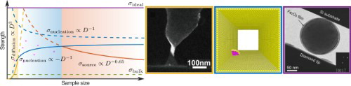
An example of this can be illustrated using small-volume samples that were specially made. Specifically, in addition to a pristine internal structure that is almost dislocation free, Han et al. used Fe nanoparticles passivated by a 4-nm smooth surface layer of its oxide, γ-Fe2O3 [Citation37], as shown in Figure A. This smooth and hard shell suppressed dislocation nucleation at edges, corners, and notches on the free surfaces [Citation12,Citation38–40]. Also, the samples were spherical particles with sizes from 80 to 430 nm. Based on contact mechanics, the spherical geometry ensures that the maximum stress site is located inside the sample when it is loaded, instead of at the contact interface [Citation41–43]. Loading, unloading prior to plastic yielding, and subsequent reloading experiments confirmed that the particles underwent large elastic deformation before the catastrophic collapse into a pancake-like shape. This latter dramatic shape change in a very short time signals a burst of copious defect (dislocation) production throughout the sample volume. The debris were detected in post-mortem TEM examinations of the deformed particles. For ∼10 pristine particles below 210 nm, the maximum contact pressure appears to reach a plateau of 10.7 GPa, see Figure B. Using interatomic potential finite element model to monitor the nonlinear anisotropic elastic deformation, the corresponding local maximum shear stress on is 9.4 GPa. This is very close to the ideal shear strength estimated from the Frenkel model, 8.5 GPa for the
shear, and that predicted by the density function theory calculation [Citation44]. In other words, under the triaxial stress loading condition (possible confined pressure effect) and the confinement by the oxide layer, the upper bound of experimentally measured critical shear stress for yielding has indeed happened at a stress level very close to that corresponding to homogeneous dislocation nucleation in the lattice, plateauing at a level approaching the theoretical shear strength of iron.
Figure 4. The strength of Fe nano-particles is saturated near the ideal strength, when the sample diameter is below 200 nm [Citation37]. (a) A spherical iron nanoparticle with the diameter of 200 nm. (b) The measured maximum contact pressure for Fe nano-particles with different diameters.
![Figure 4. The strength of Fe nano-particles is saturated near the ideal strength, when the sample diameter is below 200 nm [Citation37]. (a) A spherical iron nanoparticle with the diameter of 200 nm. (b) The measured maximum contact pressure for Fe nano-particles with different diameters.](/cms/asset/58bbb0c6-c9ad-4c9d-a16e-80dd329731d4/tmrl_a_1446192_f0004_c.jpg)
There have also been other experiments reporting observations of yielding approaching ideal strength in micro- or nano- scale samples. For example, in situ annealing of Mo pillars (prepared using focused ion beam milling) in TEM produces spherically capped Mo pillars [Citation45], which allows the maximum stress to occur in the interior of the spherical cap, resulting in an observed yielding stress very close to the calculated theoretical strength of Mo. Early Fe whiskers exhibited strong strengthening behavior along with decreasing sample diameter [Citation46,Citation47]. The measured tensile strength increased to ∼13.4 GPa when the sample diameter was reduced to ∼1.6 µm [Citation46]. This stress is very close to the theoretical de-bonding strength of bcc Fe (∼14.2 GPa) based on density functional theory [Citation48]. However, there was large scatter in the data, and a strength plateau was not observed. Experiments on Au nanowires and nanopillars have been summarized in ref. [Citation2,Citation49], showing a trend approaching ideal strength at very small sizes. In the experiment of Bei et al. [Citation50] Mo alloy pillars prepared using chemical etching method yielded without exception at a critical resolved shear stress of 4.3 GPa for the size regime from 360 to 1000 nm. However, this shear stress is about 3 times lower than the ideal shear strength predicted by theoretical calculations [Citation51]. Perhaps surface dislocation nucleation was not fully suppressed in their case, despite the pristine nature of their as-synthesized samples.
Concluding remarks and outlook
The three sets of examples above serve to illustrate that when it comes to sample size effects on strength, it is not always ‘smaller is stronger’ in the deep sub-micron regime (<∼102 nm). Under certain loading conditions (e.g. temperature, strain rate, loading mode etc.), surface effects including surface stresses, surface diffusion and surface structure may truncate the ‘smaller is stronger’ trend and turn it into ‘smaller is weaker’ or ‘saturated plateau’. These size effects are schematically summarized in . While the well-known ‘smaller is stronger’ trend due to truncated dislocation sources follows a power law with the exponent α between zero and one (e.g. 0.65 for fcc metals [Citation52]), the strength of pristine metals controlled by surface dislocation nucleation or surface diffusion are proportional to D−1 (see Equation 1) and D3, respectively. When sample size is sufficiently small such that bulk dislocation sources (e.g. Frank-Read source or single-arm source) become unstable due to image force attraction, surface dislocation sources (e.g. thermally activated surface dislocation nucleation [Citation12,Citation13,Citation16] or surface rebound of high-speed dislocations [Citation14,Citation15]) and surface diffusion would take over the plastic deformation, thus the well-received ‘smaller is stronger’ trend may switch to a ‘smaller is weaker’ regime. However, due to uncertainties coming from surface configuration, surface diffusion and thermal fluctuations [Citation32], the measurement of strength in this regime may suffer from large scatter. For those initially pristine metals with larger sample sizes, their strength may stay constant as surface dislocation nucleation now becomes almost size independent (i.e. the 1/D term in Equation 1 becomes negligible). This might explain the experimental observations on Mo [Citation50]. Ideal strength may be reached for pristine metals (e.g. with a spherical shape) as long as surface dislocation nucleation is suppressed and surface stress effects are small. Note that Figure is a schematic meant to illustrate the potential cross-over of the various trends. Exactly at what sample sizes the cross-over points reside is subject to change, depending on the material, homologous temperature, strain rate and sample/loading conditions. The determination as to exactly when a particular mechanism takes over must await systematic future research that builds on the limited case studies we cited in this Perspective.
Figure 5. Schematic summary of the trend of strength, for various dependences on crystal/sample size. Red curve represents the well-known ‘smaller is stronger’ trend (), where the exponent α is taken to be 0.65 for fcc metallic crystals [Citation52], due to truncated dislocation sources. This dependence does not necessarily continue to hold when the sample size decreases into the blue regime. Blue curves denote the sample size effects on strength controlled by surface dislocation nucleation (
) [Citation16]. When the surface-stress-induced internal stress has the same sign with the applied stress, it is ‘smaller is weaker’ (the lower solid blue curve). When the surface-stress-induced internal stress is opposite to the applied stress, it becomes ‘smaller is stronger’ (the upper dashed blue curve [Citation16]). Surface diffusion dominated strength (
) decreasing with size is shown with the yellow curve. The bulk material strength (
), and the ceiling set by the ideal strength (
) of a perfect crystal, are indicated by the dashed green line and purple line, respectively. Due to uncertainties arising from the as-synthesized surface conditions (including contamination, passivation and roughness) and influence from surface diffusion events and thermal fluctuations, the measured strength may exhibit considerable scatter in experiments, when the strength is predicted to be controlled by surface dislocation nucleation (magenta dots).
![Figure 5. Schematic summary of the trend of strength, for various dependences on crystal/sample size. Red curve represents the well-known ‘smaller is stronger’ trend (), where the exponent α is taken to be 0.65 for fcc metallic crystals [Citation52], due to truncated dislocation sources. This dependence does not necessarily continue to hold when the sample size decreases into the blue regime. Blue curves denote the sample size effects on strength controlled by surface dislocation nucleation () [Citation16]. When the surface-stress-induced internal stress has the same sign with the applied stress, it is ‘smaller is weaker’ (the lower solid blue curve). When the surface-stress-induced internal stress is opposite to the applied stress, it becomes ‘smaller is stronger’ (the upper dashed blue curve [Citation16]). Surface diffusion dominated strength () decreasing with size is shown with the yellow curve. The bulk material strength (), and the ceiling set by the ideal strength () of a perfect crystal, are indicated by the dashed green line and purple line, respectively. Due to uncertainties arising from the as-synthesized surface conditions (including contamination, passivation and roughness) and influence from surface diffusion events and thermal fluctuations, the measured strength may exhibit considerable scatter in experiments, when the strength is predicted to be controlled by surface dislocation nucleation (magenta dots).](/cms/asset/1cf0ce1e-4b65-4015-ba0b-e5adc10b8eb3/tmrl_a_1446192_f0005_c.jpg)
It should be pointed out that although there is already some evidence over the past several years that intrinsically the strength can drop or saturate in nanoscale metals, the data available are so far rather limited and there are experimental difficulties to overcome, in order to pin down the assertions above. For example, surface defects such as roughness and steps on free surfaces can facilitate dislocation nucleation, in addition to the dislocation nucleation sites on edges and corners. So there is a myriad of possible energy barriers, for dislocation nucleation at various stress levels. As such, the various trends depicted in Section 2 can be difficult to ascertain. The same can also be said for attaining the ideal strength; the spurious defects often render the measured strength much lower, even when nominally the sample size is already in the plateau region. The large variations in data can also be due to crystal orientation dependence, and minor misalignments between the samples and the diamond probe as well as the silicon substrate are inevitable, and there can be some internal defects that may be undetectable under TEM, which can dramatically affect the yield strength of the tested samples. All the aforementioned factors act to scatter the measured strength, having the common effect of indicating a lower strength for the tested material. Only the highest value found for each given sample size represents the condition that is most likely to correspond to the intrinsic strength of the tested samples. This was the practice used in Ref [Citation37]. In addition, small-volume metals are sometimes made to contain grain boundaries and twin boundaries [Citation53–64]. These pre-existing defects may also contribute to the observed strength. For example, nanocrystalline Pt nanopillars with a fixed grain size show a ‘smaller is weaker’ trend under compressive loading [Citation53], while Au nanowires containing angstrom-scale twins [Citation57] or both nanotwins and zigzag facets [Citation63] display near-ideal strength under tensile loading.
As for the diffusional mechanism, at what sample size the weakening effect becomes clearly visible depends on a number of factors, such as the homologous temperature, surface contamination/passivation, sample morphology and loading parameters (e.g. strain rate). The mechanical test data collected thus far in the community are mostly for samples not sufficiently small for surface diffusion to dominate. Moreover, for the materials tested, RT is usually not a sufficiently high homologous temperature. Therefore, the weakening mechanisms such as surface diffusive events are not pronounced enough to reveal a clear weakening trend, and can be obscured by other factors mentioned above, in particular surface conditions and loading geometry. It is therefore not surprising to observe large data scatter in this sample size regime. Last but not least, so far the samples have been tested and examined under electron beam inside a microscope. The surface diffusion may be accelerated by the energetic electron beam, and sensitive to beam heating induced local temperature rise. All these contribute to the uncertainty (scatter) of the sample size (see Figure ) at which surface diffusion could be observed to take over the plasticity. In general, we expect that low bulk melting point, significant melting point depression, low strain rate and large (clean) surface over volume ratio would lead to more dramatic surface diffusion contributing to the overall plasticity.
The ‘inverse Hall-Petch’ type of strength behavior in these isolated small-volume single crystals may have relevance to bulk nanocrystalline metals, but a direct connection remains to be established, to bridge with polycrystalline behavior. The latter metals rely on grain boundary mechanisms rather than mass transport on free surfaces, and there can be other grain boundary mediated deformation mechanisms at play that are difficult to tell apart: examples include grain boundary sliding, grain rotation and change of Frank-Bilby dislocation content in the grain boundary [Citation65], grain boundary migration, and segregation and effects of solutes in grain boundaries, which are all absent in the single-crystal experiments discussed in this paper. Given the complexity of that subject, the inverse Hall-Petch relationship in polycrystalline nanocrystals [Citation66–76] is beyond the scope of this paper and deserves a careful and separate review elsewhere.
Disclosure statement
No potential conflict of interest was reported by the authors.
ORCID
Qing-Jie Li http://orcid.org/0000-0003-2833-5374
Additional information
Funding
References
- Parthasarathy TA, Rao SI, Dimiduk DM, et al. Contribution to size effect of yield strength from the stochastics of dislocation source lengths in finite samples. Scr Mater. 2007;56:313–316. doi: 10.1016/j.scriptamat.2006.09.016
- Greer JR, De Hosson JTM. Plasticity in small-sized metallic systems: intrinsic versus extrinsic size effect. Prog Mater Sci. 2011;56:654–724. doi: 10.1016/j.pmatsci.2011.01.005
- Uchic MD, Shade PA, Dimiduk DM. Plasticity of micrometer-scale single crystals in compression. Annu Rev Mater Res. 2009;39:361–386. doi: 10.1146/annurev-matsci-082908-145422
- Pande CS, Cooper KP. Nanomechanics of Hall–Petch relationship in nanocrystalline materials. Prog Mater Sci. 2009;54:689–706. doi: 10.1016/j.pmatsci.2009.03.008
- Greer JR, Nix WD. Nanoscale gold pillars strengthened through dislocation starvation. Phys Rev B. 2006;73:245410. doi: 10.1103/PhysRevB.73.245410
- Shan ZW, Mishra RK, Syed Asif SA, et al. Mechanical annealing and source-limited deformation in submicrometre-diameter Ni crystals. Nat Mater. 2008;7:115–119. doi: 10.1038/nmat2085
- Wang Z-J, Li Q-J, Shan Z-W, et al. Sample size effects on the large strain bursts in submicron aluminum pillars. Appl Phys Lett. 2012;100:071906. doi: 10.1063/1.3681582
- Richter G, Hillerich K, Gianola DS, et al. Ultrahigh strength single crystalline nanowhiskers grown by physical vapor deposition. Nano Lett. 2009;9:3048–3052. doi: 10.1021/nl9015107
- Seo J-H, Yoo Y, Park N-Y, et al. Superplastic deformation of defect-free Au nanowires via coherent twin propagation. Nano Lett. 2011;11:3499–3502. doi: 10.1021/nl2022306
- Chen LY, Richter G, Sullivan JP, et al. Lattice anharmonicity in defect-free Pd nanowhiskers. Phys Rev Lett. 2012;109:125503. doi: 10.1103/PhysRevLett.109.125503
- Roos B, Kapelle B, Richter G, et al. Surface dislocation nucleation controlled deformation of Au nanowires. Appl Phys Lett. 2014;105:201908. doi: 10.1063/1.4902313
- Zhu T, Li J, Samanta A, et al. Temperature and strain-rate dependence of surface dislocation nucleation. Phys Rev Lett. 2008;100:025502. doi: 10.1103/PhysRevLett.100.025502
- Weinberger CR, Jennings AT, Kang K, et al. Atomistic simulations and continuum modeling of dislocation nucleation and strength in gold nanowires. J Mech Phys Solids. 2012;60:84–103. doi: 10.1016/j.jmps.2011.09.010
- Li Q-J, Li J, Shan Z-W, et al. Strongly correlated breeding of high-speed dislocations. Acta Mater. 2016;119:229–241. doi: 10.1016/j.actamat.2016.07.053
- Li Q-J, Li J, Shan Z-W, et al. Surface rebound of relativistic dislocations directly and efficiently initiates deformation twinning. Phys Rev Lett. 2016;117:165501. doi: 10.1103/PhysRevLett.117.165501
- Li Q-J, Xu B, Hara S, et al. Sample-size-dependent surface dislocation nucleation in nanoscale crystals. Acta Mater. 2018;145:19–29. doi: 10.1016/j.actamat.2017.11.048
- Hara S, Izumi S, Sakai S. Reaction pathway analysis for dislocation nucleation from a Ni surface step. J Appl Phys. 2009;106:093507. doi: 10.1063/1.3254178
- Wang Z-J, Li Q-J, Cui Y-N, et al. Cyclic deformation leads to defect healing and strengthening of small-volume metal crystals. Proc Natl Acad Sci. 2015;112:13502–13507. doi: 10.1073/pnas.1518200112
- Mousseau N, Béland LK, Brommer P, et al. The activation-relaxation technique: ART nouveau and kinetic ART. J At Mol Opt Phys. 2012;2012:925278.
- Marian J, Knap J. Breakdown of self-similar hardening behavior in Au nanopillar microplasticity. Int J Multiscale Comput Eng. 2007;5:287–294. doi: 10.1615/IntJMultCompEng.v5.i3-4.100
- Gan Y, Chen JK. Molecular dynamics study of size, temperature and strain rate effects on mechanical properties of gold nanofilms. Appl Phys A. 2009;95:357–362. doi: 10.1007/s00339-008-4970-8
- Ho DT, Kwon S-Y, Park HS, et al. Metal nanoplates: smaller is weaker due to failure by elastic instability. Phys Rev B. 2017;96:184103. doi: 10.1103/PhysRevB.96.184103
- Lührs L, Zandersons B, Huber N, et al. Plastic poisson’s ratio of nanoporous metals: a macroscopic signature of tension–compression asymmetry at the nanoscale. Nano Lett. 2017;17:6258–6266. doi: 10.1021/acs.nanolett.7b02950
- Tian L, Li J, Sun J, et al. Visualizing size-dependent deformation mechanism transition in Sn. Sci Rep. 2013;3:2113. doi: 10.1038/srep02113
- Merkle AP, Marks LD. Liquid-like tribology of gold studied by in situ TEM. Wear. 2008;265:1864–1869. doi: 10.1016/j.wear.2008.04.032
- Yue Y, Chen N, Li X, et al. Crystalline liquid and rubber-like behavior in Cu nanowires. Nano Lett. 2013;13:3812–3816. doi: 10.1021/nl401829e
- Lu Y, Huang JY, Wang C, et al. Cold welding of ultrathin gold nanowires. Nat Nanotechnol. 2010;5:218–224. doi: 10.1038/nnano.2010.4
- Sun J, He L, Lo Y-C, et al. Liquid-like pseudoelasticity of sub-10-nm crystalline silver particles. Nat Mater. 2014;13:1007–1012. doi: 10.1038/nmat4105
- Zhong L, Sansoz F, He Y, et al. Slip-activated surface creep with room-temperature super-elongation in metallic nanocrystals. Nat Mater. 2017;16:439–445. doi: 10.1038/nmat4813
- Castro T, Reifenberger R, Choi E, et al. Size-dependent melting temperature of individual nanometer-sized metallic clusters. Phys Rev B. 1990;42:8548–8556. doi: 10.1103/PhysRevB.42.8548
- Chen LY, He M, Shin J, et al. Measuring surface dislocation nucleation in defect-scarce nanostructures. Nat Mater. 2015;14:707–713. doi: 10.1038/nmat4288
- Li J. Diffusive origins. Nat Mater. 2015;14:656–657. doi: 10.1038/nmat4326
- Joachim C, Gimzewski JK, Aviram A. Electronics using hybrid-molecular and mono-molecular devices. Nature. 2000;408:541–548. doi: 10.1038/35046000
- Strachan DR, Johnston DE, Guiton BS, et al. Real-time TEM imaging of the formation of crystalline nanoscale gaps. Phys Rev Lett. 2008;100:56805. doi: 10.1103/PhysRevLett.100.056805
- Reed MA, Zhou C, Muller CJ, et al. Conductance of a molecular junction. Science. 1997;278:252–254. doi: 10.1126/science.278.5336.252
- Strachan DR, Smith DE, Fischbein MD, et al. Clean electromigrated nanogaps imaged by transmission electron microscopy. Nano Lett. 2006;6:441–444. doi: 10.1021/nl052302a
- Han W-Z, Huang L, Ogata S, et al. From “smaller is stronger” to “size-independent strength plateau”: towards measuring the ideal strength of iron. Adv Mater. 2015;27:3385–3390. doi: 10.1002/adma.201500377
- Zuo L, Ngan AHW, Zheng GP. Size dependence of incipient dislocation plasticity in Ni3Al. Phys Rev Lett. 2005;94:95501. doi: 10.1103/PhysRevLett.94.095501
- Zuo L, Ngan AHW. Molecular dynamics study on compressive yield strength in Ni3Al micro-pillars. Philos Mag Lett. 2006;86:355–365. doi: 10.1080/09500830600803890
- Bei H, Gao YF, Shim S, et al. Strength differences arising from homogeneous versus heterogeneous dislocation nucleation. Phys Rev B. 2008;77:060103. doi: 10.1103/PhysRevB.77.060103
- Johnson KL. Contact mechanics. Cambridge: Cambridge University Press; 1984.
- Valentini P, Gerberich WW, Dumitrică T. Phase-transition plasticity response in uniaxially compressed silicon nanospheres. Phys Rev Lett. 2007;99:175701. doi: 10.1103/PhysRevLett.99.175701
- Zhu T, Li J, Van Vliet K, et al. Predictive modeling of nanoindentation-induced homogeneous dislocation nucleation in copper. J Mech Phys Solids. 2004;52:691–724. doi: 10.1016/j.jmps.2003.07.006
- Ogata S, Li J, Hirosaki N, et al. Ideal shear strain of metals and ceramics. Phys Rev B. 2004;70:104104. doi: 10.1103/PhysRevB.70.104104
- Lowry MB, Kiener D, LeBlanc MM, et al. Achieving the ideal strength in annealed molybdenum nanopillars. Acta Mater. 2010;58:5160–5167. doi: 10.1016/j.actamat.2010.05.052
- Brenner SS. Tensile strength of whiskers. J Appl Phys. 1956;27:1484–1491. doi: 10.1063/1.1722294
- Brenner SS. Growth and properties of “whiskers”: further research is needed to show why crystal filaments are many times as strong as large crystals. Science. 1958;128:569–575. doi: 10.1126/science.128.3324.569
- Clatterbuck DM, Chrzan DC, Morris JW. The inherent tensile strength of iron. Philos Mag Lett. 2002;82:141–147. doi: 10.1080/095008302317262642
- Gall K, Diao J, Dunn ML. The strength of gold nanowires. Nano Lett. 2004;4:2431–2436. doi: 10.1021/nl048456s
- Bei H, Shim S, George EP, et al. Compressive strengths of molybdenum alloy micro-pillars prepared using a new technique. Scr Mater. 2007;57:397–400. doi: 10.1016/j.scriptamat.2007.05.010
- Krenn CR, Roundy D, Morris JW, et al. Ideal strengths of bcc metals. Mater Sci Eng A. 2001;319–321:111–114. doi: 10.1016/S0921-5093(01)00998-4
- Dimiduk DM, Uchic MD, Parthasarathy TA. Size-affected single-slip behavior of pure nickel microcrystals. Acta Mater. 2005;53:4065–4077. doi: 10.1016/j.actamat.2005.05.023
- Gu XW, Loynachan CN, Wu Z, et al. Size-dependent deformation of nanocrystalline Pt nanopillars. Nano Lett. 2012;12:6385–6392. doi: 10.1021/nl3036993
- Tucker GJ, Aitken ZH, Greer JR, et al. The mechanical behavior and deformation of bicrystalline nanowires. Model Simul Mater Sci Eng. 2013;21:015004. doi: 10.1088/0965-0393/21/1/015004
- Ramachandramoorthy R, Gao W, Bernal R, et al. High strain rate tensile testing of silver nanowires: rate-dependent brittle-to-ductile transition. Nano Lett. 2016;16:255–263. doi: 10.1021/acs.nanolett.5b03630
- Wu ZX, Zhang YW, Jhon MH, et al. Nanostructure and surface effects on yield in Cu nanowires. Acta Mater. 2013;61:1831–1842. doi: 10.1016/j.actamat.2012.11.053
- Wang J, Sansoz F, Huang J, et al. Near-ideal theoretical strength in gold nanowires containing angstrom scale twins. Nat Commun. 2013;4:1742. doi: 10.1038/ncomms2768
- Filleter T, Ryu S, Kang K, et al. Nucleation-controlled distributed plasticity in penta-twinned silver nanowires. Small. 2012;8:2986–2993. doi: 10.1002/smll.201200522
- Jang D, Li X, Gao H, et al. Deformation mechanisms in nanotwinned metal nanopillars. Nat Nanotechnol. 2012;7:594–601. doi: 10.1038/nnano.2012.116
- Qin Q, Yin S, Cheng G, et al. Recoverable plasticity in penta-twinned metallic nanowires governed by dislocation nucleation and retraction. Nat Commun. 2015;6:5983. doi: 10.1038/ncomms6983
- Narayanan S, Cheng G, Zeng Z, et al. Strain hardening and size effect in five-fold twinned Ag nanowires. Nano Lett. 2015;15:4037–4044. doi: 10.1021/acs.nanolett.5b01015
- Wang Z-J, Li Q-J, Li Y, et al. Sliding of coherent twin boundaries. Nat Commun. 2017;8:1108. doi: 10.1038/s41467-017-01234-8
- Deng C, Sansoz F. Near-ideal strength in gold nanowires achieved through microstructural design. ACS Nano. 2009;3:3001–3008. doi: 10.1021/nn900668p
- Jang D, Greer JR. Size-induced weakening and grain boundary-assisted deformation in 60 nm grained Ni nanopillars. Scr Mater. 2011;64:77–80. doi: 10.1016/j.scriptamat.2010.09.010
- Wang L, Teng J, Liu P, et al. Grain rotation mediated by grain boundary dislocations in nanocrystalline platinum. Nat Commun. 2014;5:4402.
- Giga A, Kimoto Y, Takigawa Y, et al. Demonstration of an inverse Hall–Petch relationship in electrodeposited nanocrystalline Ni–W alloys through tensile testing. Scr Mater. 2006;55:143–146. doi: 10.1016/j.scriptamat.2006.03.047
- Quek SS, Chooi ZH, Wu Z, et al. The inverse hall–petch relation in nanocrystalline metals: a discrete dislocation dynamics analysis. J Mech Phys Solids. 2016;88:252–266. doi: 10.1016/j.jmps.2015.12.012
- Shen TD, Schwarz RB, Feng S, et al. Effect of solute segregation on the strength of nanocrystalline alloys: inverse Hall–Petch relation. Acta Mater. 2007;55:5007–5013. doi: 10.1016/j.actamat.2007.05.018
- Carlton CE, Ferreira PJ. What is behind the inverse Hall–Petch effect in nanocrystalline materials? Acta Mater. 2007;55:3749–3756. doi: 10.1016/j.actamat.2007.02.021
- Hahn EN, Meyers MA. Grain-size dependent mechanical behavior of nanocrystalline metals. Mater Sci Eng A. 2015;646:101–134. doi: 10.1016/j.msea.2015.07.075
- Argon AS, Yip S. The strongest size. Philos Mag Lett. 2006;86:713–720. doi: 10.1080/09500830600986091
- Schiøtz J, Tolla FDD, Jacobsen KW. Softening of nanocrystalline metals at very small grain sizes. Nature. 1998;391:561–563. doi: 10.1038/35328
- Schiøtz J, Jacobsen KW. A maximum in the strength of nanocrystalline copper. Science. 2003;301:1357–1359. doi: 10.1126/science.1086636
- Padmanabhan KA, Dinda GP, Hahn H, et al. Inverse Hall–Petch effect and grain boundary sliding controlled flow in nanocrystalline materials. Mater Sci Eng A. 2007;452–453:462–468. doi: 10.1016/j.msea.2006.10.084
- Tang Y, Bringa EM, Meyers MA. Inverse Hall–Petch relationship in nanocrystalline tantalum. Mater Sci Eng A. 2013;580:414–426. doi: 10.1016/j.msea.2013.05.024
- Zhu YT, Langdon TG. Influence of grain size on deformation mechanisms: an extension to nanocrystalline materials. Mater Sci Eng A. 2005;409:234–242. doi: 10.1016/j.msea.2005.05.111