
ABSTRACT
Phosphorylation is a dynamic post-translational modification that can alter protein structure, localization, protein-protein interactions and stability. All of the identified tight junction transmembrane proteins can be multiply phosphorylated, but only in a few cases are the consequences of phosphorylation at specific sites well characterized. The goal of this review is to highlight some of the best understood examples of phosphorylation changes in the integral membrane tight junction proteins in the context of more general overview of the effects of phosphorylation throughout the proteome. We expect as that structural information for the tight junction proteins becomes more widely available and the molecular modeling algorithms improve, so will our understanding of the relevance of phosphorylation changes at single and multiple sites in tight junction proteins.
KEYWORDS:
Introduction
Tight junctions form the paracellular epithelial barrier to ions and solutes and thus separate tissue spaces and enable directional transcellular absorption and secretion.Citation1 Many proteins have been localized to tight junctions,Citation2 but the critical barrier components include transmembrane proteins that physically form the sealing contacts, including members of the claudin (cldn), tight junction-associated MARVEL proteins (TAMPS) and junctional adhesion molecule (JAM or CTX, for cortical thymocyte marker in Xenopus)Citation3 family of proteins. The transmembrane proteins bind to scaffolding proteins, including ZO-1, -2 and -3 among others, that interact with cytoskeletal elements to regulate junctional integrity. Much information is available on in vitro binding interactions between these protein components,Citation4-10 but there is considerably less information available on how these binding interactions might be regulated in vivo. One mechanism likely to modulate protein-protein interactions is phosphorylation status.
Most proteins in mammalian cells are phosphorylated;Citation11 it is a dynamic post translational modification that can regulate protein folding, protein interactions, localization and stability.Citation12,13 Phosphorylation results in addition of two negative charges at physiological pH, which will alter the electrostatic environment and can alter the strength of protein-protein interactions, both within binding sites and through longer range changes in protein conformation.Citation12,13 Although the estimated stability of most protein complexes is not altered by phosphorylation, about a third are expected to be significantly stabilized or destabilized by phosphorylation.Citation12 The predominant influence is destabilization, but a significant minority of protein-protein interactions result in stabilization of protein complexes.Citation12 In spite of the fact that thousands of phosphorylation sites have been identified by mass spectrometry (MS),Citation14 relatively few of these have been analyzed in terms of how they affect structure and function.
All of the transmembrane tight junction proteins are reported to be phosphoproteins. In some studies, phosphorylation has been associated with functional changes, but only in a couple of cases has there been any detailed analysis of the relevant mechanisms. Rather than include an exhaustive and exhausting list of phosphorylation changes, the goal of this review is to focus on the best understood examples of how phosphorylation alters interactions or functions in the limited set of tight junction integral membrane proteins. In some cases, studies of closely related proteins will be included if they appear to provide relevant insights. By focusing on relatively few different approaches and examples, we hope to highlight some general themes and ongoing challenges as well as identify areas that are particularly promising for future study.
Cldn phosphorylation and PDZ-dependent interactions
PDZ-dependent interactions are a central feature of tight junction organization. Many tight junction scaffolding proteins, including ZO proteins,Citation15-17 MUPP1,Citation18 afadin/AF6,Citation19 MAGI-1, 2, 3,Citation20–22 PARD3,Citation23 PALS124 and PATJCitation25 among others, contain multiple PDZ domains. PDZ domains form small globular structures of 80–90 residues;Citation26 which typically bind the 4–10 carboxy-terminal residues (PDZ binding motifs)Citation27,28 of transmembrane proteins and anchor them to the junction. The basic model for interaction is that the terminal hydrophobic residue of the PDZ binding motif-containing protein (numbered amino acid 0) inserts into a hydrophobic pocket in the PDZ domain, and then the upstream amino acids of the binding motif (-1 to -5) form an antiparallel beta strand to a beta strand in the PDZ domain.Citation29 Both the tight junction strand forming cldnsCitation6 and the adhesion proteins, the JAMsCitation8,9 end in PDZ binding motifs and have been shown to interact with the PDZ domain-containing scaffolding proteins.
Cldns are small, tetraspan proteins, with short cytoplasmic tails that vary in length from 26-70 amino acid residues; these tail regions contain abundant serines, threonines and tyrosines (). All but three of the 23 (human) cldn family members end in a characteristic PDZ binding motif (for most cldns -XYV) and interact with the first PDZ domains of ZO-1, -2 and -36 and MUPP1,Citation30 among others. Although specificity of interaction between binding motif and binding domain is likely to be primarily regulated by primary amino acid sequence and protein localization,Citation29 regulation of this interaction is not well understood. One attractive possible regulatory mechanism is phosphorylation.
Figure 1. Selected cldn cytoplasmic domains. Red text, phosphorylation sites identified by MSCitation14; blue highlighted residues are referred to in text, purple highlight identified ubiquitination site blocked by adjacent phosphorylation. Double underline identified regions predicted to be disordered (see text).
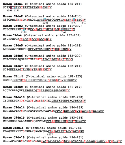
Mass Spectrometry data from tissues and cultured cells shows cldn C-terminal regions are abundantly phosphorylated,Citation14 including within the canonical C-terminal three amino acid PDZ binding motif. For example, much MS dataCitation14 identifies phosphorylation of Y(-1) in cldn1-7, -9, -10 and -18.Citation14,31 Surprisingly, phosphorylation of this site might not affect interaction with ZO-1, since by analogy, tyrosine phosphorylation of the -1 residue in the PDZ binding motif of syndecan can be accommodated in the PDZ binding pocket in Tiam1 and does not alter binding affinity.Citation32 Syndecan, like cldns, can bind to a variety of PDZ containing proteins. The authors suggest that syndecan phosphorylation at this site might provide regulatory specificity, since both phosphorylated and unphosphorylated syndecan can bind Tiam1, but only unphosphorylated syndecan can bind to the syntenin PDZ domain. Additionally, binding of phosphorylated syndecan to Tiam1 dampened PDZ domain dynamics, and the authors suggested it might result changes in binding affinities at other sites in Tiam1.Citation32 Indirect evidence suggests that tyrosine phosphorylation of cldn4 at the -1 position decreases binding to ZO-1,Citation33 but the possibility that differentially phosphorylated cldns might have differing interactions with ZO-1 PDZ1 is intriguing but unexplored.
Cldn phosphorylation sites upstream from the C-terminal motif could also affect PDZ domain binding. For example, although only the last four residues of cldn1 are involved in interaction with PDZ1 of ZO-1, seven terminal residues of cldn2 contact PDZ1 of ZO-1, and Y-6 phenol group of cldn2 contributes to higher affinity binding to ZO-1 PDZ1 compared with that for cldn1.Citation34 We recently found that tyrosine phosphorylation of cldn2 at this -6 position decreased affinity for binding to PDZ1 of ZO-1,Citation34 suggesting a potential regulatory mechanism for their interactions. Similarly, phosphorylation of the analogous site in cldn3 increases its mobility in the membrane as determined by Fluorescence Recovery After Photobleaching (FRAP),Citation35 suggesting the possibility that phosphorylation of cldn3 Y -6 decreased scaffolding to ZO-1 at the tight junction. Along with possibly altering the affinity for PDZ1 of ZO-1, phosphorylation at other sites in cldn tails may promote binding specificity, which could be important given the presence of multiple PDZ domain-containing junctional proteins.Citation2
Other cldn phosphorylation sites and tight junction association
There are many examples of cldn phosphorylation apart from the PDZ domain interaction that are thought to be important for tight junction localization.Citation31,36–41 For example, phosphorylation of cldn1(T191)Citation39 and cldn2(S208)Citation31 is associated with enhanced tight junction strand formation or localization; Fujii et al.Citation39 found that hypotonic stress resulted in dephosphorylation at these sites and junctional removal by clathrin-dependent endocytosis. In support of a role for cldn1(T191) phosphorylation in enhancing junction localization, Shiomi et al.Citation40 found that AMPK activation by AICAR resulted in phosphorylation of T191 in cldn1 and stimulated the formation of ectopic tight junction formation on lateral membrane of Eph4 cells; ectopic fibril formation is also seen with a phosphomimetic mutant of the analogous site in cldn3(T192)Citation37 and after cldn4 phosphorylation by protein kinase C epsilon.Citation36 Shiomi et al.Citation40 further found that stimulated phosphorylation by AMPK activation was associated with decreased cldn1 ubiquitination at a nearby lysine (K189). These authors interpreted these findings to mean that cldn1 ubiquitination is normally required for turnover; blocking turnover by forced phosphorylation thus resulted in ectopic fibril formation. Many cldns have similar arrangements of serines/threonines close to ubiquitinylated lysines identified by MS,Citation14 so it is possible that this could be a common theme regulating cldn turnover.
Phosphorylation/ubiquitinoylation may also be important in cldn16 removal from the tight junction. Phosphorylation of S217 in cldn16 by protein kinase A pathway results in tight junction localization.Citation41,42 This group found that phosphorylation at this site is required for interaction with syntaxin-8 and recycling to the membraneCitation42 and that dephosphorylated cldn16 was associated with the E3 ubiquitin ligase, PDZRN3, a protein containing both PDZ and ring finger domains.Citation43 Marunaka et al. suggest this interaction was likely involved in the endocytosis of dephosphorylated cldn16; interestingly, PDZRN3 has also been implicated in regulation of the stability of endothelial cell junctions through targeting the multiPDZ protein MUPP1 for proteasomal degradation.Citation44
Cldn phosphorylation has also, although less frequently, been reported to play a role in removal from the tight junction. For example, Cong et al. found that carbachol stimulated ERK phosphorylation of cldn4 and its removal from tight junctions in a rat submandibular epithelial cell line, by promoting its interaction with β-arrestin2 and clathrin endocytosis.Citation45
JAM family phosphorylation
The JAM family of proteins are single span integral membrane proteins that are members of the immunoglobulin superfamily; they are broadly distributed in immune cells as well as epithelial and endothelial cells.Citation46 JAM-A is the principal form expressed on mucosal epithelial cells and is known to be an important regulator of cell polarization, migration, cell-cell adhesion and paracellular permeability. It, like the other JAMs, has a fairly short cytoplasmic C-terminal tail (∼40 amino acids) that contains conserved consensus phosphorylation sites (). Like the cldns, JAM cytoplasmic tails end in PDZ binding motifs that interact with PDZ3 of ZO1, as well as with other PDZ domain containing proteins, including afadin, MUPP1 and PAR3.Citation47
Figure 2. JAM-A, CAR and CLMP cytoplasmic domains. Red text, phosphorylation sites identified by MS (14; blue highlighted residues are referred to in text, double underline identified regions predicted to be disordered (see text).
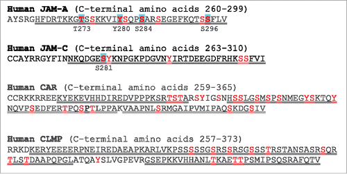
The carboxyl terminal of human JAM-A, -B and -C all end in the conserved sequence S(295)-S(296)-F(297)-I/V/L(298)-V/I(299). Structural analysis demonstrated that S(296) is a critical component of interaction with ZO-148 and high throughput MS has identified this as a phosphorylation site. Although there is no evidence for an effect of phosphorylation at S(296)Citation49 on interaction with ZO-1 or other PDZ domain containing proteins, possible phosphorylation at this site might regulated the affinity or specificity of interaction with tight junction PDZ containing protein.
In epithelial cells, JAM-A recruits atypical Protein Kinase C (aPKC) to nascent tight junctions where it phosphorylates JAM-A at S285 (S284 in human JAM-A). Total JAM-A is normally present at tight junctions and also to a variable extent along the lateral membrane, but JAM-A S285P is exclusively localized to tight junctions.Citation50 This phosphorylation is required for the full development of the paracellular barrier and is negatively regulated by Protein Phosphatase 2A (PP2A), which is also involved in de-phosphorylation of other tight junction proteins.Citation50,51 JAM-A S285 phosphorylation increases as cell contacts mature,Citation50 and Burridge and colleaguesCitation52 reasoned that tension associated with junction formation might stimulate JAM-A phosphorylation at this site. They used anti JAM-A antibody-coated (to the extracellular domain) paramagnetic beads to apply tension to JAM-A and found tension increased JAM-A phosphorylation at S284; further, tension applied to JAM-A activated RhoA in a phosphorylation-dependent fashion. Inhibition of RhoA activity is associated with barrier disruption,Citation53 which might explain in part how JAM-A phosphorylation regulates barrier function. However, the exact mechanism is unclear.
It seems likely that other phosphorylation sites are important in JAM-A function, but they are less well studied. For example, T273 is required for the development of hepatocyte polarityCitation49 and Y280 development of tube formation by endothelial cells [54]. In addition, tyrosine phosphorylation of JAM-A in platelets modulates integrin signaling;Citation55 it is not clear if a similar modification is relevant in epithelial cells. However, given that JAMs play myriad roles and bind to a variety of proteins, it is likely that other phosphorylation sites are important, perhaps in altering the affinity for specific binding partners and signaling pathways.
JAM-C, which has been localized both to desmosomesCitation56 and tight junctionsCitation57 is also found in a variety of tumor cell linesCitation58 where its function is unknown. Mandicourt et al.Citation59 demonstrated that exogenous expression of JAM-C in a tumor cell lacking the endogenous protein improved epithelial barrier function. Further, these authors demonstrated that phosphorylation of JAM-C S281 was required for improved tight junction function, since mutation of this residue to alanine blocked the epithelial phenotypic change.
A JAM-related protein, the Coxsackie and Adenovirus Receptor (CAR) has also been identified as a tight junction proteinCitation60 that interacts with both ZO-1Citation60 and MUPP1.Citation61 Although addition of peptides corresponding to the extracellular domain of CAR interfere with recovery of TER after junction disruption following calcium removal,Citation60 it seems likely that CAR may also play a role at the adherens junction. Morton et al.Citation62 demonstrated that unphosphorylated CAR was involved in E-cadherin endocytosis and that when two residues in the cytoplasmic tail of CAR were phosphorylated by PKCδ, this blocked the endocytosis stimulated by over-expression of wild-type CAR. These results suggested that CAR normally plays a role in E-cadherin recycling and that CAR phosphorylation might stabilize E-cadherin at the membrane. One caveat to this study is that the fluorescent tag used to follow CAR localization is likely to block interaction with PDZ domain binding and thus may alter localization. These two sites are not found in the canonical JAMs, but another JAM family member, CLMP (CAR-like membrane protein) has a serine and threonine-rich tail with potential phosphorylation sites in the same region. CLMP is required for intestinal development and its absence results in short bowel syndrome.Citation63,64 It has been shown to localize at tight junctionsCitation65 and it can mediate cell-cell adhesionCitation65 but more study is required to understand its specific functional role and the role of phosphorylation.Citation66
TAMP family phosphorylation
Occludin Phosphorylation
Occludin (Ocln) was the first identified tight junction transmembrane protein;Citation67 like cldns, it is a tetraspan protein and most phosphorylation sites are found in the C-terminal cytoplasmic domainCitation14 () It was early identified as being multiply phosphorylated.Citation68 Ocln, although normally concentrated at tight junctions, is found all along the lateral membrane; in an elegant study, Tsukita and colleagues demonstrated that ocln at the lateral membrane was phosphorylated at a much lower level than the highly phosphorylated junctional ocln.Citation68 Following this initial observation, there were a large number of studies that correlated the degree of ocln phosphorylation with its localization; in most cases, ocln dephosphorylation, as measured by changes in migration in SDS-PAGE, was correlated with barrier loss,Citation69–73 reviewed in.Citation74 A large number of kinases and phosphatases have been implicated in regulation of ocln phosphorylation, including CK2,Citation75 cYES,Citation76 CK1,Citation77 PKC,Citation78 Rho kinase,Citation79 Src,Citation80 G-coupled receptor kinases (GRK),Citation81 PP2A,Citation51 density enhanced phosphastase 1Citation82 and PTPN2 (non-receptor protein tyrosine phosphatase N2).Citation83
Figure 3. Ocln, tricellulin and marvelD3 cytoplasmic N- and C-terminal domains. Red text, phosphorylation sites identified by MSCitation14; blue highlighted residues are referred to in text, double underline identified regions predicted to be disordered (see text). Yellow highlighted area in ocln C-terminal domain identifies area with structural information.Citation89,107
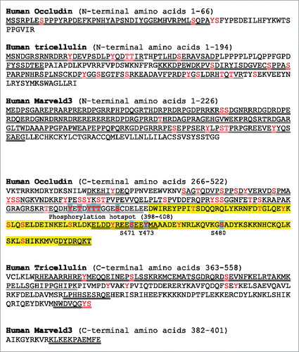
Although correlations between ocln phosphorylation with localization and with different signaling pathways were noted soon after the discovery of ocln, mechanistic studies on the importance of specific phosphorylation sites are relatively recent. An elegant pair of studies from the Antonetti laboratory identified a number of ocln phosphorylation sitesCitation84,85 and important functional consequences associated with phosphorylation of one of them. These authors found S490 was phosphorylated in response to VEGF treatment and that this phosphorylation attenuated interaction with ZO-1, suggesting a molecular mechanism for ocln dissociation from the tight junction. In addition, mutation of S490 to a non-phosphorylatable alanine suppressed VEGF-induced ubiquitination, tight junction protein trafficking and the increase in permeability associated with VEGF treatment. More recent work from this group identified PCKβ as the relevant kinase.Citation86 Unexpectedly, Antonetti and colleagues also has implicated this phosphorylation site in ocln-dependent regulation of mitotic entryCitation87 and in control of VEGF-induced neovascularization.Citation88 These data suggest that phosphorylation of ocln at S490 could be an important site in the coordination of endothelial proliferative response to injury associated with tight junction barrier changes.
Antonetti and colleagues went on to analyze the structural contacts between ZO-1 and ocln and identified a potential stabilizing phosphorylation site within the ocln tail, S471.Citation89 In a more recent study, they demonstrated that over-expression of ocln S471A blocked monolayer maturation and normal tight junction protein localization, consistent with a requirement for phosphorylation at this site in meditating normal interactions with ZO-1 and cell packing.Citation81
In addition to these phosphorylation sites, Dorfel and HuberCitation90 identified a phosphorylation hotspot within the ocln c-terminal domain, an 11 amino acid stretch from 398-408. Within this region, Raleigh et al.Citation91 had found that inhibition of CK2-mediated phosphorylation of ocln at S408 resulted in increased TER. Dephosphorylated ocln interacted with ZO-1 and through ZO-1 with cldn 2; these interactions were attenuated by S408 phosphorylation. The authors speculated that phosphorylation decreased interaction between ZO-1, cldn2 and ocln, affecting the dynamic behavior among these proteins and resulting in decreased paracellular permeability by disrupting the cation-permeable paracellular pores formed by cldn2. In contrast to the findings of Raleigh et al., ocln T400 and T404 were also identified as CK2 phosphorylation sites and when T400, T404 and S408 were mutated to phosphomimetic amino acids, Huber and colleagues found diminished interaction with ZO-2 and increased TER;Citation92 some differences were seen between the effects of the mutations on interactions with ZO-1 and ZO-2. Also within this region is T403, which with T404 has also been reported to be a phosphorylation site for PKCη;Citation93 expression of wild-type or dominant active PKCη enhances tight junction assembly, similar to what was reported by Huber and colleagues.Citation92 Phosphorylation of T403/4 was associated with tight junction assembly, since T403/4A ocln mutants fail to localize to tight junctions, while T403/4D mutants block the ability of a PKCη inhibitor to disrupt ocln localization. The reason for the different effects of phosphorylation in this small region is unclear, but may reflect differences in cell lines and/or relative cldn expression levels and requires further investigation.
Along with serine and threonine ocln phosphorylation, tyrosine phosphorylation of ocln has also been implicated in both tight junction disassembly;Citation71,94 and assemblyCitation76 however, most studies have reported that tyrosine phosphorylation is predominantly associated with diminished junctional ocln. Both Y398 and 402 phosphomimetics have been shown to block binding to ZO-1 and to destabilize ocln at tight junctions.Citation95 In one unusual example, ocln Y473P was localized to the leading edge of wounded epithelial monolayers; where is was reported to recruit the phosphoinositol-3-kinase regulatory subunit, p85α and thus activate Rac1 to promote lamellipodial formation and migration.Citation80
Tricellulin and MarvelD3 phosphorylation
Both tricellulin and marvelD3 are ocln-related proteins; marvelD3, like ocln, is distributed along bicellular tight junction contactsCitation96,97 while tricellulin is concentrated at tricellular contacts. Tricellulin is it critical for regulation of paracellular flux of larger moleculesCitation98 while the role of marvelD3 is more complex.Citation99 Both tricellulin and marvelD3 are reported to be phosphoproteins,Citation14 but in contrast to ocln, tricellulin and marvelD3 have relatively extended intracellular N-terminal domains which contain a significant number of phosphosites; in the case of marvelD3, most phosphorylation sites lie in this domain (). All phospho sites to date within both tricellulin and marvelD3 have been identified solely by high throughput MS analysis, without direct experimental verification, so that the importance of these sites remains to be explored. As an aside, tricellulin is concentrated at tricellular contacts through interaction with angulin1/LSR, an immunoglobulin superfamily transmembrane protein. Although little is known about tricellulin phosphorylation, a recent study demonstrated the requirement for JNK1/2-mediated phosphorylation of angulin 1/LSR at S288 for its localization, and thus localization of tricellulin, to the tricellular junctions.Citation100
Future approaches
Although it is possible to deduce some general rules from the above studies, for example, that serine/threonine phosphorylation of the cldn and JAM C-terminal domains is (mostly) associated with localization at the tight junction, while tyrosine phosphorylation of either cldns or ocln results in junction disruption, most findings referenced in this review are necessarily simplistic. However, these studies at a minimum identify key areas and relevant pathways that should allow us to ask increasingly sophisticated questions about the importance of tight junction protein phosphorylation. For example, the growing availability of structural information (as inCitation34,89) can be used to guide mutational analyses and interpretations about phosphorylation in well-ordered protein domains. In addition, the recognition that intrinsically disordered protein domains are enriched in phosphorylationCitation101 and are particularly important in trafficking and protein localizationCitation102 is highly relevant. The cytoplasmic domains of cldns and JAM-A, as well as regions within the ocln tail are predicted to be disordered by several algorithms;Citation103,104 phosphorylation in these regions has different implications than that in highly structured domains.Citation12 Further, it is important to remember that all of the tight junction integral membrane proteins have multiple phosphorylation sites that in combination are likely to have complex and interacting contributions to their behavior. In many cases the domains in other proteins with which these phosphorylated proteins interact are themselves likely to be variably phosphorylated; for example, it has recently been shown that threonine phosphorylation on T770/T772 in the GUK (guanylate kinase) domain of ZO-1 by PKCϵ disrupts interaction with ocln.Citation105 Phosphorylation can be difficult to detect, is dynamic and reversible and can have subtle long range as well as large local effects. As algorithms improve, molecular modeling is likely to be increasingly useful in predicting how phosphorylation might alter protein interactions.Citation106
Dynamic phosphorylation changes are clearly relevant to regulation of the interactions and stability of tight junction proteins, and better understanding of the role of this post-translational modification will provide important biologic and potentially therapeutic insights in the regulation of the paracellular barrier.
Disclosure of potential conflicts of interest
No potential conflicts of interest were disclosed.
Funding
This research was supported by the Division of Intramural Research, National Institutes of Health.
References
- Mills C, Matter K, Balda MS. Tight junctions: from simple barriers to multifunctional molecular gates. Nat Rev Mol Cell Biol. 2016;17(9):564–80. https://doi.org/10.1038/nrm.2016.80 PMID:27353478
- Van Itallie CM, Anderson JM. Architecture of tight junctions and principles of molecular composition. Semin Cell Dev Biol. 2014;36:157–65. https://doi.org/10.1016/j.semcdb.2014.08.011 PMID:25171873
- Chretien I., Robert J, Marcuz A, Garcia-Sanz JA, Courtet M, Du Pasquier L. CTX, a novel molecule specifically expressed on the surface of cortical thymocytes in Xenopus. Eur J Immunol. 1996;26(4):780–91. https://doi.org/10.1002/eji.1830260409 PMID:8625968
- Rodgers LS, Fanning AS. Regulation of epithelial permeability by the actin cytoskeleton. Cytoskeleton. 2011;68(12):653–60. https://doi.org/10.1002/cm.20547 PMID:22083950
- Furuse M, Itoh M, Hirase T, Nagafuchi A, Yonemura S, Tsukita S, Tsukita S. Direct association of occludin with ZO-1 and its possible involvement in the localization of occludin at tight junctions. J Cell Biol. 1994;127(6 Pt 1):1617–26. https://doi.org/10.1083/jcb.127.6.1617 PMID:7798316
- Itoh M, Furuse M, Morita K, Kubota K, Saitou M, Tsukita S. Direct binding of three tight junction-associated MAGUKs, ZO-1, ZO-2, and ZO-3, with the COOH termini of claudins. J Cell Biol. 1999;147(6):1351–63. https://doi.org/10.1083/jcb.147.6.1351 PMID:10601346
- Fanning AS, Jameson BJ, Jesaitis LA, Anderson JM. The tight junction protein ZO-1 establishes a link between the transmembrane protein occludin and the actin cytoskeleton. J Biol Chem. 1998;273(45):29745–53. https://doi.org/10.1074/jbc.273.45.29745 PMID:9792688
- Ebnet K, Schulz CU, Meyer Zu Brickwedde MK, Pendl GG, Vestweber D. Junctional adhesion molecule interacts with the PDZ domain-containing proteins AF-6 and ZO-1. J Biol Chem. 2000;275(36):27979–88. PMID:10856295
- Bazzoni G, Martinez-Estrada OM, Orsenigo F, Cordenonsi M, Citi S, Dejana E. Interaction of junctional adhesion molecule with the tight junction components ZO-1, cingulin, and occludin. J Biol Chem. 2000;275(27):20520–6. https://doi.org/10.1074/jbc.M905251199 PMID:10877843
- Riazuddin S., Ahmed ZM, Fanning AS, Lagziel A, Kitajiri S, Ramzan K, Khan SN, Chattaraj P, Friedman PL, Anderson JM, et al. Tricellulin is a tight-junction protein necessary for hearing. Am J Hum Genet. 2006;79(6):1040–51. https://doi.org/10.1086/510022 PMID:17186462
- Olsen JV, Vermeulen M, Santamaria A, Kumar C, Miller ML, Jensen LJ, Gnad F, Cox J, Jensen TS, Nigg EA, et al. Quantitative phosphoproteomics reveals widespread full phosphorylation site occupancy during mitosis. Sci Signal. 2010;3(104):ra3. https://doi.org/10.1126/scisignal.2000475 PMID:20068231
- Nishi H, Hashimoto K, Panchenko AR. Phosphorylation in protein-protein binding: effect on stability and function. Structure. 2011;19(12):1807–15. https://doi.org/10.1016/j.str.2011.09.021 PMID:22153503
- Narayanan A, Jacobson MP. Computational studies of protein regulation by post-translational phosphorylation. Curr Opin Struct Biol. 2009;19(2):156–63. https://doi.org/10.1016/j.sbi.2009.02.007 PMID:19339172
- Hornbeck PV, Zhang B, Murray B, Kornhauser JM, Latham V, Skrzypek E. PhosphoSitePlus, 2014: mutations, PTMs and recalibrations. Nucleic Acids Res. 2015;43(Database issue):D512–20. https://doi.org/10.1093/nar/gku1267 PMID:25514926
- Willott E, Balda MS, Fanning AS, Jameson B, Van Itallie C, Anderson JM. The tight junction protein ZO-1 is homologous to the Drosophila discs-large tumor suppressor protein of septate junctions. Proc Natl Acad Sci U S A. 1993;90(16):7834–8. https://doi.org/10.1073/pnas.90.16.7834 PMID:8395056
- Beatch M, Jesaitis LA, Gallin WJ, Goodenough DA, Stevenson BR. The tight junction protein ZO-2 contains three PDZ (PSD-95/Discs-Large/ZO-1) domains and an alternatively spliced region. J Biol Chem. 1996;271(42):25723–6. https://doi.org/10.1074/jbc.271.42.25723 PMID:8824195
- Haskins J, Gu L, Wittchen ES, Hibbard J, Stevenson BR. ZO-3, a novel member of the MAGUK protein family found at the tight junction, interacts with ZO-1 and occludin. J Cell Biol. 1998;141(1):199–208. https://doi.org/10.1083/jcb.141.1.199 PMID:9531559
- Hamazaki Y, Itoh M, Sasaki H, Furuse M, Tsukita S. Multi-PDZ domain protein 1 (MUPP1) is concentrated at tight junctions through its possible interaction with claudin-1 and junctional adhesion molecule. J Biol Chem. 2002;277(1):455–61. https://doi.org/10.1074/jbc.M109005200 PMID:11689568
- Yamamoto T, Harada N, Kano K, Taya S, Canaani E, Matsuura Y, Mizoguchi A, Ide C, Kaibuchi K. The Ras target AF-6 interacts with ZO-1 and serves as a peripheral component of tight junctions in epithelial cells. J Cell Biol. 1997;139(3):785–95. https://doi.org/10.1083/jcb.139.3.785 PMID:9348294
- Dobrosotskaya I, Guy RK, James GL. MAGI-1, a membrane-associated guanylate kinase with a unique arrangement of protein-protein interaction domains. J Biol Chem. 1997;272(50):31589–97. https://doi.org/10.1074/jbc.272.50.31589 PMID:9395497
- Wu X, Hepner K, Castelino-Prabhu S, Do D, Kaye MB, Yuan XJ, Wood J, Ross C, Sawyers CL, Whang YE. Evidence for regulation of the PTEN tumor suppressor by a membrane-localized multi-PDZ domain containing scaffold protein MAGI-2. Proc Natl Acad Sci U S A. 2000;97(8):4233–8. https://doi.org/10.1073/pnas.97.8.4233 PMID:10760291
- Wu Y, Dowbenko D, Spencer S, Laura R, Lee J, Gu Q, Lasky LA. Interaction of the tumor suppressor PTEN/MMAC with a PDZ domain of MAGI3, a novel membrane-associated guanylate kinase. J Biol Chem. 2000;275(28):21477–85. https://doi.org/10.1074/jbc.M909741199 PMID:10748157
- Gao L, Macara IG, Joberty G. Multiple splice variants of Par3 and of a novel related gene, Par3L, produce proteins with different binding properties. Gene. 2002;294(1-2):99–107. https://doi.org/10.1016/S0378-1119(02)00681-9 PMID:12234671
- Roh MH, Makarova O, Liu CJ, Shin K, Lee S, Laurinec S, Goyal M, Wiggins R, Margolis B. The Maguk protein, Pals1, functions as an adapter, linking mammalian homologues of Crumbs and Discs Lost. J Cell Biol. 2002;157(1):161–72. https://doi.org/10.1083/jcb.200109010 PMID:11927608
- Lemmers C, Médina E, Delgrossi MH, Michel D, Arsanto JP, Le Bivic A. hINADl/PATJ, a homolog of discs lost, interacts with crumbs and localizes to tight junctions in human epithelial cells. J Biol Chem. 2002;277(28):25408–15. https://doi.org/10.1074/jbc.M202196200 PMID:11964389
- Cho KO, Hunt CA, Kennedy MB. The rat brain postsynaptic density fraction contains a homolog of the Drosophila discs-large tumor suppressor protein. Neuron. 1992;9(5):929–42. https://doi.org/10.1016/0896-6273(92)90245-9 PMID:1419001
- Songyang Z, Fanning AS, Fu C, Xu J, Marfatia SM, Chishti AH, Crompton A, Chan AC, Anderson JM, Cantley LC. Recognition of unique carboxyl-terminal motifs by distinct PDZ domains. Science. 1997;275(5296):73–7. https://doi.org/10.1126/science.275.5296.73 PMID:8974395
- Zhang Y, Yeh S, Appleton BA, Held HA, Kausalya PJ, Phua DC, Wong WL, Lasky LA, Wiesmann C, Hunziker W, et al. Convergent and divergent ligand specificity among PDZ domains of the LAP and zonula occludens (ZO) families. J Biol Chem. 2006;281(31):22299–311. https://doi.org/10.1074/jbc.M602902200 PMID:16737968
- Luck K, Charbonnier S, Trave G. The emerging contribution of sequence context to the specificity of protein interactions mediated by PDZ domains. FEBS letters. 2012;586(17):2648–61. https://doi.org/10.1016/j.febslet.2012.03.056 PMID:22709956
- Poliak S, Matlis S, Ullmer C, Scherer SS, Peles E. Distinct claudins and associated PDZ proteins form different autotypic tight junctions in myelinating Schwann cells. J Cell Biol. 2002;159(2):361–72. https://doi.org/10.1083/jcb.200207050 PMID:12403818
- Van Itallie CM, Tietgens AJ, LoGrande K, Aponte A, Gucek M, Anderson JM. Phosphorylation of claudin-2 on serine 208 promotes membrane retention and reduces trafficking to lysosomes. J Cell Sci. 2012;125(Pt 20):4902–12. https://doi.org/10.1242/jcs.111237 PMID:22825868
- Liu X, Shepherd TR, Murray AM, Xu Z, Fuentes EJ. The structure of the Tiam1 PDZ domain/ phospho-syndecan1 complex reveals a ligand conformation that modulates protein dynamics. Structure 2013;21(3):342–54. https://doi.org/10.1016/j.str.2013.01.004 PMID:23395182
- Tanaka M, Kamata R, Sakai R. EphA2 phosphorylates the cytoplasmic tail of Claudin-4 and mediates paracellular permeability. J Biol Chem. 2005;280(51):42375–82. https://doi.org/10.1074/jbc.M503786200 PMID:16236711
- Nomme J, Antanasijevic A, Caffrey M, Van Itallie CM, Anderson JM, Fanning AS, Lavie A. Structural Basis of a Key Factor Regulating the Affinity between the Zonula Occludens First PDZ Domain and Claudins. J Biol Chem. 2015;290(27):16595–606. https://doi.org/10.1074/jbc.M115.646695 PMID:26023235
- Twiss F, Oldenkamp M, Hiemstra A, Zhou H, Matheron L, Mohammed S, de Rooij J. HGF signaling regulates Claudin-3 dynamics through its C-terminal tyrosine residues. Tissue barriers. 2013;1(4):e27425. https://doi.org/10.4161/tisb.27425 PMID:24665413
- D'Souza T, Indig FE, Morin PJ. Phosphorylation of claudin-4 by PKCepsilon regulates tight junction barrier function in ovarian cancer cells. Exp Cell Res. 2007;313(15):3364–75. https://doi.org/10.1016/j.yexcr.2007.06.026 PMID:17678893
- D'Souza T, Agarwal R, Morin PJ. Phosphorylation of claudin-3 at threonine 192 by cAMP-dependent protein kinase regulates tight junction barrier function in ovarian cancer cells. J Biol Chem. 2005;280(28):26233–40. https://doi.org/10.1074/jbc.M502003200 PMID:15905176
- Aono S, Hirai Y. Phosphorylation of claudin-4 is required for tight junction formation in a human keratinocyte cell line. Exp Cell Res. 2008;314(18):3326–39. https://doi.org/10.1016/j.yexcr.2008.08.012 PMID:18786529
- Fujii N, Matsuo Y, Matsunaga T, Endo S, Sakai H, Yamaguchi M, Yamazaki Y, Sugatani J, Ikari A. Hypotonic Stress-induced Down-regulation of Claudin-1 and -2 Mediated by Dephosphorylation and Clathrin-dependent Endocytosis in Renal Tubular Epithelial Cells. J Biol Chem. 2016;291(47):24787–24799. https://doi.org/10.1074/jbc.M116.728196 PMID:27733684
- Shiomi R, Shigetomi K, Inai T, Sakai M, Ikenouchi J. CaMKII regulates the strength of the epithelial barrier. Sci Rep. 2015;5:13262. https://doi.org/10.1038/srep13262 PMID:26281891
- Ikari A, Matsumoto S, Harada H, Takagi K, Hayashi H, Suzuki Y, Degawa M, Miwa M. Phosphorylation of paracellin-1 at Ser217 by protein kinase A is essential for localization in tight junctions. J Cell Sci. 2006;119(Pt 9):1781–9. https://doi.org/10.1242/jcs.02901 PMID:16608877
- Ikari A, Tonegawa C, Sanada A, Kimura T, Sakai H, Hayashi H, Hasegawa H, Yamaguchi M, Yamazaki Y, Endo S, et al. Tight junctional localization of claudin-16 is regulated by syntaxin 8 in renal tubular epithelial cells. J Biol Chem. 2014;289(19):13112–23. https://doi.org/10.1074/jbc.M113.541193 PMID:24659781
- Marunaka K, Furukawa C, Fujii N, Kimura T, Furuta T, Matsunaga T, Endo S, Hasegawa H, Anzai N, Yamazaki Y, et al. The RING finger- and PDZ domain-containing protein PDZRN3 controls localization of the Mg2+ regulator claudin-16 in renal tube epithelial cells. J Biol Chem. 2017;292(31):13034–13044. https://doi.org/10.1074/jbc.M117.779405 PMID:28623232
- Sewduth RN, Kovacic H, Jaspard-Vinassa B, Jecko V, Wavasseur T, Fritsch N, Pernot M, Jeaningros S, Roux E, Dufourcq P, et al. PDZRN3 destabilizes endothelial cell-cell junctions through a PKCzeta-containing polarity complex to increase vascular permeability. Sci Signal. 2017;10(464). https://doi.org/10.1126/scisignal.aag3209 PMID:28143902
- Cong X, Zhang Y, Li J, Mei M, Ding C, Xiang RL, Zhang LW, Wang Y, Wu LL, Yu GY. Claudin-4 is required for modulation of paracellular permeability by muscarinic acetylcholine receptor in epithelial cells. J Cell Sci. 2015;128(12):2271–86. https://doi.org/10.1242/jcs.165878 PMID:25948584
- Luissint AC, Nusrat A, Parkos CA. JAM-related proteins in mucosal homeostasis and inflammation. Semin Immunopathol. 2014;36(2):211–26. https://doi.org/10.1007/s00281-014-0421-0 PMID:24667924
- Ebnet K, Suzuki A, Ohno S, Vestweber D. Junctional adhesion molecules (JAMs): more molecules with dual functions? J Cell Sci. 2004;117(Pt 1):19–29. https://doi.org/10.1242/jcs.00930 PMID:14657270
- Nomme J, Fanning AS, Caffrey M, Lye MF, Anderson JM, Lavie A. The Src homology 3 domain is required for junctional adhesion molecule binding to the third PDZ domain of the scaffolding protein ZO-1. J Biol Chem. 2011;286(50):43352–60. https://doi.org/10.1074/jbc.M111.304089 PMID:22030391
- Braiterman LT, Heffernan S, Nyasae L, Johns D, See AP, Yutzy R, McNickle A, Herman M, Sharma A, Naik UP, et al. JAM-A is both essential and inhibitory to development of hepatic polarity in WIF-B cells. Am J Physiol Gastrointest Liver Physiol. 2008;294(2):G576–88. https://doi.org/10.1152/ajpgi.00159.2007 PMID:18096610
- Iden S, Misselwitz S, Peddibhotla SS, Tuncay H, Rehder D, Gerke V, Robenek H, Suzuki A, Ebnet K. aPKC phosphorylates JAM-A at Ser285 to promote cell contact maturation and tight junction formation. J Cell Biol. 2012;196(5):623–39. https://doi.org/10.1083/jcb.201104143 PMID:22371556
- Nunbhakdi-Craig V, Machleidt T, Ogris E, Bellotto D, White CL 3rd, Sontag E. Protein phosphatase 2A associates with and regulates atypical PKC and the epithelial tight junction complex. J Cell Biol. 2002;158(5):967–78. https://doi.org/10.1083/jcb.200206114 PMID:12196510
- Scott DW, Tolbert CE, Burridge K. Tension on JAM-A activates RhoA via GEF-H1 and p115 RhoGEF. Mol Biol Cell. 2016;27(9):1420–30. https://doi.org/10.1091/mbc.E15-12-0833 PMID:26985018
- Hopkins AM, Li D, Mrsny RJ, Walsh SV, Nusrat A. Modulation of tight junction function by G protein-coupled events. Adv Drug Deliv Rev. 2000;41(3):329–40. https://doi.org/10.1016/S0169-409X(00)00050-8 PMID:10854690
- Naik MU, Mousa SA, Parkos CA, Naik UP. Signaling through JAM-1 and alphavbeta3 is required for the angiogenic action of bFGF: dissociation of the JAM-1 and alphavbeta3 complex. Blood. 2003;102(6):2108–14. https://doi.org/10.1182/blood-2003-04-1114 PMID:12750158
- Naik MU, Caplan JL, Naik UP, Junctional adhesion molecule-A suppresses platelet integrin alphaIIbbeta3 signaling by recruiting Csk to the integrin-c-Src complex. Blood. 2014;123(9):1393–402. https://doi.org/10.1182/blood-2013-04-496232 PMID:24300854
- Zen K, Babbin BA, Liu Y, Whelan JB, Nusrat A, Parkos CA. JAM-C is a component of desmosomes and a ligand for CD11b/CD18-mediated neutrophil transepithelial migration. Mol Biol Cell. 2004;15(8):3926–37. https://doi.org/10.1091/mbc.E04-04-0317 PMID:15194813
- Chavakis T, Keiper T, Matz-Westphal R, Hersemeyer K, Sachs UJ, Nawroth PP, Preissner KT, Santoso S. The junctional adhesion molecule-C promotes neutrophil transendothelial migration in vitro and in vivo. J Biol Chem. 2004;279(53):55602–8. https://doi.org/10.1074/jbc.M404676200 PMID:15485832
- Santoso S, Orlova VV, Song K, Sachs UJ, Andrei-Selmer CL, Chavakis T. The homophilic binding of junctional adhesion molecule-C mediates tumor cell-endothelial cell interactions. J Biol Chem. 2005;280(43):36326–33. https://doi.org/10.1074/jbc.M505059200 PMID:16118203
- Mandicourt G, Iden S, Ebnet K, Aurrand-Lions M, Imhof BA. JAM-C regulates tight junctions and integrin-mediated cell adhesion and migration. J Biol Chem. 2007;282(3):1830–7. https://doi.org/10.1074/jbc.M605666200 PMID:17099249
- Cohen CJ, Shieh JT, Pickles RJ, Okegawa T, Hsieh JT, Bergelson JM. The coxsackievirus and adenovirus receptor is a transmembrane component of the tight junction. Proc Natl Acad Sci U S A. 2001;98(26):15191–6. https://doi.org/10.1073/pnas.261452898 PMID:11734628
- Coyne CB, Voelker T, Pichla SL, Bergelson JM. The coxsackievirus and adenovirus receptor interacts with the multi-PDZ domain protein-1 (MUPP-1) within the tight junction. J Biol Chem. 2004;279(46):48079–84. https://doi.org/10.1074/jbc.M409061200 PMID:15364909
- Morton PE, Hicks A, Nastos T, Santis G, Parsons M. CAR regulates epithelial cell junction stability through control of E-cadherin trafficking. Sci Rep. 2013;3:2889. https://doi.org/10.1038/srep02889 PMID:24096322
- Alves MM, Halim D, Maroofian R, de Graaf BM, Rooman R, van der Werf CS, Van de Vijver E, Mehrjardi MY, Aflatoonian M, Chioza BA, et al. Genetic screening of Congenital Short Bowel Syndrome patients confirms CLMP as the major gene involved in the recessive form of this disorder. Eur J Hum Genet. 2016;24(11):1627–1629. https://doi.org/10.1038/ejhg.2016.58 PMID:27352967
- Van Der Werf CS, Wabbersen TD, Hsiao NH, Paredes J, Etchevers HC, Kroisel PM, Tibboel D, Babarit C, Schreiber RA, Hoffenberg EJ, et al. CLMP is required for intestinal development, and loss-of-function mutations cause congenital short-bowel syndrome. Gastroenterology. 2012;142(3):453–462 e3. https://doi.org/10.1053/j.gastro.2011.11.038 PMID:22155368
- Raschperger E, Engstrom U, Pettersson RF, Fuxe J. CLMP, a novel member of the CTX family and a new component of epithelial tight junctions. J Biol Chem. 2004;279(1):796–804. https://doi.org/10.1074/jbc.M308249200 PMID:14573622
- van der Werf CS, Hsiao NH, Conroy S, Paredes J, Ribeiro AS, Sribudiani Y, Seruca R, Hofstra RM, Westers H, van Ijzendoorn SC. CLMP is essential for intestinal development, but does not play a key role in cellular processes involved in intestinal epithelial development. PloS one. 2013;8(2):e54649. https://doi.org/10.1371/journal.pone.0054649 PMID:23460781
- Furuse M, Hirase T, Itoh M, Nagafuchi A, Yonemura S, Tsukita S, Tsukita S. Occludin: a novel integral membrane protein localizing at tight junctions. J Cell Biol. 1993;123(6 Pt 2):1777–88. https://doi.org/10.1083/jcb.123.6.1777 PMID:8276896
- Sakakibara A, Furuse M, Saitou M, Ando-Akatsuka Y, Tsukita S. Possible involvement of phosphorylation of occludin in tight junction formation. J Cell Biol. 1997;137(6):1393–401. https://doi.org/10.1083/jcb.137.6.1393 PMID:9182670
- Wong V. Phosphorylation of occludin correlates with occludin localization and function at the tight junction. Am J Physiol. 1997;273(6 Pt 1):C1859–67. PMID:9435490
- Cordenonsi M, Mazzon E, De Rigo L, Baraldo S, Meggio F, Citi S. Occludin dephosphorylation in early development of Xenopus laevis. J Cell Sci. 1997;110(Pt 24):3131–9. PMID:9365283
- Tsukamoto T, Nigam SK. Role of tyrosine phosphorylation in the reassembly of occludin and other tight junction proteins. Am J Physiol. 1999;276(5 Pt 2):F737–50. PMID:10330056
- Farshori P, Kachar B. Redistribution and phosphorylation of occludin during opening and resealing of tight junctions in cultured epithelial cells. J Membr Biol. 1999;170(2):147–56. https://doi.org/10.1007/s002329900544 PMID:10430658
- Antonetti DA, Barber AJ, Hollinger LA, Wolpert EB, Gardner TW. Vascular endothelial growth factor induces rapid phosphorylation of tight junction proteins occludin and zonula occluden 1. A potential mechanism for vascular permeability in diabetic retinopathy and tumors. J Biol Chem. 1999;274(33):23463–7. https://doi.org/10.1074/jbc.274.33.23463 PMID:10438525
- Feldman GJ, Mullin JM, Ryan MP. Occludin: structure, function and regulation. Adv Drug Deliv Rev. 2005;57(6):883–917. https://doi.org/10.1016/j.addr.2005.01.009 PMID:15820558
- Cordenonsi M, Turco F, D'atri F, Hammar E, Martinucci G, Meggio F, Citi S. Xenopus laevis occludin. Identification of in vitro phosphorylation sites by protein kinase CK2 and association with cingulin. Eur J Biochem. 1999;264(2):374–84. https://doi.org/10.1046/j.1432-1327.1999.00616.x PMID:10491082
- Chen YH, Lu Q, Goodenough DA, Jeansonne B. Nonreceptor tyrosine kinase c-Yes interacts with occludin during tight junction formation in canine kidney epithelial cells. Mol Biol Cell. 2002;13(4):1227–37. https://doi.org/10.1091/mbc.01-08-0423 PMID:11950934
- McKenzie JA, Riento K, Ridley AJ. Casein kinase I epsilon associates with and phosphorylates the tight junction protein occludin. FEBS letters. 2006;580(9):2388–94. https://doi.org/10.1016/j.febslet.2006.03.048 PMID:16616143
- Andreeva AY, Krause E, Müller EC, Blasig IE, Utepbergenov DI. Protein kinase C regulates the phosphorylation and cellular localization of occludin. J Biol Chem. 2001;276(42):38480–6. https://doi.org/10.1074/jbc.M104923200 PMID:11502742
- Yamamoto M, Ramirez SH, Sato S, Kiyota T, Cerny RL, Kaibuchi K, Persidsky Y, Ikezu T. Phosphorylation of claudin-5 and occludin by rho kinase in brain endothelial cells. Am J Pathol. 2008;172(2):521–33. https://doi.org/10.2353/ajpath.2008.070076 PMID:18187566
- Du D, Xu F, Yu L, Zhang C, Lu X, Yuan H, Huang Q, Zhang F, Bao H, Jia L, et al. The tight junction protein, occludin, regulates the directional migration of epithelial cells. Dev Cell. 2010;18(1):52–63. https://doi.org/10.1016/j.devcel.2009.12.008 PMID:20152177
- Bolinger MT, Ramshekar A, Waldschmidt HV, Larsen SD, Bewley MC, Flanagan JM, Antonetti DA. Occludin S471 Phosphorylation Contributes to Epithelial Monolayer Maturation. Mol Cell Biol. 2016;36(15):2051–66. https://doi.org/10.1128/MCB.00053-16 PMID:27185880
- Sallee JL, Burridge K. Density-enhanced phosphatase 1 regulates phosphorylation of tight junction proteins and enhances barrier function of epithelial cells. J Biol Chem. 2009;284(22):14997–5006. https://doi.org/10.1074/jbc.M901901200 PMID:19332538
- Siddiqui MR, Mayanil CS, Kim KS, Tomita T. Angiopoietin-1 Regulates Brain Endothelial Permeability through PTPN-2 Mediated Tyrosine Dephosphorylation of Occludin. PloS one. 2015;10(6):e0130857. https://doi.org/10.1371/journal.pone.0130857 PMID:26090670
- Sundstrom JM, Tash BR, Murakami T, Flanagan JM, Bewley MC, Stanley BA, Gonsar KB, Antonetti DA. Identification and analysis of occludin phosphosites: a combined mass spectrometry and bioinformatics approach. J Proteome Res. 2009;8(2):808–17. https://doi.org/10.1021/pr7007913 PMID:19125584
- Murakami T, Felinski EA, Antonetti DA. Occludin phosphorylation and ubiquitination regulate tight junction trafficking and vascular endothelial growth factor-induced permeability. J Biol Chem. 2009;284(31):21036–46. https://doi.org/10.1074/jbc.M109.016766 PMID:19478092
- Murakami T, Frey T, Lin C, Antonetti DA. Protein kinase cbeta phosphorylates occludin regulating tight junction trafficking in vascular endothelial growth factor-induced permeability in vivo. Diabetes. 2012;61(6):1573–83. https://doi.org/10.2337/db11-1367 PMID:22438576
- Runkle EA, Sundstrom JM, Runkle KB, Liu X, Antonetti DA. Occludin localizes to centrosomes and modifies mitotic entry. J Biol Chem. 2011;286(35):30847–58. https://doi.org/10.1074/jbc.M111.262857 PMID:21757728
- Liu X, Dreffs A, Díaz-Coránguez M, Runkle EA, Gardner TW, Chiodo VA, Hauswirth WW, Antonetti DA. Occludin S490 Phosphorylation Regulates Vascular Endothelial Growth Factor-Induced Retinal Neovascularization. Am J Pathol. 2016;186(9):2486–99. https://doi.org/10.1016/j.ajpath.2016.04.018 PMID:27423695
- Tash BR, Bewley MC, Russo M, Keil JM, Griffin KA, Sundstrom JM, Antonetti DA, Tian F, Flanagan JM, The occludin and ZO-1 complex, defined by small angle X-ray scattering and NMR, has implications for modulating tight junction permeability. Proc Natl Acad Sci U S A. 2012;109(27):10855–60. https://doi.org/10.1073/pnas.1121390109 PMID:22711802
- Dorfel MJ, Huber O. A phosphorylation hotspot within the occludin C-terminal domain. Ann N Y Acad Sci. 2012;1257:38–44. https://doi.org/10.1111/j.1749-6632.2012.06536.x PMID:22671587
- Raleigh DR, Boe DM, Yu D, Weber CR, Marchiando AM, Bradford EM, Wang Y, Wu L, Schneeberger EE, Shen L, et al. Occludin S408 phosphorylation regulates tight junction protein interactions and barrier function. J Cell Biol. 2011;193(3):565–82. https://doi.org/10.1083/jcb.201010065 PMID:21536752
- Dorfel MJ, Westphal JK, Bellmann C, Krug SM, Cording J, Mittag S, Tauber R, Fromm M, Blasig IE, Huber O. CK2-dependent phosphorylation of occludin regulates the interaction with ZO-proteins and tight junction integrity. Cell Commun Signal. 2013;11(1):40. https://doi.org/10.1186/1478-811X-11-40 PMID:23758859
- Suzuki T, Elias BC, Seth A, Shen L, Turner JR, Giorgianni F, Desiderio D, Guntaka R, Rao R. PKC eta regulates occludin phosphorylation and epithelial tight junction integrity. Proc Natl Acad Sci U S A. 2009;106(1):61–6. https://doi.org/10.1073/pnas.0802741106 PMID:19114660
- Kale G, Naren AP, Sheth P, Rao RK. Tyrosine phosphorylation of occludin attenuates its interactions with ZO-1, ZO-2, and ZO-3. Biochem Biophys Res Commun. 2003;302(2):324–9. https://doi.org/10.1016/S0006-291X(03)00167-0 PMID:12604349
- Elias BC, Suzuki T, Seth A, Giorgianni F, Kale G, Shen L, Turner JR, Naren A, Desiderio DM, Rao R. Phosphorylation of Tyr-398 and Tyr-402 in occludin prevents its interaction with ZO-1 and destabilizes its assembly at the tight junctions. J Biol Chem. 2009;284(3):1559–69. https://doi.org/10.1074/jbc.M804783200 PMID:19017651
- Raleigh DR, Marchiando AM, Zhang Y, Shen L, Sasaki H, Wang Y, Long M, Turner JR. Tight junction-associated MARVEL proteins marveld3, tricellulin, and occludin have distinct but overlapping functions. Mol Biol Cell. 2010;21(7):1200–13. https://doi.org/10.1091/mbc.E09-08-0734 PMID:20164257
- Steed E, Rodrigues NT, Balda MS, Matter K. Identification of MarvelD3 as a tight junction-associated transmembrane protein of the occludin family. BMC Cell Biol. 2009;10:95. https://doi.org/10.1186/1471-2121-10-95 PMID:20028514
- Krug SM, Amasheh S, Richter JF, Milatz S, Günzel D, Westphal JK, Huber O, Schulzke JD, Fromm M. Tricellulin forms a barrier to macromolecules in tricellular tight junctions without affecting ion permeability. Mol Biol Cell. 2009;20(16):3713–24. https://doi.org/10.1091/mbc.E09-01-0080 PMID:19535456
- Steed E, Elbediwy A, Vacca B, Dupasquier S, Hemkemeyer SA, Suddason T, Costa AC, Beaudry JB, Zihni C, Gallagher E, et al. MarvelD3 couples tight junctions to the MEKK1-JNK pathway to regulate cell behavior and survival. J Cell Biol. 2014;204(5):821–38. https://doi.org/10.1083/jcb.201304115 PMID:24567356
- Nakatsu D, Kano F, Taguchi Y, Sugawara T, Nishizono T, Nishikawa K, Oda Y, Furuse M, Murata M. JNK1/2-dependent phosphorylation of angulin-1/LSR is required for the exclusive localization of angulin-1/LSR and tricellulin at tricellular contacts in EpH4 epithelial sheet. Genes to Cells: Devoted to Molecular & Cellular Mechanisms. 2014;19(7):565–81. https://doi.org/10.1111/gtc.12158
- Collins MO, Yu L, Campuzano I, Grant SG, Choudhary JS. Phosphoproteomic analysis of the mouse brain cytosol reveals a predominance of protein phosphorylation in regions of intrinsic sequence disorder. Mol Cell Proteomics. 2008;7(7):1331–48. https://doi.org/10.1074/mcp.M700564-MCP200 PMID:18388127
- Kjaergaard M, Kragelund BB. Functions of intrinsic disorder in transmembrane proteins. Cell Mol Life Sci. 2017;74(17):3205–3224. https://doi.org/10.1007/s00018-017-2562-5 PMID:28601983
- Linding R, Jensen LJ, Diella F, Bork P, Gibson TJ, Russell RB. Protein disorder prediction: implications for structural proteomics. Structure. 2003;11(11):1453–9. https://doi.org/10.1016/j.str.2003.10.002 PMID:14604535
- Ishida T, Kinoshita K, PrDOS: prediction of disordered protein regions from amino acid sequence. Nucleic Acids Res. 2007;35(Web Server issue):W460–4. https://doi.org/10.1093/nar/gkm363 PMID:17567614
- Chattopadhyay R, Dyukova E, Singh NK, Ohba M, Mobley JA, Rao GN. Vascular endothelial tight junctions and barrier function are disrupted by 15(S)-hydroxyeicosatetraenoic acid partly via protein kinase C epsilon-mediated zona occludens-1 phosphorylation at threonine 770/772. J Biol Chem. 2014;289(6):3148–63. https://doi.org/10.1074/jbc.M113.528190 PMID:24338688
- Audagnotto M, Dal Peraro M, Protein post-translational modifications: In silico prediction tools and molecular modeling. Comput Struct Biotechnol J. 2017;15:307–319. https://doi.org/10.1016/j.csbj.2017.03.004 PMID:28458782
- Li Y, Fanning AS, Anderson JM, Lavie A. Structure of the conserved cytoplasmic C-terminal domain of occludin: identification of the ZO-1 binding surface. J Mol Biol. 2005;352(1):151–64. https://doi.org/10.1016/j.jmb.2005.07.017 PMID:16081103