
 ?Mathematical formulae have been encoded as MathML and are displayed in this HTML version using MathJax in order to improve their display. Uncheck the box to turn MathJax off. This feature requires Javascript. Click on a formula to zoom.
Abstract
Silver myconanosomes prepared from Alternaria brassicae may exhibit potential antimicrobial and immunomodulatory activity due to their inimitable character. The prepared myconanosomes were characterized by using differential light scattering, zeta potential, UV–visible spectroscopy and transmission electron microscopic analyses. Mycologically produced AgNPs were found as spherical and irregular shaped measuring size range between 55.4 and 70.23 nm. The antimicrobicidal activity of these AgNPs against pathogenic microbes was evaluated by agar well diffusion method. Results showed that AgNPs inhibit the growth of various bacteria and fungi, which may be due to the disruption of cell membranes, leakage of cytoplasm and DNA degradation. Cytotoxicity analysis of AgNPs on cell lines revealed its dose dependent effect. Moreover, significant increase of intracellular reactive oxygen species was characterized in AgNPs treated cells after 4 h of incubation. Thus, AgNPs may have a significant advantage over conventional antibiotics as microorganisms are acquiring resistance against the broad range of available antibiotics.
Introduction
Myco-nanotechnology is an emerging scientific discipline aimed towards studying the production of nanomaterials or nanostructures with desirable shapes and sizes by fungi. Potential appliances of myco-nanotechnology have fascinated scientist and researchers to contribute in providing incremental solutions through green chemistry approaches in the field of agriculture, crop protection [Citation1] and for targeted drug delivery [Citation2]. Thus, nano-sized particles have attracted worldwide attention due to their unusual photo-electrochemical, electronic chemical and optical properties.
AgNPs offer unique optical, catalytic and disinfectant properties gaining high scientific and commercial interest. They are used in broad range of different products ranging from acting as an antimicrobial agent, coatings of surgical instruments, contraceptive devices, wound dressing and prostheses [Citation3], cancer diagnosis [Citation4] to the use in food container systems. Besides that, they are highly attractive for creation of advanced functional materials. To meet the wide scope of nanostructures, number of procedure like electrochemical methods, laser ablation method, microwave irradiation method, thermal decomposition may successfully produce pure well-defined nanoparticles. However, these procedures are capital-intensive, dangerous, energy use and often create health hazards due to the usage of toxic chemicals, which lead to production of harmful by-products [Citation5,Citation6] that makes their use inappropriate within biological systems.
The safety of nanomaterials marketed during past decades does not appear to be adequately addressed. There is a persistent requirement to develop new methods which should be safe, nontoxic, reliable, cost effective and ecofriendly. The new methods should also be able to assess the cytotoxicological safety of current and future nanomaterials under different scenarios. Thus, synthesis of nanoparticles by using bio-organisms like bacteria [Citation7,Citation8], fungi [Citation9,Citation10] and plant extracts [Citation11–13] is compatible with the green chemistry principles [Citation14]. Biosynthesis of metal nanoparticles is a kind of bottom up approach where the main reaction is the reduction/oxidation reaction. The enzymes from these microbes possess high redox potential which is usually responsible for reduction of metal compounds into their respective nanoparticulates () and synthesized nanomaterials would be intervened by biological systems. Recently, resistance to commercially available antimicrobial agents by pathogenic bacteria and fungi has been rising at an alarming rate and has become a severe problem. The biological synthesis of AgNPs has offered a consistent, non-toxic and eco-friendly approach for disease management due to their strong antimicrobial properties [Citation15].
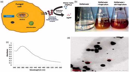
Figure 1. (a) Schematic representation of reduction of metal ions by fungal secretary proteins (b) Nanoparticles formation on the basis of colour change due to surface plasmon resonance (c) UV–visible spectrophotometry of AgNPs and (d) Transmission electron microscopic analysis of AgNPs.
In this study, a phosphate solubilizing fungi was preferred for nanostructure formulation due to their ability to produce large amount of secretary proteins [Citation16]. Numbers of reports have showed the biological synthesis of metal nanoparticles but the potential of a phosphate solubilizing fungi, Alternaria brassicae has not yet been demonstrated. The present investigation is an effort to synthesize AgNPs from A. brassicae (Accession No. KF934409). Moreover, antimicrobials of mycological origin have enormous therapeutic potential; therefore, it is the need of time to develop new antimicrobial drugs for the treatment of infectious diseases.
To the best of our knowledge it is the first ever report on synthesis of silver nanoparticles in the presence of A. brassicae followed by evaluation of its biological potential. The basis for selecting this fungus was that few reports have mentioned the antimicrobial as well as cancer chemo-preventive potency of its secretory proteins apart from exhibiting phosphate solubilizing activity [Citation12,Citation13,Citation17–19]. Keeping this in mind the green synthesis, characterization, antimicrobial, cytotoxity and immunomodulatory activity of silver-myconanosomes was studied using A. brassicae isolated from rhizospheric region. The synthesis of AgNPs was examined by UV–visible, DLS and TEM which delivered an important information about the particle size and shape. Antimicrobial efficacy of AgNPs was determined by agar well diffusion assay using pathogenic bacterial and fungal strains The cytotoxic and immunomodulatory potential was assessed on human and mouse macrophages cells by mitochondrial synthetase enzyme inhibition and estimation of ROS generation. The results obtained indicate that the AgNPs hereby synthesized may have a significant advantage over or in addition with conventional antibiotics as well as an immunomodulatory therapeutic agent.
Material and methods
Isolation, screening and characterization of A. brassicae (KF934409)
The fungus was isolated from rhizospheric region of Central Institute of Medicinal And Aromatic Plants (CIMAP), Lucknow, India and screened by its phosphate solubilizing ability on Pikovskaya’s medium by using plate assay method [Citation20]. The fungal colonies were subculture on fresh petriplates containing potato dextrose agar (PDA) media and the plates were incubated in inverted position for 72 h at 28 ± 3 °C and examine for the halo zones around the colony. Further, the biochemical characterization of the fungus were tested on the basis of morphology of colony, conidial microscopic analysis, solubilization index (SI), cellulose hydrolysis test and starch hydrolysis test according to standard protocols [Citation21].
The fungus was molecularly characterized by using 18 S rRNA sequencing where genomic DNA was isolated and subjected to high-fidelity PCR using universal primers, i.e. forward primer (5′-GGAAGTAAAAGTCGTAACAAGG-3′) and reverse primer (5′-TCCTCCGCTTATTGATATGC-3′) and analysed on 1% agarose gel. The PCR products were sequenced bi-directionally using the forward and reverse primers. Homology between this 18 S rDNA sequence and the strains available at the public databases (Genbank, EMBL and DDBJ) was determined using BLASTN sequence match routines. The unweighted pair group mathematical average (UPGMA) algorithm was used to perform hierarchical cluster analysis [Citation22]. The sequences were aligned using CLUSTALW2 program and its phylogenetic and molecular evolutionary analysis were conducted. Sequences analyses were performed by alignment of the partial 18 S rRNA gene sequences to those from the GenBank database, using the program BLAST (NCBI BLAST® homepage, Bethesda, MD). The nucleotide sequences of 18 S rRNA gene segments determined in this study have been deposited in GenBank database under accession number KF934409.
Myco-production of AgNPs and its characterization
The isolated fungus was grown in MGYP (maltose, glucose, yeast potato broth which comprises of malt extract (0.5%), glucose (1%), yeast extract (0.3%) and peptone (0.5%). The culture was incubated at 27 °C and harvested after 5 d of growth by sieving followed by extensive washing with sterile double-distilled water. Initially 20 g of biomass (wet weight) was transferred to 100 ml deionized water for 48 h at 27 °C in an Erlenmeyer flask and agitated at 150 × rpm for release of secretory proteins. Silver nitrate (1 mM) was added to the Erlenmeyer flasks and the reaction was allowed to progress under dark conditions for production of AgNPs. Time-dependent formation of AgNPs was observed by using ultraviolet-visible spectrophotometer Beckman DU-20 spectrophotometer (Range: 200–900 nm; Wavelength: ±2 nm). The scanning range was 350–650 nm for AgNPs at a scan speed of 420 nm/min. The data was recorded and analysed using “UVWinlab” software (PerkinElmer, Waltham, MA). The obtained AgNPs were freeze dried and stored for further use.
Differential light scattering (DLS)
The suspensions of AgNPs were prepared in distilled water (dH2O) by using a bath-sonicator (ULTRAsonik 57 X, 50/60 Hz, CA) prior to size measurements. Dynamic light scattering size measurements were performed with the aid of a Malvern Zeta Sizer Nano ZS (Malvern Instruments, Worcestershire, UK) operating with version 5.03 of the systems Dispersion Technology Software (DTS Nano, Letchworth, UK). The samples for DLS were equilibrated at 25 °C for 3 min before each measurement. The refractive index (RI) of AgNP.dH2O was 1.330. The aliquots of the samples were prepared and stored at 4 °C. The DLS spectra was determined fortnightly for a period of 120 d.
Transmission electron microscopy (TEM)
The synthesized AgNPs were characterized by TEM analysis. The samples were prepared by placing a drop of synthesized nanoparticles over gold-coated negative grid followed by evaporation of the solvent [Citation23]. TEM analysis was performed on Perkin–Elmer model [Resolution: 0.23 nm; Lattice: 0.14 nm] which was operated at an accelerating voltage of 1000 kV.
Antibacterial efficacy
The antibacterial efficacy of synthesized AgNPs was determined by using the agar well diffusion assay method [Citation24]. Pure cultures of five pathogenic bacteria namely, B. subtilis, B. cereus, S. aureus, E. coli and E. aerogenes were procured from National Chemical Laboratory, Pune. Bacterial stock cultures were maintained at 4 °C on nutrient agar media slants and subculture on nutrient broth media for antibacterial analysis of NPs. The stock solution of 15 wt % silver nanoparticles (AgNPs) was prepared and mixed with distilled water followed by sonication to facilitate dispersion. The working solutions were prepared from this stock solution as per the required concentration. Of 5 mm wells in diameter were prepared and filled with AgNPs with a range of concentrations (10, 50, 100 and 150 µM). Each experiment was performed in triplicate and the average zone of inhibition, excluding well was recorded. Of 1 mM AgNO3 were used as negative controls. The diameter of such zones of inhibition was measured for each organism and expressed in centimetre.
Antifungal efficacy
Disk diffusion method was used to evaluate the antifungal activity of Terconazole against A. niger, Trichoderma sp. and F. semitectum on PDA. The standard terconazole disks (Fu10; 10 μg/disk) were purchased from Hi-Media (Bangalore, India). To determine the combined effect, each standard paper disk was further imbued with 25 μl of freshly prepared AgNPs with a concentrations range of 10, 50, 100 and 200 µM. PDA plates were inoculated with a spore suspension (20 μl) of the test fungi. Standard antifungal terconazole disks were used as positive control. The terconazole disks were impregnated with AgNPs, placed onto the PDA medium and inoculated with tested fungi. The fungal cell filtrate, used for the synthesis of AgNPs was used as negative control. The cultured plates were incubated at 28 ± 4 °C for 7 d. The average inhibition zone, excluding well, for each case was measured.
Cell lines
Human macrophage cell line THP1α and mouse macrophage cell line J774 was procured from the National Centre of Cell Sciences, Pune, India and maintained at Animal Tissue Culture facility of Central Drug Research Institute (CDRI). Cells were cultured in Dulbecco’s modified Eagle’s medium (DMEM) supplemented with 10% Foetal bovine serum (FBS) and 1% antibiotic and antimycotic solution (50,000 units/l of penicillin and 50 mg/l of streptomycin) and 2 mM glutamine. Cultures were held in 75 cm culture flasks, at 37 °C and 5% CO2 using standard cell culture methods.
Cytotoxicity assay
Cell viability was assessed by using the MTT [3-(4,5-dimethylthiazol-2-yl)-2, 5- diphenyltetrazolium bromide] conversion assay [Citation25]. Thus, 1 × 10⁴ cells/ml were seeded on 96-well culture plates and incubated with increasing concentrations of nanoparticles (10, 25, 50, 100, 150 and 200 μM) for 24 h at 37 °C in CO2 incubator. The MTT dye was added to each well and plate was incubated at 37 °C for 4 h. The absorbance of insoluble formazan salts was assessed at 550 nm using Powerwave XS (BIOTEK, Winooski, VT) spectrophotometer [Resolution: 200–999 nm] [Citation26]. Data produced were used to plot a dose-reaction curve and the concentration of these metal nanoparticles required to kill 50% of cell population (IC50) was determined.
Intracellular reactive oxygen species estimation
Intracellular oxidative stress was calculated with the help of 2', 7'-dichlorofluorescin diacetate (DCFH-DA), a well-accepted fluorescent marker for study of intracellular hydroperoxides. The experiment was performed according to the protocol described [Citation27,Citation28] with slight modifications. Primarily, healthy confluent cells were harvested and seeded (1000 cells/well) into black bottomed 96 well plates (Nunc, Roskilde, Denmark) and allowed to adhere for a period of 24 h prior to exposure. For ROS quantification, both mouse and human macrophage cells were plated and distributed in triplicates. A working stock of 25 µM DCFH-DA in phosphate buffered saline (PBS) was prepared and all test concentrations, unexposed negative controls and positive controls were prepared and exposed to the cells in this working stock. The negative control consisted of the working stock solely a 25 µM DCFH-DA solution in PBS, the positive control consisted of 1 µM hydrogen peroxide (H2O2) in 20 µM DCFH-DA/PBS working stock solution and finally the test concentrations consisted of a continuous range of AgNPs. Lipopolysaccharide (1 µg/ml) was used as a mitogen for stimulation of macrophages as well as comparison of phagocytic activity of stimulated and non-stimulated macrophages. The test concentration for AgNPs was 10 µg/ml and the incubation period ranged from 2 to 6 h. The rate of intracellular oxidative stress was monitored by measuring their fluorescence intensity via fluorometre (BIOTEK-FLX800, Winooski, VT) [Wavelength Range: 340–650 nm] emission at 520 nm (by 485 nm excitation).
Statistical analysis
All the experiments were conducted in triplicates and results were expressed as mean ± SD. One-way analysis of variance (ANOVA) with a Dunnett’s test was performed for the multiple comparisons for normally distributed samples with homogenous variance. Statistically significant differences were set at p < .05.
Results
Screening and fungal characterization
The rhizospheric sample was collected, serially diluted and plated on Pikovskaya’s media to check their phosphate solubilization efficiency. After 7 d of incubation, a fungal colony was observed, identified, as darkash colour, looking like cotton having irregular edges and 1.2 cm in diameter. On Pikovskaya’s medium, the transparency of the media is a primary indicator of phosphate solubilization to be visualized as halo zone formation around the colony. Thus among all, the fungal isolate FCK 20 was found as an efficient fungus on the basis of SI, pH and phosphatase enzyme production. This fungus was further characterized morphologically, biochemically and at molecular level. The SI of selected fungus was recorded with size of 2.9 cm in comparison with others strains. Further, colony morphology was analysed on the Sabouraud agar medium and filamentous structural pattern was analysed microscopically after lactophenol staining. Moreover, this fungus gave a negative test for starch hydrolysis and cellulose degrading ability.
For molecular characterization, the genomic DNA was amplified using 18 S rRNA specific primers (ITS 4 and ITS 5). An amplicon of 500 bp was observed on 1% agarose gel. The DNA sequencing of the amplified product and its BLAST analysis confirmed the fungus as A. brassicae (KF934409). The phylogenetic analysis (S-Figure 1) revealed the fungus to belong to Pleosporaceae family and the genus was Alternaria.
Synthesis and size estimation of AgNPs
AgNPs were mycological produced from A. brassicae (KF934409). Of 1 mM of silver nitrate were added to the fungal filtrate under magnetic stirring which was used as reducing and stabilizing agent for 1 mM of silver nitrate. Appearance of a brownish colour in solution () reflected the formation of AgNPs in the reaction mixture. The control sets did not demonstrate any colour change under similar experimental conditions.
Ultraviolet–visible spectrophotometric analysis
UV–visible spectrophotometer showed no evidence of absorption in the range of 380–750 nm for the fungal extract. Whereas, the AgNO3 exposed fungal extract showed a distinct absorption at around 350 and 650 nm, with a peak at 370 nm for AgNPs ().
Dynamic light analysis (DLS)
This technique enables the particle size determination by measuring the random changes in the intensity of light scattered from a suspension or solution. The DLS spectra showed that average diameter of AgNPs was 92.7 nm (S-Figure 2) with polydispersity index (PDI) 0.281. No difference was observed in the size as well as PDI after the storage of 60 or 120 d of storage. The zeta potential of the biosynthesized AgNPs was found as a sharp peak at −0.30 mV (S-Figure 3). It is reported that the surface of the silver nanoparticles is negatively charged and dispersed in the medium which confirms the repulsion among the nanoparticles and thus proves that they are very stable.
Transmission electron microscopic (TEM) analyses
The morphology of AgNPs is spherical and the TEM micrographs suggest that particle diameters ranged from 55.4 to 70.23 nm (). The dimensions of AgNPs were small enough and imaged as poly dispersed small and large spherical nanoparticles with variable diameter. This was further confirmed by the representative images recorded from the uniformly dispersed drop-coated film of the AgNPs on grid.
Antibacterial activity of AgNPs
The bactericidal activity of AgNPs were studied using the pathogenic strains of bacteria as S. aureus, E. coli, B. subtilis, B. cereus and E. aerogenes using agar well diffusion method. After the incubation time, zone of inhibition (clear zones) were observed against all the tested microbes. The results recorded in centimetres for AgNPs are shown in respectively.
Figure 2. Comparative analysis of (a) antibacterial activity and (b) antifungal activity of AgNP for different pathogenic bacteria and fungi. Results are presented in relative units compared with controls (Ab) which is not shown in the graph. Different signs (* and **) letters indicate significant differences (p < .05).
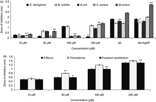
The effectiveness of AgNPs could be attributed to the fact that their larger surface area enabled them to have a better contact with the microbial cell wall. This is further supported by the observation that size dependent interaction of AgNPs with bacteria leads to its antibacterial activity [Citation29]. The exact mechanism by which the reaction occurs is still largely unknown. However, some literature proposes that Ag interact with the thiol groups of proteins, which is necessary for microbial respiration ability [Citation30]. Ag might also interact with phosphorus containing compounds like DNA disturbing the replication process or preferably by their attack on the respiratory chain. The direct interaction of Ag with cell membrane of bacteria has also been shown which consequently breaks the membrane. Earlier experimental evidences have also advocated the failure of replication ability by the DNA after treatment with silver ions [Citation31]. The comparative histogram demonstrated that the best antibacterial efficacy of AgNPs was against B. cereus followed by S. aureus. The marked increase in antibacterial activity was demonstrated with increasing concentration of AgNPs. In addition, the efficacy of AgNPs was also found to be enhanced in combination with the antibiotic rather than alone. Thus nano-silver is an effective and a fast-acting microbicide against a broad spectrum of pathogenic bacteria [Citation32].
Antifungal activity of AgNPs
The colloidal AgNPs inhibited the growth of the fungus (A. niger, Trichoderma sp. and F. semitectum) which was seeded in the Muller Hinton agar plate and formed a zone of inhibition around the central cavity. The zone of inhibition with diameter of 1 cm was recorded in case of A. niger, 1.2 cm in Trichoderma sp. and 1.1 cm in case of F. semitectum (). The antifungal activity is due to the formation of insoluble compounds by inactivation of sulfhydryl groups in the fungal cell wall and disruption of membrane bound enzymes and lipids, which causes cell lysis [Citation33]. We have observed that Trichoderma sp. and F. semitectum are susceptible to the lethal effects of the prepared silver due to the smaller size of AgNPs.
Cell lines
To determine the biomaterial capability of nanoparticles with no toxic effects, the cell viability and cytotoxicity assays were performed. Cell viability was determined by a standard MTT conversion assay.
In vitro cytotoxicity assay
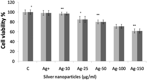
Treatment of AgNPs to J774 and THP1α cell lines exhibited mild cytotoxicity in a dose dependant manner. At low doses (10 µg) no cytotoxic effects were observed whereas at high doses of 100–150 µg of AgNPs mild cytotoxicity was observed. Long-time exposure resulted in additional toxicity to the cells and reached to maximum dead cells after 24 h of incubation, which might be due to over-accumulation of metal nanoparticles within the cell ().
Figure 3. Dose-dependent effect of AgNPs over cell viability using MTT assays on J774 and THP1α cells. Results are presented in relative units compared with controls. Different signs (* and **) letters indicate significant differences (p < .05).
Intracellular reactive oxygen species estimation
The ROS estimation has shown that AgNP exposure to J774 and THP1α cell caused time dependant increased production of ROS at respective concentration indicating macrophage stimulation. The maximum amount of ROS was produced up till 4 h of incubation, which declined at 6 h (). Here, the comparable ROS generation by AgNPs, from non-stimulated cells to mitogen-stimulated cells was observed which indicates the immunomodulatory potential of the synthesized AgNPs. This may be due to the small size of AgNPs as these could easily penetrate through the cell wall in an appreciable number [Citation34].
Figure 4. ROS estimation in (a) J774 cell lines and (b) THP1α cell lines after incubation with AgNPs at various time points (2, 4 and 6 h). Results are presented in relative units compared with controls. Different signs (* and **) letters indicate significant differences (p < .05).
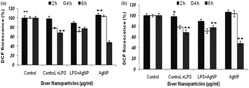
The maximum free radical activity was obtained by the J774 cells () and THP1α cells () after 4 h of incubation with AgNPs. Free radical generation and analogous encapsulation efficacy capability of AgNPs revealed the potential role of AgNPs as drug/vaccine delivery vehicle for macrophages as well as also indicate towards their immunomodulatory activity.
Discussion
Myconanotechnology has become an extensive field of study involving chemistry, physics, engineering, computing, electronics, energy, agriculture and biomedicine. In the realm of biomedicine, nanotechnology is widely touted as one of the next promising and important approaches to diagnose and treat various ailments [Citation4,Citation35,Citation36]. Since physical and chemical methods of metal nanoparticle production are expensive and involve incorporation of toxic chemicals therefore their biological synthesis (bacterial, fungal and plant extract) would be preferred [Citation37–39]. This owes to their ease of availability, nontoxic nature and quicker synthesis, which prompted us to use this phosphate solubilizing fungus A. brassicae for the synthesis of AgNPs. In best of our knowledge this is the pioneer study showed the use of fungus for the production of nanoparticles.
In this study, a phosphate solubilizing fungal isolate, FCK 20 was isolated and subjected to its characterization. The sequence of 18 S rRNA of the fungal strain FCK 20 was submitted to GenBank with an accession number of (KF934409). The homology search using BLAST indicated a close genetic relatedness of the strain FCK 20 with the rRNA sequence of A. brassicae (18 S: 100% similarity with the reference sequence BankIt1680606 Seq5) in NCBI database. Such a higher identical value confirmed the strain FCK 20 to be A. brassicae. The confirmation of synthesis of AgNPs was based on surface plasmon resonance involving colour alteration. Further, in the UV–visible absorption, a strong peak located at 370 nm was observed. The peak obtained at 370 nm confirmed the formation of silver nanoparticles after reduction from the fungal enzymes. The earlier reports on AgNPs have also reported the maximum absorbance of silver nanoparticles at 360–375 nm [Citation40,Citation41]. Average particle size calculated from DLS data was found to be 92.7 nm for AgNPs whereas the zeta potential of the biosynthesized AgNPs was found as a sharp peak at −0.30 mV. Furthermore, the TEM image of the synthesized AgNPs has validated the formation of spherical, nanoparticles, which was 50 nm in diameter. This indicated the reduction of Ag+ to elemental silver (Ag). Additionally, the synthesized AgNPs were stable in solution over a time of three months at room temperature.
Nano-silver is an effective and a fast-acting microbicide against a broad spectrum of pathogenic bacteria and fungi thus can be utilized in various processes in the medical field. Our findings are in agreement to the previous reports and synthesized AgNPs have showed the considerable antibacterial activities along with the standard antibiotic (tetracycline) against B. cereus. The antibacterial activity of AgNPs is due to the permeability of the cell membrane [Citation42] or formation of free radicals [Citation43] or interaction of AgNPs with the thiol groups of many enzymes thus inactivating them. Beside this, the moderate antifungal activity of AgNPs against Trichoderma sp. and F. semitectum was due to formation of insoluble compounds by inactivation of sulfhydryl groups in the fungal cell wall and disruption of membrane bound enzymes and lipids, causing cell lysis [Citation44,Citation45].
The MTT assay determines the mitochondrial activity of the cells which reduce the soluble, yellow MTT into an insoluble, purple formazan. The reduction of MTT to formazan indicates the decrease in mitochondrial metabolism of the cells. Therefore, the absorption of formazan formed, directly co relates to the number of cells whose mitochondrial metabolism is intact even after the exposure of AgNPs. Dose response of AgNPs in J774 cells and THP1α cells showed the decrease in the reduction of MTT to formazan with the increasing concentration and time of exposure. However, AgNPs were found to be significantly more toxic to THP1α cells as compared to J774 cells, which could be attributed to the intrinsic anticancer property of AgNPs. This might be due to small particle size of AgNPs with enormous specific surface area, which facilitated further expression and dissolution of ions, leading to increased toxicity. AgNPs are highly reactive, exhibit oxidative potential, contains the ability to bind with biomolecules like proteins and DNA and consequently caused the disturbance in the functioning of biomolecules [Citation34,Citation46]. In both cell lines J774 and THP1α, significant increase in the levels of ROS was observed in comparison to the unexposed controls. The maximum amount of ROS was produced after 4 h of incubation with AgNPs, which declined at 6 h. ROS, plays an important role in triggering cellular pathways that can lead to cellular death by either causing nuclear damage or by contributing in cell membrane rupture mechanisms. Exposure of AgNPs to cells caused increased production of ROS, suggested their potential use as an anti-microbicide, occupying its application in agriculture. Moreover, these nanoparticles can also act as immunomodulatory agent alone or in combination with established therapeutic immunomodulatory agents. As these NPs are simply engulfed by the macrophages, they also pose themselves as targeted drug/vaccine delivery vehicle to macrophages thereby a boom for development of a potent chemotherapeutic vehicle for diseases involving macrophages viz., leishmaniasis, tuberculosis, etc. Hence, care has to be taken to utilize this marvel well and in a good, efficient and effective ways. Researchers should also understand its limitations and taking excessive care for its effect on environment and human health.
In future, these bio-synthesized nanomaterials (encapsulation) may lead to enhancement of agricultural productivity like slow release of phosphorus from fertilizers and its effective uptake by plants. Fungus A. brassicae contains phosphatase enzyme whose main function is to solubilize the insoluble form of phosphorus in to soluble one. Thus, the bio-nanoparticles prepared from this fungus may display slow release of encapsulated enzyme and hence may improve phosphate solubilization. These synthesized myco-nanoparticles may be mixed with fertilizers to increase the uptake of phosphorus nutrient. The AgNPs thereby synthesized by this route using A. brassicae might be developed as a novel antimicrobial immunostimulatory therapeutic agent for human ailments as it contains some important secondary metabolites which are present as a protein coating on AgNPs. However, in future, this strategy needs to be validated.
Conclusion
This study showed the mycogenic production of AgNPs with particular emphasis on its potential applications in human welfare as well as in other allied sectors. Findings showed that synthesized AgNPs were spherical in shape and have a promising antimicrobial agent against both gram-positive/gram-negative bacteria and pathogenic fungi also. However, higher doses of AgNPs exhibited the cytotoxicity and ROS production in J774 and THP1α cells in time dependant manner. Observations have showed that these efficacies of synthesized nanoparticles can be implemented as novel antimicrobial immunostimulatory therapeutic agent. However, further studies are required to fully characterize the mechanistic and toxicity aspects of synthesized nanoparticles.
Supplementary_figures.doc
Download MS Word (210.5 KB)Acknowledgements
The authors are highly thankful to Vice Chancellor, Integral University for his support and encouragement. We sincerely thank Mr. Sharma ITRC, Lucknow for carrying out TEM analysis of nanoparticles.
Disclosure statement
No potential conflict of interest was reported by the authors.
Related Research Data
References
- Chowdappa P, Gowda SK. Review article on nanotechnology in crop protection: status and scope. Pest Manag Hort Ecosyst. 2013;19:131–151.
- Chahardoli A. Green approach for synthesis of gold nanoparticles from Nigella arvensis leaf extract and evaluation of their antibacterial, antioxidant, cytotoxicity and catalytic activities. Artif Cells Nanomed Biotechnol. 2017;45. Forthcoming. [cited 2017 May 25]. doi: 10.1080/21691401.2017.1332634
- Alexander JW. History of the medical use of silver. Surg Infect (Larchmt). 2009;10:289–292.
- Popescu M, Velea A, Lorinczi A. Biogenic production of nanoparticles. Dig J Nanomater Bios. 2010;5:1035–1040.
- Thakkar KN, Mhatre SS, Parikh Y, et al. Biological synthesis of metallic nanoparticles. Nanomed Nanotech Biol Med. 2009;6:257–262.
- Reddy GAK, Joy JM, Mitra T, et al. Nano silver – a review. Int J Adv Pharm. 2012;2:9–15.
- Singh T, Jyoti K, Patnaik A, et al. Biosynthesis, characterization and antibacterial activity of silver nanoparticles using an endophytic fungal supernatant of Raphanus sativus. J Genet Eng Biotechnol. 2017;15:31–39.
- Jo JH, Singh P, Kim YJ, Wang C, et al. Pseudomonas deceptionensis DC5-mediated synthesis of extracellular silver nanoparticles. Artif Cells Nanomed Biotechnol. 2016;44:1576–1581.
- Rahim KA, Mahmoud SY, Ali AM, et al. Extracellular biosynthesis of silver nanoparticles using Rhizopus stolonifer. Saudi J Biol Sci. 2017;4:208–216.
- Korbekandi H, Mohseni S, Jouneghani RM, et al. Biosynthesis of silver nanoparticles using Saccharomyces cerevisiae. Artif Cells Nanomed Biotechnol. 2016;44:235–239.
- Arokiyaraj S, Savariar V, Muthupandian S, et al. Green synthesis of silver nanoparticles using Rheum palmatum root extract and their antibacterial activity against Staphylococcus aureus and Pseudomonas aeruginosa. Artif Cells Nanomed Biotechnol. 2017;45:372–379.
- Al Shmgani HSA, Mohammed WH, Sulaiman GM, et al. Biosynthesis of silver NPs from Catharanthus roseus leaf extract and assessing their antioxidant, antimicrobial, and wound-healing activities. Artif Cells Nanomed Biotechnol. 2017;45:1234–1240.
- Sharma S, Sayyed RZ, Trivedi MH, et al. Phosphate solubilizing microbes: sustainable approach for managing phosphorus deficiency in agricultural soils. Springerplus. 2013;2:587.
- Calle LC, López MLE. Green synthesis of silver nanoparticles using green coffee bean extract. IFMBE Proceedings. 2017;60:217–218.
- Ge L, Li Q, Wang M, et al. Nanosilver particles in medical applications: synthesis, performance, and toxicity. Int J Nanomedicine. 2014;9:2399–2407.
- Mohanpuria P, Rana NK, Yadav SK. Biosynthesis of nanoparticles: technological concepts and future applications. J Nanopart Res. 2008;10:507–517.
- Horiuchi M, Tokuda H, Ohnishi K, et al. Porritoxins, metabolites of Alternaria porri, as anti-tumor-promoting active compounds. Nat Prod Res. 2006;20:161–166.
- Monneret C. Histone deacetylase inhibitors. Eur J Med Chem. 2005;40:1–13.
- Lakshmi AI, Madhusudhan T, Kumar PD, et al. Histone deacetylase inhibitors in cancer therapy: an update. Int J Pharm Sci Rev Res. 2011;10:38–44.
- Pikovskaya I. Mobilization of phosphate in soil in connection with their vital activities of some microbial species. Microbiology. 1948;17:362–370.
- Grant WD, Kamekura M, McGenity TJ, et al. Order I halobacteriales grant and larsen 1989b. In: Boone DR, Calstenholz RW, Garrity GM, editors. Vol. 2. Bergey’s manual of systematic bacteriology. Baltimore (MD): Williams & Wilkins; 2001. p. 294–334.
- Dias LAS. Fundamentos e aplicações em plantas e microrganismos, análises multidimensionais. In: Alfenas AC, editor. Eletroforese de isoenzimas e proteínas afin. Viçosa: UFV; 1998. p. 405–473.
- Germain V, Li JD, Ingert Z, et al. Stacking faults in formation of silver nanodisks. J Phys Chem B. 2003;107:8717–8720.
- Perez C, Pauli M, Bazerque P. An antibiotic assay by agar-well diffusion method. Acta Biologiae Et Medecine Experimentaalis. 1990;15:113–115.
- Mosmann F. Rapid calorimetric assay for cellular growth and survival: application to proliferation and cytotoxicity assay. J Immunol. 1983;65:55–63.
- Lam C, James J, Mccluskey R. Pulmonary toxicity of single-wall carbon nanotubes in mice 7 and 90 days after intra-tracheal instillation. Toxicol Sci. 2004;77:126–134.
- Cathcart R, Schwiers E, Ames BN. Detection of picomole levels of hydroperoxides using a fluorescent dichlorofluorescin assay. Anal Biochem. 1983;134:111–116.
- Goswami P, Gupta S, Biswas J, et al. Endoplasmic reticulum stress plays a key role in rotenone-induced apoptotic death of neurons. Mol Neurobiol. 2014;53:285–298.
- Singh T, Jyoti K, Patnaik A, et al. Biosynthesis, characterization and antibacterial activity of silver nanoparticles using an endophytic fungal supernatant of Raphanus sativus. J Genet Eng Biotechnol. 2017;15:31–39.
- Arokiyaraj S, Choi SH, Lee Y, et al. Characterization of ambrette seed oil and its mode of action in bacteria. Molecules. 2014;20:384–395.
- Liau S, Pugh DW, Russell F. Interaction of silver nitrate with readily identifiable groups: relationship to the antibacterial action of silver ions. Lett Appl Microbiol. 1997;25:279–283.
- Fatima F, Bajpai P, Pathak N, et al. Antimicrobial and immunomodulatory efficacy of extracellularly synthesized silver and gold nanoparticles by a novel phosphate solubilizing fungus Bipolaris tetramera. BMC Microbiol. 2015;15:1–10.
- Dorau B, Arango R, Green F, editors. Proceedings of the 2nd Wood-Frame Housing Durabili and Disaster Issues Conference; Las Vegas (NV): Forest Products Society; 2004. p. 133.
- Reidy B, Haase A, Luch A, et al. Mechanisms of silver nanoparticles release, transformation and toxicity: a critical review of current knowledge and recommendations for future studies and applications. Materials. 2013;6:2295–2350.
- Bertrand N, Wu J, Xu X, et al. Cancer nanotechnology: the impact of passive and active targeting in the era of modern cancer biology. Adv Drug Deliv Rev. 2014;66:2–25.
- Asadian-Birjand M, Biglione C, Bergueiro J, et al. Transferrin decorated thermoresponsive nanogels as magnetic trap devices for circulating tumor cells. Macromol Rapid Commun. 2016;37:439–445.
- Kalpana D, Lee YS. Synthesis and characterization of bactericidal silver nanoparticles using cultural filtrate of simulated microgravity grown Klebsiella pneumoniae. Enzyme Microb Technol. 2013;52:151–156.
- Ottoni CA, Simões MF, Fernandes S, et al. Screening of filamentous fungi for antimicrobial silver nanoparticles synthesis. AMB Expr. 2017;7:1–10.
- Johnson I, Prabu HJ. Green synthesis and characterization of silver nanoparticles by leaf extracts of Cycas circinalis, Ficus amplissima, Commelina benghalensis and Lippia nodiflora. Int Nano Lett. 2015;5:43–51.
- Sastry M, Patil V, Sainkar SR. Electrostatically controlled diffusion of carboxylic acid derivatized silver colloidal particles in thermally evaporated fatty amine films. J Phys Chem B. 1988;102:1404–1410.
- Henglein A. Physicochemical properties of small metal particles in solution: microelectrode reactions, chemisorption, composite metal particles, and the atom-to-metal transition. J Phys Chem. 1993;97:5457–5471.
- Gavrieli Y, Sherman Y, Bensasson S. Identification of programmed cell death in situ via specific labeling of nuclear DNA fragmentation. J Cell Biol. 1992;119:493–501.
- Hussain S, Hess K, Gearhart J, et al. In vitro toxicity of nanoparticles in BRL 3A rat liver cells. Toxicol in Vitro. 2005;19:975–983.
- Khatoon N, Mishra A, Alam H, et al. Biosynthesis, Characterization and antifungal activity of the silver nanoparticles against pathogenic Candida species. Bio Nano Sci. 2015;5:65–74.
- Elgorban AM, El-Samawaty AEM, Yassin MA, et al. Antifungal silver nanoparticles: synthesis, characterization and biological evaluation. Biotechnol Biotechnol Equip. 2016;30:56–62.
- Li Y, Bhalli JA, Ding W, et al. Cytotoxicity and genotoxicity assessment of silver nanoparticles in mouse. Nanotoxicology. 2014;8:36–45.