
Abstract
Nanoflowers are a newly developed class of nanoparticles showing structure similar to flower and gaining much attention due to their simple method of preparation, high stability and enhance efficiency. This article focuses on advantages, disadvantages, method of synthesis, types and applications of nanoflowers with futuristic approaches. The applications of nanoflower include its use as a biosensor for quick and precise detection of conditions like diabetes, Parkinsonism, Alzheimer, food infection, etc. Nanoflowers have been revealed for site-specific action and controlled delivery of drugs. The extended applications of nanoflowers cover purification of enzyme, removal of dye and heavy metal from water, gas-sensing using nickel oxide. Recent investigation shows 3 D structure of nanoflowers for enhancing surface sensitivity using Raman spectroscopy. This nanoflower system will act as a smart material in the near future due to high surface–to-volume ratio and enhance adsorption efficiency on its petals.
Introduction
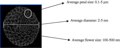
Among different nanoparticulate systems, nanoflower, a newly developed class of tiny particles showing structural similarity to plant flowers in a nanoscale range of 100–500 nm [Citation1–4]. Research on nanoflower is of interest nowadays because of its simple method of synthesis from organic, inorganic materials and sometimes a combination of both organic and inorganic materials to improve stability and efficiency of surface reaction. Nanoflowers are composed of several layers of petals to encompass a larger surface area in a small structure for multiple applications in catalysis, biosensors and delivery of drugs ().
Figure 1. Structure of nanoflower.
Advantages
Nanoflower shows high surface to volume ratio to enhance surface adsorption for accelerating the kinetics of reactions. The zinc oxide nanoflowers revealed a higher number of adsorption sites, which plays a major role in determination of surface-enhanced Raman spectroscopy (SERS).
Nanoflowers demonstrated better charge transfer and carrier immobility due to large surface area. The silver-coated zinc oxide nanoflowers possessed better charge transfer and carrier immobility for SERS sensitivity.
The efficiency of surface reaction is increased in the 3D structure of nanoflowers. The 3D structure of zinc oxide nanoflowers enhanced number of adsorption sites due to which their efficiency was increased for SERS sensing [Citation1].
Synthetic methods like ionotropic gelation, precipitation method and green synthesis for the preparation of nanoflowers are simple, non-toxic and cost-effective [Citation2].
Due to instability of proteins and enzymes, their immobilization on the surface of the metal enhances the stability. The enzyme-like glucose oxidase immobilized onto the surface of concanavalin A using calcium as a metal ion to increase its stability [Citation3].
Disadvantages
Structural features of nanoflower like its petals and dimensions are very difficult to control during and completion of reaction [Citation4].
In synthetic reactions, nanoflowers are prepared in extreme conditions like 80–550 °C where toxic elements or byproducts might be formed. Hydrothermal method for nanoflowers preparation required high temperature in comparison to other methods like ionotropic gelation method, precipitation method, green synthesis method, etc [Citation5].
Due to immobilization of protein and peptide, their therapeutic activities are decreased in comparison to free forms. The immobilization of glucose oxidase may reduce its therapeutic activity for determination of E. coli as food pathogen.
Comparative aspects of microflower and nanoflower
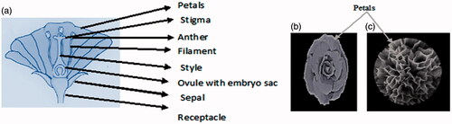
Flowers are usually floral part of plant, which mainly facilitate reproduction. The flower is classified into the following parts:
Petals: Colored part of the flower, also called modified leaf and when in bunch called corolla.
Stigma: Part of a flower where pollen grains germinate.
Anther: Part of the flower where pollens are produced.
Filament: Thread-like portion of the flower that supports the anther.
Style: Part which joins stigma and ovary.
Ovule: Reproductive part of a flower.
Sepal: Outermost leaf-like part of a flower.
Receptacle: Part of the stalk where whole flower is attached.
Among these different parts of plant, flower, petal-like structures are observed microscopically in microflowers and nanoflowers. The size of the flowers is basically arranged in the order of plant flower > microflowers > nanoflowers ()
Figure 2. Comparison of (a) flower; (b) microflower; and (c) nanoflower.
Microflowers are micro-dimensional particulate system,which resemble to the structure of the plant flower. The synthetic birnessite delta-magnesium dioxide microflower prepared by facile one-pot method was used for oxidative degradation of rhodamine-B [Citation6], whereas hydrothermally synthesized vanadium peroxide microflower was used for gas sensing [Citation7].
On the basis of characterization parameters like size, diameter, petal size, etc., the micro- and nano-flowers are compared in .
Table 1. Comparative aspects of micro- and nano-flowers.
History
Since the inceptive investigations of new nanomaterials in the mid-2000 s, different nanomaterials have been created with topographical morphologies like core-shell structure and Janus molecule [Citation5]. In recent years, researchers developed nanoplates and nanoparticles of lead hexaferrite using maltose and metal nitrate as a reductant by simple, quick and efficient sol–gel auto-combustion method. It was found that the size and morphology of nanoplates were dependent on concentration of maltose and calcination temperature. These nanoplates were used as a material for storing data in electronic devices [Citation8]. Nanocomposites of copper ferrite and iron (II) oxide were prepared using chitosan and onion as a reductant to control the surface morphology of the nanoparticle. Surface coating with chitosan prevented direct exposure of magnetic nanoparticles in the body, efficiently resisted adsorption of non-specific protein and circulated for a longer time. Due to absence of organic solvents and non-toxic reactants in reaction, this method was found to be easy, cost-effective and eco-friendly [Citation9]. Nanocomposites of M-type hexaferrite/lead titanate were prepared using sol-gel auto-combustion technique. To maintain the stability of product, better morphology of nanoparticle was obtained by adjusting calcination temperature and the method proved to be quick and efficient [Citation10]. Further in 2016, researchers synthesized copper hexaferrite nanoparticles using maltose and surfactant to control size and morphology of the nanoparticles. In the absence of surfactant, bean-shaped nanoparticles were formed and thus the property of nanostructure depends upon the morphology and particle size. This method proved to be simple and eco-friendly [Citation11]. In 2015, lead hexaferrite was synthesized by decomposition technique using different carboxylic acids like oxalic acid, malonic acid, succinic acid and maleic acid, which acted as a fuel and reducing and capping agents. It was found that the maleic acid was an efficient capping agent and fuel as compared to other carboxylic acids and maintains chemical stability of lead hexaferrite. Morphology of product was controlled by varying calcination temperature like 50 °C, 100 °C and 900 °C [Citation12]. Later in 2016, an economic, novel, rapid, green synthesis method was developed to synthesize lead hexaferrite nanoparticles using cherry juice and sol-gel auto combustion route, without the use of surfactant. Cherry juice acted as a green capping agent and prevented aggregation of particles and also helped to maintain the chemical stability of lead hexaferrite. The size of formed nanoparticles increased with the decreasing cherry juice content [Citation13]. In 2016, nickel ferrite nanomaterial was prepared using sol-gel technique and onion acted as a reducing agent and green capping agent. Nickel ferrite nanoparticles can be used as semiconductors, microwave systems and sensors. Onion maintained the chemical stability and also prevented the particle aggregation. It was observed that the amount of onion used also affected the morphology and purity of the product [Citation14]. Among these new nanostructures, the geographical elements of nanoflowers have increased the interest of researchers on the basis of higher surface-to-volume proportion in comparison with round nanoparticles to improve the efficiency of nanomaterials surface. Accordingly, sophisticated instrumental examinations like scanning electron microscopy (SEM), transmission electron microscopy (TEM) and X-ray diffraction spectroscopy (XRD) () are required for the characterization of the nanoflowers. In spite of the expanding interest for nanoflowers, preparation methods require extreme conditions of toxic natural solvents, high temperature and pressure. Consequently, it is difficult to control their morphological components in the structures of nanoflowers in the first approach. Chemicals are organic species with brilliant action and substrate specificity, yet their utilization is constrained by specific disadvantages like high affectability to the encompassing condition, low reproducibility of trial results and the prerequisite for complex purging procedures. However, another approach is easy to use and safe method for cross-breed nanomaterials to overcome the constraints of traditional techniques. Since it is remarkable to create a nanomaterial by adding protein to metal ions, this technique need not require any toxic components. The natural substance required in the process is subjected to less control as compared to other regular techniques to maintain the movement of the immobilized compound.
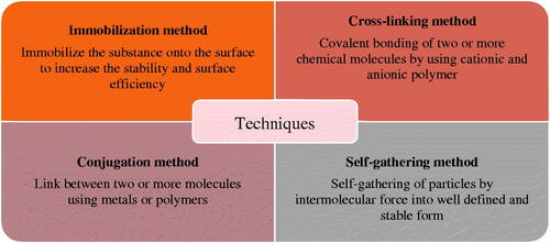
Current strategies for the arrangement of nanobiomaterials in the form of nanoflowers can be grouped into four classes as mentioned in .
Figure 3. Techniques used for arrangement of nanoflowers.
Types of nanoflowers
Nanoflowers are classified on the basis of their size and structure with metal ions [Citation15–26]. The synthesis of nanoflowers using different atoms like copper, calcium, magnesium, protein, DNA and their applications are mentioned in .
Table 2. Types of nanoflower based on metal atoms.
Size of nanoflower and its petals
Microscopic size of different formulations of nanoflower are discussed in .
Table 3. Size of nanoflower and its petal.
Methods for synthesis of nanoflower and their functions
Various nanoflowers like dual-functional nanoflower, zinc oxide nanoflower, concanavalin A nanoflower, etc. are discussed in with respect to their different synthetic methods and functions.
Table 4. Methods for synthesis of nanoflower and their function.
Characterization parameters for nanoflower
Different characterization parameters for nanoflowers are mentioned in .
Table 5. Parameters for characterization of nanoflowers.
Applications of nanoflower
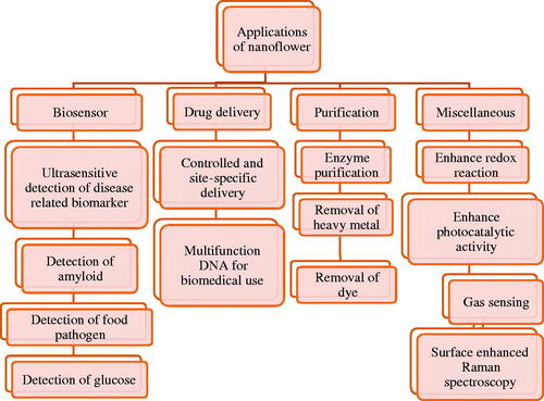
Applications of nanoflower in different fields are enlisted in .
Figure 4. Applications of nanoflowers.
Biosensors
Biosensors are biologically derived analytical device, which are combined with traducers for recognition unit and signal converting system to detect various diseases and disorders [Citation40–44]. Nanoflowers in the form of biosensor showed the following applications:
Dual functional nanoflowers for ultrasensitive detection of disease biomarker
Colorimetric sensors are widely used for the detection of biomarkers related to conditions like diabetes, Parkinson’s disorder, etc. Protein biomarkers were used at low concentration due to which it showed difficulty in estimation. Henceforth, various attempts are made to increase the sensitivity of colorimetric sensor by using different amplification signals. Enzyme-based amplification signals are mostly differentiated for bioassay using ELISA (enzyme-linked immunosorbent assay). Dual function nanoflowers were employed to construct a colorimetric sensor for ultrasensitive detection of disease using biomarker. In this method, enzymes were used as protein due to which activity, stability and durability of protein was improved [Citation27]. Dual function nanoflowers were prepared by green synthesis method where streptavidin (SA), a protein, is used as a biological recognition unit and horseradish peroxidase(HRP), an enzyme used as a signal amplification unit and these are combined together using copper ions in phosphate buffer solution at room temperature and SA-HRP-Cu3(PO4)2 nanoflower were obtained. These nanoflowers like structures are formed mainly for obtaining a link between protein, i.e. streptavidin and copper ion. This method was performed without toxic elements and extreme conditions. SA has an affinity for biotin and HRP acts as a catalyst, which catalyzes the oxidation of 3, 3′, 5, 5′-tetramethylbenzidine (TMB) to a blue-colored substance in the presence of hydrogen peroxidase. Thus, introduction of SA and HRP showed dual functional nanoflowers to build easy and highly sensitive technique for colorimetric sensing for assay of alpha-fetoprotein (AFP). These nanoflowers also enhances enzymatic activity and stability for producing a signal for amplification.
Zinc oxide nanoflower for amyloid detection
Amyloids are protein aggregates that are folded into shape and stick together to form fibrils. Amyloids are found in conditions like amyloidosis and neurodegenerative disorder like Parkinsonism, Alzheimer, etc. Fluorescent dyes were commonly used for detection of amyloid but the major disadvantage was photo stability and low intensity. Detection of amyloid at very low concentration is a difficult and hence a sensitive technique is required for detection of amyloid. Thioflavin T is a benzothaiazole salt (dye). Thioflavin T is widely used to visualize these folded proteins, i.e. amyloids, which bind to beta sheet showed enhanced fluorescence and red shift. Thioflavin T when bound to insulin amyloid beta sheet showed poor fluorescence, this problem can be overcome by preparing biosensor of zinc oxide nanoflowers and thioflavin T dye adsorbed onto its surface and grown over nano silver film, which was coated on the glass surface. It showed enhanced fluorescence during the detection of insulin amyloid [Citation28].
Concanavalin A nanoflower for detection of E.coli in food
Contaminated food is the most common source of E.coli infection. E.coli is very harmful microorganism as it infects the red blood cells, causes irritation and vomiting. Hence to detect this harmful microorganism, biosensors are prepared. Concanavalin A (con A) is also called lectin, which is carbohydrate-binding protein. Glucose oxidase is an enzyme, which is immobilized on to the con A with the help of metal, i.e. calcium ions. This complex of Concanavalin A, glucose oxidase and calcium ion were used as a biosensor to detect the E.coli in food [Citation3].
Microfluidic paper-based analytic devices (µPADs) biosensor for detection of glucose
Biosensor was fabricated by Zhu (2017) for sensitive and visual detection of glucose. A novel microfluidic paper-based analytic devices (µPADs) was fabricated with a hybrid nanocomplex composed of dual enzymes of glucose oxidase (GOx) and horseradish peroxidase (HRP) with copper (II) phosphate inorganic nanocrystals incorporated in the detection zones using wax printing technique. The formed complex was found to resemble the structure of a flower, which allowed co-immobilization of GOx and HRP in a biocompatible environment. The prepared nanoflowers facilitated the transport between enzyme and its substrate and also preserved the activity and stability of enzymes. Biosensor enabled rapid and sensitive detection of glucose in 0.1–10 mM concentration range with limit of detection (LOD) of 25 μM [Citation45].
Drug delivery
Cationic cyclodextrin, chitosan and alginate-based nanoflowers for site-specific drug delivery system
Cyclodextrins are used widely as carriers for poorly water soluble drugs of BCS class II and IV [Citation46–48]. 5-Flurouracil is an anticancer drug, which is poorly water soluble and used to treat colon cancer since many years. Main disadvantage of 5-flurouracil is it has a short half-life and also has several toxic side effects. Hence to deliver this drug orally, it requires certain properties such as increased biocompatibility, controlled release pattern of drug, reduction of toxicity of the drug and increase therapeutic efficiency. To achieve oral drug delivery, cationic β-cyclodextrin is used as a carrier due to its unique structure and also non-hydrolysable nature in the stomach [Citation31]. When cationic β-cyclodextrin conjugates with 5-flurouracil, it delivered drug specifically in the colon. Lyophilization method was used to prepare inclusion complex of cation-beta-cyclodextrin and 5-flurouracil. Petals of nanoflower prepared from sodium alginate and chitosan were used to deliver the drug in sustained and controlled manners.
Multifunctional and programmable DNA for biomedical application
DNA is biocompatible exogenous material, which is naturally soluble in water. DNA utilized as a building block to develop different DNA nanoparticles, which have inherent functionalities for biomedical applications. Grouping programmability and robotized controllable combination additionally make DNA nanostructures more reasonable biomedical nanotools. Combination of DNA strands for preparation of DNA nanostructure is tedious and expensive. DNA hybridization generated scratches in the DNA spine, which can diminish the bio-stability of conventional DNA nanostructures. The steric block of DNA strands, hydrogen bond-based DNA nanoassemblies frequently shows free inside structures, which are troublesome for therapeutic and bio-imaging applications. Conventional DNA nanostructures loose structural features during denaturation. The DNA nanoflower makes them perfect candidate for drug transport [Citation32]. In this regards, color atoms, ideally tranquilize related groupings (e.g. two-fold stranded 5′-GC-3′ or 5′-CG-3′ arrangements for anthracycline tranquilizes in this convention), and aptamers (sgc8c for CEM and HeLa cell lines) are consolidated into the format arrangements to functionalize nanoflower as ‘shrewd nanotherapists’, which are capable to explicitly convey antineoplastic medications. DNA nanoflower showed clear points of interest, for example, high biocompatibility, adaptable programmability, strength against physiological impedance, and also high medication stacking limit and traceability.
Purification
Self-assembled nanoflower for enzyme purification
Enzyme-enclosed nanoparticles are gaining more attention due to their special structural and functional features. Nanomaterials with higher surface area are used as a substrate in enzyme immobilization due to which activity of enzyme is enhanced. Soyabean peroxidase (SBP) is an enzyme found in the root, leaf and seeds. SBP catalyzes oxidation of various organic and inorganic substrates. SBP are gaining more attention these days due to their various advantages in various fields like diagnostic tests, waste water treatment, biosensor, etc. Traditional means of purification of SBP are costly, limits the applicability and is also time consuming complex process. Hence, to simplify purification method, reduce cost and enhance the activity, self-enclosed nanoflowers were prepared [Citation33]. SBP inorganic hybrid nanoflowers were prepared using copper sulfate solution and saline phosphate buffer. In comparison to SBP, SBP enclosed with hybrid nanoflower showed enhanced enzymatic activity.
Titanate nanoflower for removal of heavy metal from water
Waste water contains heavy metals like zinc, lead, chromium, copper, nickel, etc. which are harmful to the environment. In order to protect environment, removal of metals from water is essential. There are various methods of removal of metals from water [Citation30,Citation36,Citation37,Citation39–41] but among all, adsorption is the most effective technique. Activated carbon is mostly used as an adsorbent, but it has the issue of desorption and regeneration. To encounter this issue, titanates [Citation49,Citation50] and silicates [Citation51] were used, which had special surface properties. Titanate nanoflowers were prepared by using thin nanosheets and concentrated sodium hydroxide solution. Due to the increase in surface area efficiency, the removal of toxic metals from waste water is enhanced [Citation34].
Graphene oxide enzyme nanoflower for efficient removal of dye
Enzyme technology is booming nowadays as it is a highly efficient technique, requires very less amount of energy and no harm to the environment. Biocatalysts are used for removal of pollution, bio-sensing, synthesis of drug and processing of food. Major drawback of bio entity is their stability and reusability issue. Enzyme immobilization enhances the advancement of biocatalysts. Immobilization of enzyme increases the stability and reusability. Graphene oxide and carbon nanotubes showed enzyme immobilization property but when compared to traditional metal catalyst demonstrated low-catalytic mass per unit volume per unit area. To encounter this limitation, 3 D structure is produced [Citation35]. Graphene oxide and carbon nanotube were modified to build 3 D structure by using copper sulfate, which enhances stability, enzyme immobilization and loading property.
Miscellaneous
Platinum cobalt nanoflowers with enhanced redox reaction
Direct menthol fuel cells (DMFCs) are gaining attention these days due to their high-power density and easy method of handling [Citation52,Citation53]. Two main reactions are involved in this process, i.e. oxidation of methanol on anode and reduction of oxygen on the cathode. In this system, platinum is used as anode and platinum-based catalyst is used as a cathode. In this method, durability of platinum-based catalyst is diminished due to Ostwald’s ripening and also the intermediates developed during the process, whichleads to poisoning during the process. Besides all these, the biggest disadvantage is the high cost of platinum. The best way to improve catalytic activity of platinum metal is alloying platinum with cheaper metal and prepare 3 D transition metal due to which stability is increased. Catalysis always occurs on catalyst surface, hence catalyst fabricated with large surface area shows enhanced catalytic activity [Citation36]. Platinum cobalt nanoflowers prepared by co-reduction leads to increase stability and electro-catalytic activity due to its large surface area.
Zinc oxide nanoflower showing enhanced photocatalytic activity
Zinc oxide (ZnO) nanoflowers nanoparticle is a semiconductor having lower band width and lower cost. ZnO has higher stability, photosensitivity and it only absorbs the UV-rays. Hence, the range of photo-response and photo-quantum activity is low. Doping is performed to add intentional impurity into semiconductor for changing their electrical properties [Citation54]. ZnO-doped with cadmium works to capture electrons, which are photogenerated and creates cavity on surface and restrains recombination process of carrier. Doping of ZnO with cadmium increases the spectral response of ZnO in the visible region and utilization of solar energy is also increased, thus improving the photocatalytic activity of zinc oxide.
Nickel oxide nanoflower for gas sensing of nanoneedle and nanosheet
Nickel oxide (NiO) is a semiconductor having higher band width (3.6–4 ev) and is highly thermostable. NiO shows various applications in gas sensing, glucose sensing, battery cathoding, catalysis and supercapacitor electrode [Citation55–61] and semiconductor as it has electrical, magnetic and optical properties. Nanoflowers showed unique physicochemical properties in contrast with conventional nanoparticles. Structure in the form of 1 D and 2 D have less impact on gas sensing than 3 D structure. To enhance gas sensing, nanoflowers of NiO was prepared using nanosheet and nanoneedle graded assembly by hydrothermal method and using calcination [Citation29].
Zinc oxide (ZnO) and silver nanoflower for surface-enhanced Raman spectroscopy (SERS)
Surface-enhanced Raman spectroscopy has gained attention due to its high resolution methods for detecting very less concentration of substances [Citation62–66]. SERS has various applications such as bio-sensing [Citation67], drug delivery [Citation68] and detection of chemical agents. Scattering was observed by two ways, i.e. surface resonance and chemical enhancement. Elements of noble metal and semiconductor when combined together showed increased scattering rate and also exhibited electromagnetical and chemical enhancement [Citation1]. Three-dimensional structured nanoflower composed of zinc oxide and silver demonstrates increased degree of detection of small quantity of substance due to their larger surface area.
Titanium dioxide (TiO2) nanoflower as photoanodes for dye sensitized solar cell
Dye sensitizing solar cell (DSSC) are solar cell belonging to the family of thin film solar cell. These are formed based on semiconductor formed between anode and electrolyte called photo-electrochemical system and of low-cost [Citation69–76]. DSSC are gaining much attention presently due to eco-friendly nature, easy to fabricate and good performance in weak light. TiO2 are generally used as photo anode in DSSC as it has high band width, higher stability and low cost [Citation77]. Earlier TiO2 nano-powders were prepared to coat on electric substrate and the photoanodes were prepared using annealing method but the main disadvantage with this method were weak adhesion of TiO2 and higher internal resistance of the solar cells. To overcome this problem, TiO2 nanoflowers were prepared by directly growing on electrical substrate due to which efficiency of photoelectric conversion is enhanced and adhesion of TiO2 is also enhanced [Citation30]. 3 D nanoflowers of TiO2 can also be prepared, which will increase the dye absorption for DSSC due to its large surface area.
Toxicity
Analytical techniques for determination of bacterial toxicity are as follows [Citation78].
Plate count method
This method is used to calculate cell density and gives information about number of viable bacteria in formulation.
No. of bacteria/ml = No. of CFU (colony forming units)/dilution × amount plated.
Matrix-assisted laser desorption/ionization mass spectrometry (MALDI-MS)
MALDI-MS is an ionization technique used for identifying microorganism such as bacteria in formulation. Mass spectra are generated using software and compared with standard profiles.
Fluorescence microscopy
This method is used for determining the number of viable bacteria by staining cell with fluorescent dyes like dye- 4', 6-diamidino-2-phenylindole (DAPI), Hoechst, Acridine orange, etc.
Future scope
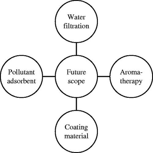
Nanoflower has gained immense success in multiple fields like water purification, photo-anode, enhance redox reaction and photocatalytic activity. Nanoflowers are under research and have tremendous potential in pharmaceuticals for protein drug delivery, anti-cancer therapy, multidrug delivery, diabetes management, waste water treatment, detection of toxicity and impurities. We proposed nanoflowers have enormous application and scope in the future in the field of bio sensing, bio-catalysis and related devices, nanocomposites, doping, nanoclusters, bio-nanocomposite and artificial intelligence. They can be used in electronics to save energy for decorative drug delivery. Eco-friendly method for synthesizing nanoflower are being developed to increase the durability and stability of enzymes and proteins. Nanoflower in the near future may be used as a photodetector ().
Figure 5. Future scope for nanoflowers.
Conclusions
Nanoflowers show potential application in the protein drug delivery system, removal of heavy metal from water, improving surface reaction, enhance immobilization efficiency, increase the stability of proteins, enzyme purification and bio-sensing. Different methods like hydrothermal method, precipitation method, ionotropic gelation method, green synthesis method, liquid crystallization method and bio-mineralization are used for the synthesis of nanoflowers, which are economical and eco-friendly. They can be synthesised without any addition of capping and reducing agents, hence eliminates the extra step, which is involved in physiochemical synthesis. Due to the ease of downstream processing, nanoflowers can be exploited to scale-up production at a large scale. The biosynthesized nanoflowers have the potential for multifunctional use, as compared to other nanoparticulate systems. Various metals like copper, magnesium, calcium can be used for fabrication of nanoflowers. Due to large surface-volume ratio, nanoflowers are advanced materials for futuristic trend in multiple applications.
Disclosure statement
No potential conflict of interest was reported by the authors.
Related Research Data
References
- Zhang G, Deng C, Shi H, et al. ZnO/Ag composite nanoflowers as substrates for surface-enhanced Raman scattering. Appl Opt. 2016;55:9105–9112.
- Wang X, Shi J, Li Z, et al. Facile one-pot preparation of chitosan/calcium pyrophosphate hybrid microflowers. Acs Appl Mater Interfaces. 2014;6:14522–14532.
- Ye R, Zhu C, Song Y, et al. Bioinspired synthesis of all-in-one organic-inorganic hybrid nanoflowers combined with a handheld pH meter for on-site detection of food pathogen. Small. 2016;12:3094–3100.
- Lee SW, Cheon SA, Kim MI, et al. Organic–inorganic hybrid nanoflowers: types, characteristics, and future prospects. J Nanobiotechnol. 2015;13:54.
- Kharisov IB. A review for synthesis of nanoflowers. Recent Pat Nanotechnol. 2008;2:190–200.
- Qin M, Zhao H, Yang W, et al. A facile one-pot synthesis of three-dimensional microflower birnessite (δ-MnO2) and its efficient oxidative degradation of rhodamine B. RSC Adv. 2016;6: 23905–23912.
- Yang XH, Xie H, Fu HT, et al. Synthesis of hierarchical nanosheet-assembled V2O5 microflowers with high sensing properties towards amines. RSC Adv. 2016;6:87649–87655.
- Fatemeh A, Azam S, Masoud SN. Sol–gel auto-combustion synthesis of PbFe12O19 using maltose as a novel reductant. RSC Adv. 2014;4:63946–63950.
- Fatemeh A, Azam S, Masoud SN. Green synthesis of magnetic chitosan nanocomposites by a new sol-gel auto-combustion method. J Magn Magn Mater. 2016;410:27–33.
- Fatemeh A, Azam S, Masoud SN. PbTiO3/PbFe12O19 nanocomposites: Green synthesis through an eco-friendly approach. Composites Part B. 2016;85:170–175.
- Fatemeh A, Azam S, Masoud SN. Facile synthesis, characterization and magnetic property of CuFe12O19 nanostructures via a sol-gel auto-combustion process. J Magn Magn. 2016;401:362–369.
- Fatemeh A, Faezeh S, Masoud SN. Utilizing maleic acid as a novel fuel for synthesis of PBFe12O19 nanoceramics via sol-gel auto-combustion route. Mater Charact. 2015;103:11–17.
- Fatemeh A, Masoud SN. Simple sol-gel auto-combustion synthesis and characterization of lead hexaferrite by utilizing cherry juice as a novel fuel and green capping agent. Adv Powder Technol. 2016;27:2025–2031.
- Fatemeh A, Mehdi B, Masoud SN. NiTiO3/NiFe2O4nanocomposites: simple sol-gel auto combustion synthesis and characterization by utilizing onion extract as a novel fuel and green capping. Mater Sci Semicond Process. 2016;43:34–40.
- Ge J, Lei J, Zare RN. Protein-inorganic hybrid nanoflowers. Nat Nanotechnol. 2012;7:428–432.
- Zhu L, Gong L, Zhang Y, et al. Rapid detection of phenol using a membrane containing laccase nanoflowers. Chem Asian J. 2013;8:2358–2360.
- Sun J, Ge J, Liu W, et al. Multi-enzyme coembedded organic–inorganic hybrid nanoflowers: synthesis and application as a colorimetric sensor. Nanoscale. 2014;6:255–262.
- Lin Z, Xiao Y, Wang L, et al. Facile synthesis of enzyme-inorganic hybrid nanoflowers and their application as an immobilized trypsin reactor for highly efficient protein digestion. RSC Adv. 2014;4:413888–413891.
- Lin Z, Xiao Y, Yin Y, et al. Facile synthesis of enzyme–inorganic hybrid nanoflowers and its application as a colorimetric platform for visual detection of hydrogen peroxide and phenol. ACS Appl Mater Interfaces. 2014;6:10775–10782.
- Manesh KM, Santhosh P, Uthayakumar S, et al. One-pot construction of mediatorless bi-enzymatic glucose biosensor based on organic-inorganic hybrid. Biosens Bioelectron. 2010;25:1579–1586.
- Delaittre G, Reynhout IC, Cornelissen JJ, et al. Cascade reactions in an all-enzyme nanoreactor. Chemistry. 2009;15:12600–12603.
- Fan Z, Wagschal K, Chen W, et al. Multimeric hemicellulases facilitate biomass conversion. Appl Environ Microbiol. 2009; 75:1754–1757.
- Wang LB, Wang YC, He R, et al. A new nanobiocatalytic system based on allosteric effect with dramatically enhanced enzymatic performance. J Am Chem Soc. 2013;135:1272–1275.
- Zhang Z, Zhang Y, Song R, et al. Manganese(II) phosphate nanoflowers as electrochemical biosensors for the high-sensitivity detection of ractopamine. Sens Actuat B Chem. 2015;211:310–317.
- Hu R, Zhang X, Zhao Z, et al. DNA nanoflowers for multiplexed cellular imaging and traceable targeted drug delivery. Angew Chem Int Ed Engl. 2014;53:5821–5826.
- Shi J, Zhang S, Wang X, et al. Preparation and enzymatic application of flower-like hybrid microcapsules through a biomimetic mineralization approach. J Mater Chem B. 2014;2:4289–4296.
- Liu Y, Chen J, Du M, et al. The preparation of dual-functional hybrid nanoflower and its application in the ultrasensitive detection of disease-related biomarker. Biosens Bioelectron. 2017; 92:68–73.
- Akhtar N, Metkar SK, Girigoswami A, et al. ZnO nanoflower based sensitive nano-biosensor for amyloid detection. Mater Sci Eng C Mater Biol Appl. 2017;78:960–968.
- Zhang Y, Zeng W. New insight into gas sensing performance of nanoneedle-assembled and nanosheet-assembled hierarchical NiO nanoflowers. Mater Lett. 2017;195:217–219.
- Ma J, Ren W, Zhao J, et al. Growth of TiO2 nanoflowers photoanode for dye-sensitized solar cells. Nano Lett. 2010;10:2562–2567.
- Lakkakula RJ, Matshaya T, Maçedo RW, et al. Cationic cyclodextrin/alginate chitosan nanoflowers as 5-fluorouracil drug delivery system. Mater Sci Eng C Mater Biol Appl. 2017;70:169–177.
- Lv Y, Hu R, Zhu G, et al. Preparation and biomedical applications of programmable and multifunctional DNA nanoflowers. Nat Protoc. 2015;10:1508–1524.
- Yu Y, Fei X, Tian J, et al. Self-assembled enzyme-inorganic hybrid nanoflower and their application to enzyme purification. Colloids Surf B Biointerfaces. 2015;130:299–304.
- Huang J, Cao Y, Liu Z, et al. Efficient removal of heavy metal ions from water system by titanate nanoflowers. Chem Eng J. 2012;180:75–80.
- Li H, Hou J, Duan L, et al. Graphene oxide-enzyme hybrid nanoflowers for efficient water soluble dye removal. ACS Nano. 2010;4:7358–7362.
- Zheng JN, He LL, Chen C, et al. One-pot synthesis of platinum3cobalt nanoflowers with enhanced oxygen reduction and methanol oxidation. J Power Sources. 2014;268:744–751.
- Miao R, Zeng W, Gao Q. Hydrothermal synthesis of novel NiO nanoflowers assisted with CTAB and SDS respectively and their gas-sensing properties. Mater Lett. 2017;186:175–177.
- Singh P, Yeon JK, Wang C, et al. Microbial synthesis of flower-shaped gold nanoparticles. Artif Cells Nanomed Biotechnol. 2016;44:1469–1474.
- Wu ZF, Wang Z, Zhang Y, et al. Amino acids-incorporated nanoflowers with an intrinsic peroxidase-like activity. Sci Rep. 2016;6:22412.
- Yoo EH, Lee SY. Glucose biosensors: an overview of use in clinical practice. Sensors. 2010;10:4558–4576.
- Turner AP. Tech.Sight. Biochemistry. Biosensors-sense and sensitivity. Science. 2000;290:1315–1317.
- Clark LC, Lyons JR. Electrode systems for continuous monitoring in cardiovascular surgery. Ann N Y Acad Sci. 1962;102:29–45.
- Updike SJ, Hicks GP. The enzyme electrode. Nature. 1967; 214:986–988.
- Hiratsuka A, Fujisawa K, Muguruma H. Amperometric biosensor based on glucose dehydrogenase and plasma-polymerized thin films. Anal Sci. 2008;24:483–486.
- Xueli Z, Jin H, Jinwen L, et al. Dual enzyme-inorganic hybrid nanoflowers incorporated microfluidic paper-based analytic devices (µPADs) biosensor for sensitive visualized detection of glucose. Nanoscale. 2017;9:5658–5663.
- Ekberg H, Tranberg KG, Persson B, et al. Intraperitoneal infusion of 5-FU in liver metastases from colorectal cancer. J Surg Oncol. 1988;37:94–99.
- Gamelin EC, Danquechin-Dorval EM, Dumesnil YF, et al. Relationship between 5-fluorouracil (5-FU) dose intensity and therapeutic response in patients with advanced colorectal cancer receiving infusional therapy containing 5-FU. Cancer. 1996; 77:441–451.
- Taylor IVSG, Murthy AK, Griem KL, et al. Concomitant cisplatin/5-FU infusion and radiotherapy in advanced head and neck cancer: 8- year analysis of results. Head Neck. 1997;19:684–691.
- Yang DJ, Zheng ZF, Zhu HY, et al. Titanate nanofibres as intelligent absorbents for the removal of radioactive ions from water. Adv Mater. 2008;20:2777–2781.
- Huang JQ, Cao YG, Deng ZH, et al. Formation of titanate nanostructures under different NaOH concentration and their application in wastewater treatment. J Solid State Chem. 2011; 184:712–719.
- Wang Y, Wang G, Wang H, et al. Chemical- template synthesis of micro-nanoscale magnesium silicate hollow spheres for waste-water treatment. Chem Eur J. 2010;16:3497–3503.
- Lu Y, Jiang Y, Wu H, et al. Nano-PtPd Cubes on graphene exhibit enhanced activity and durability in methanol electrooxidation after CO stripping–cleaning. J Phys Chem C. 2013;117:2926–2938.
- Song P, Li-Li H, Ai-Jun W, et al. Surfactant-free synthesis of reduced graphene oxide supported porous PtAu alloyed nanoflowers with improved catalytic activity. J Mater Chem A. 2015;3:5321–5327.
- Zhai YJ, Li JH, Fang X, et al. Preparation of cadmium-doped zinc oxide nanoflowers with enhanced photocatalytic activity. J MSSP. 2014;26:225–230.
- Lai X, Shen G, Xue P, et al. Ordered mesoporous NiO with thin pore walls and its enhanced sensing performance for formaldehyde. Nanoscale. 2015;7:4005–4012.
- Wang C, Zhou E, He W, et al. NiCo2O4-Based supercapacitor nanomaterials. Nanomaterials. 2017;7:41.
- Jiangwen X, Na X, Xuming Z, et al. In situ fabrication of Ni nanoparticles on N-doped TiO2 nanowire arrays by nitridation of NiTiO3 for highly sensitive and enzyme-free glucose sensing. J Mater Chem B. 2017;5:1779–1786.
- Wang SX, Jin CC, Qian WJ. Bi2O3 with activated carbon composite as a supercapacitor electrode. J Alloys Compd. 2014;165:12–11.
- Skoufa Z, Heracleous E, Lemonidou AA. Redox catalysis in fossil and bio-based olefins production routes. J Catal. 2015;322: 118–129.
- Lu Y, Zhu T, Zhang G, et al. Influence of magnetic fields on the morphology and pseudocapacitive properties of NiO on nickel foam. RSC Adv. 2015;5:99745–99753.
- Yuhao W, Ping H, Xiaomei Z, et al. Coal tar residues-based nanostructured activated carbon/Fe3O4 composite electrode materials for supercapacitors. 2014;18:665–672.
- Nie S, Emory SR. Probing single molecules and single nanoparticles by surface-enhanced Raman scattering. Science. 1997;275:1102–1106.
- Sharma B, Frontiera R, Henry AI, et al. SERS: materials, applications and the future. Mater Today. 2012;15:16–25.
- Andrea L, Alessandro V, Fabrizio G. Microfluidic electrochemical growth of vertically aligned TiO2 nanotubes for SERS optofluidic devices. RSC Adv. 2015;5:105484–105488.
- Sha MY, Xu H, Natan MJ, et al. Surface-enhanced Raman scattering tags for rapid and homogeneous detection of circulating tumor cells in the presence of human whole blood. J Am Chem Soc. 2008;130:17214–17215.
- Pallaoro A, Hoonejani MR, Braun GB, et al. Rapid identification by surface-enhanced Raman spectroscopy of cancer cells at low concentrations flowing in a microfluidic channel. ACS Nano. 2015;9:4328–4336.
- Halvorson RA, Vikesl PJ. Surface-enhanced Raman spectroscopy (SERS) for environmental analyses. Environ Sci Technol. 2010; 44:7749–7755.
- Lin X, Hai-Xin G, Shou-Qi Y, et al. Facile fabrication of silver nanoparticle-coated silica-C18 core–shell microspheres and their applications in SERS detection. RSC Adv. 2017;7:19262.
- Song CB, Qiang YH, Zhao YL, et al. Adhesion of TiO2 nanotube arrays on transparent conducting substrates using CNT-Ti02 composite pastes. Appl Surf Sci. 2014;305:792–796.
- Yu J, Fan J, Zhao L. Dye-sensitized solar cells based on hollow anatase TiO2spheres prepared by self-transformation method. Electrochimica Acta. 2010;55:597–602.
- Hauch A, Georg A. Diffusion in the electrolyte and charge-transfer reaction at the platinum electrode in dye-sensitized solar cells. Electrochimica Acta. 2001;46:3457–3466.
- Thomas S, Deepak TG, Anjusree GS, et al. A review on counter electrode materials in dye-sensitized solar cells. J Mater Chem A. 2014;2:4474–4490.
- Gao Y, Chu L, Wu M, et al. Improvement of adhesion of Pt-free counter electrodes for low-cost dye-sensitized solar cells. J Photochem Photobiol A Chem. 2012;245:66–71.
- Shalan AE, Rashad MM, Yu YH, et al. A facile low-temperature synthesis of TiO2 nanorods for high-efficiency dye sensitized solar cells. Appl Phys A Mater Sci Process. 2013;110:111–122.
- Georg M, Alexandre G, Robert V. Plasma spraying of efficient photoactive TiO2 Coatings. Surf Coat Technol. 2013;220:40–43.
- O'Regan B, Gratzel M. A low-cost, high-efficiency solar cell based on dye-sensitized colloidal TiO2 films. Nature. 1991; 353:737–740.
- Park KH, Dhayal M. Simultaneous growth of rutile TiO2 as 1D/3D Nanorod/nanoflower on FTO in one-step process enhances electrochemical response of photoanode in DSSC. Electrochem Commun. 2014;49:47–50.
- Gopal J, Hasan N, Manikandan M, et al. Bacterial toxicity/compatibility of platinum nanospheres, nanocuboids and nanoflowers. Sci Rep. 2013;3:1260.