
Abstract
In this study, we synthesised the zinc oxide nanoparticles from Vernonia amygdalina and evaluated its anti-inflammatory and antinociceptive potentials against the different inflammation and pain induced mice model. The synthesised zinc oxide nanoparticles were characterised by UV, SEM, XRD and FTIR techniques. The anti-nociceptive effects of V. amygdalina were examined by different stimuli e.g. acetic acid, glutamate, capsaicin, and formalin-induced nociception in mice. The anti-inflammatory effects of synthesised zinc oxide nanoparticles were assessed by air sack assessment and the level of inflammatory cytokines were studied. The muscle tension of animals were studied through open field assessment. The present study exhibited proficient antinociceptive and anti-inflammatory actions of the synthesised Zinc oxide nanoparticles from V. amygdalina. The sormulated zinc oxide nanoparticles were appreciably reduced the acetic acid, glutamate, capsaicin, and formalin-induced nociceptive responses in mice. Further the zinc nanoparticles were exhibited the potent anti-inflammatory actions via reducing the inflammatory response and pro-inflammatory cytokines level in the mice. In conclusion, the findings of this study proved the beneficial effects of zinc oxide nanoparticles from V. amygdalina against the different pain and inflammation-induced mice. Hence, it was clear that the zinc nanoparticles from V. amygdalina could be promising antinociceptive and anti-inflammatory agent in the future.
Introduction
The evolving of nanotechnology in an ecofriendly manner has become a part of greenery biotechnology [Citation1] with crucial study of dimension and their shape and structure [Citation2]. The ultimate intention of the nano medicine domain is to develop drugs with precise targets [Citation3] and these nanoparticles can be manufactured by means of physical, chemical and natural process. Among these, the opting of biosynthesis is an ideal way in the terms of ecological friendly [Citation4], which possess outstanding properties of antiseptic and antimicrobial and further are supposed to reveal substantial phototoxicity on budding flora [Citation5].
Nanoparticles can be used as nanofertilizer for emergent of plants but although a number of researches utter nanoparticles prevent the enlargement of some plants, few others stated the development of some other plants [Citation3,Citation6]. The techniques comprise in the fusion of ZnO nanoparticles (ZnO NPs) are chemical steam toppling, gas stage method, drenched decomposition, hydrothermal mixture, micro suspension, electrochemical technique, thumped laser deposit, microwave blend, and the gel processing [Citation7]. The significant of the produced ZnO has possible characteristics such as antimicrobial, gas detector, photoreactions, and deprivation of organic dyes [Citation8–11]. Vernonia amygdalina is generally known as sour leaf by its bitter flavour, which usually grow in the tropical Africa as a small bush with 22 cm to 5 m long and belongs to the family of Asteraceae kin [Citation12]. These leaves can be consumed as a cocktail snack and its water as a digestive stimulant [Citation13]. The foliage are extensively worned for agitation and moreover as an alternate for quinine in Nigeria and other few African countries [Citation14]. This herbal medicine can widely used as antiparasitic antimalarial, purgative, expectorant, enema and an inducer of fertility in women (subfertile) [Citation15,Citation16]. And it found to be a notorious plant to treat diabetes, fever, and possess many undocumented pharmacological properties for body pain and joints pain [Citation17].
Inflammation or irritation is a multifactorial biological phenomenon with various processes, which formed by stimulus such as microorganisms injured cells in vascular tissues [Citation18,Citation19]. Preceding results demonstrated that the biosynthesis of zinc oxide nanoparticles from the V. amygdalina leaf extracts would be used as nano level fertilisers. It also possesses therapeutic principles by holding bio molecules with reduction capability. When in combination, it is used to treat inflammation, annoyance, pneumonia, swollen veins, common colds and infant tremor [Citation20,Citation21]. With all these supportive evidences, the present study evaluated the diverse anti-inflammation effect of V. amygdalina in dissimilar nociceptive mice models.
Materials and methods
Chemicals and reagents
Chemicals such as dexamethasone, diclofenac sodium, carrageenan, capsaicin, formalin, and dexamethasone were obtained from Sigma Aldrich, USA. All other analytical grade reagents used in this study were purchased from Himedia, USA.
Experimental animals
Swiss Albino male mice weighing about 25–30 g were selected for this study. The animals segregated in disinfected cages (plastic) at warmth of 20–25 °C by regular light and dark sequence for 12 h. The humidity of the mice domicile was maintained for about 50–60%. The mice were allowed to take feed and water. In the beginning, the animals were adapted for 10 days at regular laboratory circumstances. Then the animals were fasted in the night prior to the behavioural examination and the analysis was executed in the morning sessions (8.00 am to 12.00 am). All the experiments were approved by the institutional ethical committee, The Second Affiliated Hospital of Soochow University, Jiangsu, 215004, China.
Collection and preparation of V. amygdalina
The fresh and matured leaves of V. amygdalina was collected from the Classical Gardens of Suzhou (31°19′ N, 120°27′ E), Jiangsu province, China, and washed with the distilled water to remove the dust particles. Then the leaves were shade dried and powdered by using mechanical grinder. The resulted leaf powder was used to prepare the aqueous extract. The 15 g of the leaf powder was soaked in 100 ml of double distilled water and then heat macerated for 30 min at 75 °C. After that, the suspension was filtered and then resulted extract was used to synthesis the zinc oxide nanoparticles.
Nanoparticles synthesis and preparation
Illustration of zinc oxide nanoparticles was biosynthesized using 20% leaf extracts of V. amygdalina with zinc acetate dihydrate and sodium hydroxide as predecessor resources [Citation22]. Then the amalgamation was kept in dark condition at room temperature and the mixture was centrifuged for 15 min at 10,000 rpm. Then the centrifugation pellet was allowed to dry and it was placed in a water bath at 45 °C for 10 h subsequent to centrifugation and dehydrated in a hot air oven for 6 h. The samples of V. amygdalina ZnO nanoparticles were synthesised and characterised.
Assessment by UV-Visible spectroscopy
The refuse of uncontaminated metal ions was recognised via shaping the amalgamation of retorting assortment by UV-Visible Spectrophotometer (1700 series Shimadzu model) as of 400 to 800 nm [Citation23].
Investigation of Fourier transform infrared (FTIR)
FTIR is an important device to categorise the purposeful cluster to metal constituent part and organic molecules. The spectrums were established at 1 cm-1 movement by FTIR spectrophotometer (FTIR-8400S Shimadzu Corporation) by means of Potassium Bromide shot process [Citation24]. It displayed the incidence in the range between 3500 and 500 cm−1.
Scanning electron microscopy
The formed nanoparticles were dotted in water and the suspension was detected by ultra sonicator for about 3 h. Very few drops of the nano suspension were situated on a segment of micro glass slide, which offered metal lattice enclosed by carbon cover and dried out gradually at room temperature. The discharge was then covered and envisaged with a JEOL-JSM 6480 Scanning Electron Microscope to assess the dimension, outline and proportion of the particle [Citation25].
X-ray diffraction examination
The X-ray diffraction (XRD) was used to determine the disposition and range of the ZnO nanoparticles, the section shot was suspended in sterile water and continued the procedure constant for 3 times by the identical solution pursued by centrifugation. The pellet was sealed and allowable to dry. The fine particles getting from the section was enclosed with XRD lattice, where the spectrum were recognised in 35 kV and a current of 30 mA with K alpha and K beta radiation with XRD (XRD-7000-Shimadzu). The thinned potency was established from 10 to 70 °C at 2 theta angles. The crystal surroundings of intermingled nanoparticles were examined from the wideness of the XRD peak by means of Debye formula [Citation26].
Acetic acid persuaded nociception examination
Experimental animals divided into five clusters consisting of six animals in each, control I was treated with tween 80 (1%), group II to IV animals were treated with ZNO-NP of V. amygdalina (2.5, 5.0, 7.5 mg/kg) and group V animals treated with diclofenac (10 mg/kg) as a standard. After the 1 h treatment of V. amygdalina and diclofenac sodium, the animals were challenged with 1% of acetic acid through intraperitoneal route and then all experimental animals were located in the observation cabin [Citation27]. The numbers of writhing like stretching movement, elongation of body and hind limbs were noted. The number of writhes done by experimental animals within 30 min was recorded.
Glutamate induced nociception check
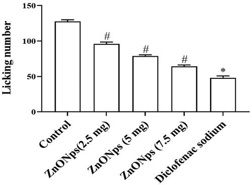
The similar experimentation was continued for glutamate-induced nociception analysis. The animals were pre-treated with ZNO-NP of V. amygdalina (2.5, 5.0, 7.5 mg/kg) and 10 mg of diclofenac sodium before 10 min of the experiment. These mice were subsequently induced with glutamate (10 μm) by injecting slowly it to the abdominal plane of left posterior foot of the animal and examined for surveillance for 20 min while control received the saline [Citation28]. The amount of licks achieved by the mice were counted and documented.
Capsaicin provoked foot licking test
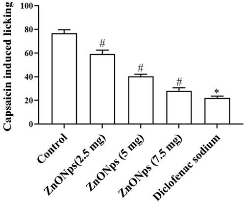
The animals were submitted to capsaicin induced nocicpetive trial to spot the consequence of ZNO-NP from V. amygdalina adjacent to neuropathic nociception. Sooner than 30 min, the experimentation performed the mice were pre-treated with dissimilar absorption of V. amygdalina and diclofenac sodium. Capsaicin (15 μl) suspended in 95% phosphate-buffered saline and ethanol (5%) was introduced to the appendage of the mice as each base with 1.5 μg capsaicin while control received only saline. The standard control animals received the diclofenac sodium [Citation29]. The animals were kept under examination for 10 min and the moment of licking on inserted paw be verified for the occurrence of nociception.
Formalin stimulated appendage licking test
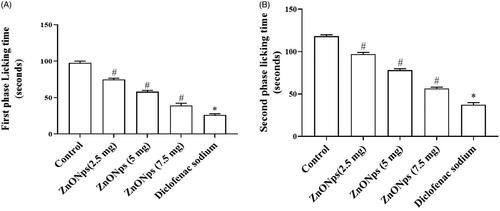
Formalin provoked foot licking tests were achieved to affirmed the antinociceptive effectiveness of ZNO-NP from V. amygdalina [Citation30]. The animals were pre-treated with normal (Tween 80) for control; V. amygdalina (2.5, 5.0, 7.5 mg/kg) & diclofenac sodium (5 mg/kg) as positive control (group V) ahead of 30 min via subcutaneous shot. Subsequently pre-treatment of 3% of formalin was injected to the right back mitt plantar plane and kept back for scrutiny cavity for half an hour. The quantity of lickings carried out by mice from the first stage to the arising of neuro ache (0–5 min) and subsequent stage (20–30 min) of tenderness was recorded.
Peritoneal hollow leukocyte permeation test
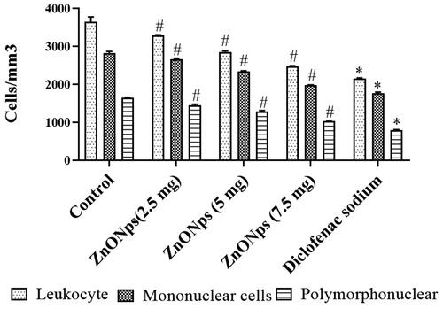
Leukocyte permeability into the peritoneal movement subsequent to induction of pre-treatment & anti-inflammation effectiveness of ZNO-NP from V. amygdalina was noticed by the technique of Vinegar et al. [Citation31]. The experimental animals were pre-initiated with Tween 80 for control mice, ZNO-NP from V. amygdalina (2.5, 5.0, 7.5 mg/kg) and last group mice were induced with diclofenac sodium, considered as positive control (5 mg/kg) previous to 30 min of experimentation. The animals were sacrificed according to ethical guidelines, and then the peritoneal void was cleaned and washed with PBS, which contains 1 mM EDTA to collect cells. The elucidation was centrifuged and scrutinised whole leukocyte in addition to discrepancy cell count up. The total leukocytes mononuclear and polynuclear cells were counted by using the Neubauer chamber in blood mixed with 3% of acetic acid and 1% of crystal violet in 5:44:1 ration. The total amount of entire leukocytes, mono & polymorphonuclear cells were documented.
Consequence of ZNO-NP from V. amygdalina on inflammation promoting cytokines
The animals were subjected with anaesthesia and the backside fur was shaved, 5 ml of disinfected space was subcutaneously infused two times at similar spot at an intermission of 3 days to shape a pocket [Citation32]. The animals with sacks were separated into 6 groups and induced with Tween 80 (1%) (Normal mice), 0.5 ml Carageenan (control), diverse absorption of V. amygdalina (2.5, 5.0, 7.5 mg/kg) and dexamethasone (optimistic control). The experimental animals were sacrificed by means of cervical displacement following 60 min, the pouch was pierced and 1.5 ml saline was injected within the hollow and sucked backside to collect the cells. Mass of cells was centrifuged and pellets were submitted to investigate proinflammatory cytokine like IL-6, IL-1β and TNF-α.
Open field assessment
The tranquiliser consequence of ZNO-NP from V. amygdalina was evaluated by executing open field test. The animals were subjected with dissimilar attention of ZNO-NP from V. amygdalina (2.5, 5.0, 7.5 mg/kg) and affirmative control, diclofenac sodium (5 mg/kg). Subsequent to 1 h, the experimental animals were introduced in to the open turf equipment which was a container with all sides of 50 cm each. Then, the package was alienated into 25 equivalent cubes. Later, the mice were allowed to ascertain the open ground device for few minutes, the quantity of squares traversed by the mice by all the foot was documented. The equipment used was kept clean by using of alcohol every time prior to performing the experimentation with new mice.
Statistics
All the obtained data were significantly examined by means of graph prism software and interpreted as mean ± standard deviation. Dunnet’s post hoc testing was executed for one way Analysis of Variance to evaluate momentous dissimilarity among the cluster. p values were represented as p < .05, p < .01 correspondingly.
Results
UV-Visible spectroscopy exploration of V. amygdalina
The reduction of zinc oxide ions in the V. amygdalina was confirmed by UV-Vis Spectrophotometer, shown in (. The UV-Vis incorporation of ZnO NPs was at the crest maximum at 350 nm that was compared to the range of zinc oxide nanoparticles. Subsequent incubation, the colour was faded because of the excited state of Surface Plasmon in the ZnO NPs. The decline of zinc was confirmed by UV-Vis Spectrophotometer. Combination spectra of ZnO NPs twisted in the response medium which has crest at 320 nm; improvement of peak state that the rudiments are detached. The promptness and width of the surface plasmon absorption depend on the quantity and shape of the nanoparticles and as well on the insulating constant of the metal itself and the adjacent intermediary [Citation33]. Allium cepa extract and zinc nitrate changed the colour of the ZnO NPs. The uppermost strength at 350 nm was tentative which designate entire refuse of zinc ions. The range of ZnO NPs by UV-Vis established a critical absorbance at 350 nm, which assign an estimated reliable accumulation of the nanoparticles.
Figure 1. (A) UV-Visible spectrum absorption pattern, (B) TEM and (C) TEM analysis of ZnO-NPs synthesised from V. amygdalina. UV-Vis absorption spectra of ZnO-NPs. The peak values for UV-VIS, plotted between ZnO-NPs/absorbance ratios. The highest absorbance peak is about 350.00. All solutions were in water. V. amygdalina extract ZnO NPs were found as nano shaped ( The organisation, segment and form of amalgamated product were examined by the SEM study.
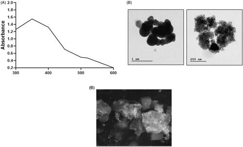
Electron microscope examination of V. amygdalina
The external shape and structure of the nanoparticles was illustrated by Scanning Electron Microscopy . Vernonia amygdalina extract mediated ZnO NPs were initiated as nano shaped (). The usual progress in the use of V. amygdalina extract provided primary time as a tumbling substance and adding external improving mediator for the fabrication of tiny ball shaped ZnO NPs. The association, section and shape of combined product were scrutinised by SEM study, which also were used to develop ZnO NPs from Ocimum tenuiflorum extract [Citation34,Citation35]. In this current study, the SEM result demonstrated the cylindrical nanoparticle shaped with thickness of about 20–40 nm.
Fourier transform infrared spectroscopy study of V. amygdalina
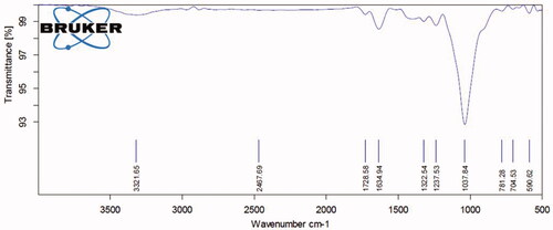
FTIR study established to distinguish the biomolecules, which responsible for diminishing metal ions into ZnO NPs in association with V. amygdalina extract and also demonstrated various integration peaks ranged from 3500 cm−1 to 500 cm−1 (). The biological constituents originated in the V. amygdalina extract were responsible for the arrangement of a mixture of nanoparticles. The FTIR array of V. amygdalina demonstrated various integration peaks ranged from 3500 cm−1 to 500 cm−1. The range of incidence from 3321 cm−1 crest represented the O–H stretching vibration as well indicated the presence of amino acids and carbohydrates. The peak of 2467 cm−1 denoted the C–H extension particularly, lipids. The occurrence of range varied from 1634 cm−1 peak represented proteins by Amide I: C = O length. Also, ranges from 1322 cm−1 peak symbolised sulphur compounds and the range from 1037 cm−1 illustrated the C–N stretching: amino acids.
Figure 2. FTIR spectra of ZnO nanoparticles of V. amygdalina. V. amygdalina extract were answerable for the configuration of a variety of nanoparticles. The FTIR range of V. amygdalina illustrated several assimilation peaks ranged from 3500 cm−1 to 500 cm−1. shows the representative FT-IR spectra obtained from V. amygdalina.
X-ray diffraction analysis
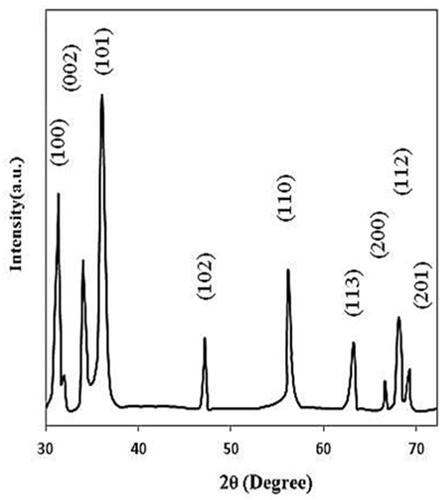
The distribution of the dissimilar peaks in the XRD model depicted crystalline zinc oxide with hexa organisation in the network parameters (). In the whole band of 2θ values, the XRD outline established different absorption peaks ranges from 10° to 70° for the V. amygdalina. The V. amygdalina produced ZnO NPs were indexing in 100, 002, 101, 102, 110, 103, 112 and 201. The zinc was subjected and elevated at 101 and 100 and the usual crystal shaped dimension was determined by Scherer’s equation. In addition, no further peaks have perceived in the section and recognised the synthesis of ZnO NPs in an elevated clarity plane. Natural amalgamation of ZnO NPs from V. amygdalina extract demonstrated sturdy diffracted peaks, which notably conformed to the JCPDS folder. Elevated limpidness and crystal figure of the ZnO NPs were also long-established.
Figure 3. X-ray diffraction analysis of ZnO-NPs synthesised from V. amygdalina. The XRD pattern showed dissimilar concentration peaks in the entire band of 2θ values between range from 25° to 75° for the V. amygdalina extract. The V. amygdalina synthesised ZnO NPs were indexed as 100, 002, 101, 102, 110, 103, 112, and 201. The zinc was indexed and excited at 101 and 100.
Antinociceptive activity of ZNO-NP from V. amygdalina
Acetic acid stimulated test
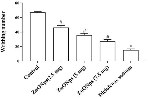
The enteric bending assessment was used to examine the efficiency of V. amygdalina to ease out pain. Animals except control were given acetic acid to persuade intestinal twisting and integer of writhes and the intestinal elongation performed by the mice was counted. The number of writhes was considerably (p < .05) reduced (34 and 46) in V. amygdalina managed mice as compared with control. But, the 7.5 mg of V. amygdalina treated animals displayed condensed twist (25) that was close to the effect of diclofenac (18) treated mice ().
Figure 4. Antinociceptive upshot of ZNO-NP in the nociception induced by acetic acid in mice. All values are illustrated as mean ± SD of six animals. The statistical significance level was calculated by one-way ANOVA followed by the Dunnet’s post hoc test; note: #p < .05 when compared with control group and *p < .05 when compared with ZnONPs administered groups.
Glutamate stimulation outcome
demonstrated the anti-nociceptive effects of V. amygdalina in glutamate induced mice. There was lessen amount of licks indicated the anti-nociceptive effects of V. amygdalina. Also, the V. amygdalina (7.5 mg) oral routed animals achieved 58.57 ± 1.11 licks (p < .05) which was equivalent to the regular diclofenac, which showed 50.38 ± 1.12 licks. But in contrast to control animals, the 2.5 mg and 5.0 mg of V. amygdalina managed mice that illustrated reduced ranges of licks subsequent to glutamate insertion.
Figure 5. Antinociceptive consequence of ZNO-NP in the nociception encouraged by glutamate in mice. All values are illustrated as mean ± SD of six animals. The statistical significance level was calculated by one-way ANOVA followed by the Dunnet’s post hoc test; note: #p < .05 when compared with control group and *p < .05 when compared with ZnONPs administered groups.
Capsaicin encouraged paw licking analysis
During the post inoculation phase, mitt licking actions took almost 5 min in the aggravated capsaicin introduced mice. By contrary, the regular drug of diclofenac (48 ± 2.3 lick) and 7.5 mg of V. amygdalina introduced mice performed fewer counts (p < .05) of licks (43 ± 1.65 licks). The highest quantity of clicks was found in normal mice, while it was appreciably reduced in 2.5 and 5.0 mg of V. amygdalina induced animals in .
Figure 6. Effect of ZNO-NP on capsaicin-induced licking in mice. All values are illustrated as mean ± SD of six animals. The statistical significance level was calculated by one-way ANOVA followed by the Dunnet’s post hoc test; note: #p < .05 when compared with control group and *p < .05 when compared with ZnONPs administered groups.
Paw licking test induced by formalin
explained the sedative pain medicine in the formalin-induced model to know the consequence of V. amygdalina and morphine. Formalin provoked mitt licking trial explicated and confirmed the anti-nociceptive upshot in V. amygdalina at two stages. In comparison to normal and all other dosage of V. amygdalina, pre-initiated mice licking discharge were extensively (p < .05) declined in two stage of study. The V. amygdalina treated mice was increased at Stage B (15–30 min) that was similar to Stage A (5 min) hammering. And the Morphine managed animals revealed remarkable drop off in licking, which contrast to control and V. amygdalina introduced mice.
Figure 7. Result of ZNO-NP on duration of formalin-induced paw-licking (seconds) in the first 5 min (first phase, A) and from 15 to 30 min (second phase, B) of nociception in mice. All values are illustrated as mean ± SD of six animals. The statistical significance level was calculated by one-way ANOVA followed by the Dunnet’s post hoc test; note: #p < .05 when compared with control group and *p < .05 when compared with ZnONPs administered groups.
Peritoneal void leukocyte permeable test
The amount of whole leukocytes, mononuclear and polymorphonuclear invaded cells in the serous membrane hollow space in control animals, V. amygdalina (2.5, 5.0, 7.5 mg) and morphine pre-trial animals were completely represented in . The V. amygdalina and morphine treated animals showed (p < .05) declined number of leukocyte invasion when compared to control mice. Also, there was negligible invasion of leukocytes was observed in the morphine pre-treated mice. As well, the minimum permeable of leukocytes were observed in 75 mg of V. amygdalina treated mice, which was similar to the morphine treated.
Figure 8. Effect of ZNO-NP on total leukocytic mononuclear and polymorphonuclear cell recruitment into the peritoneal cavity of mice. All values are illustrated as mean ± SD of six animals. The statistical significance level was calculated by one-way ANOVA followed by the Dunnet’s post hoc test; note: #p < .05 when compared with control group and *p < .05 when compared with ZnONPs administered groups.
Outcome of V. amygdalina on inflammatory cytokines
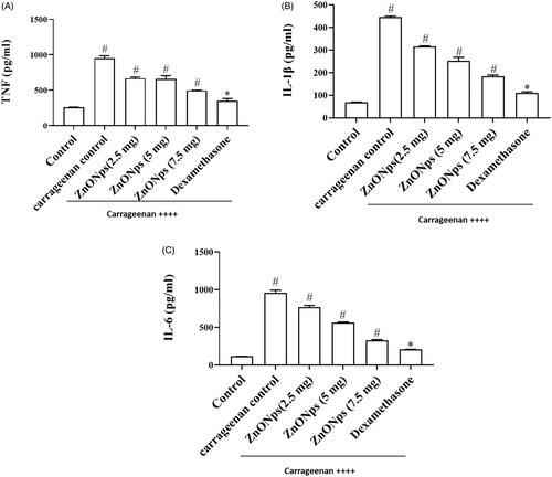
The inflammatory cytokines such as TNF-α (), IL-1β (), IL-6 () were examined in carageenan alone, V. amygdalina and dexamethasone treated mice that achieved in space sacks, In contrast, carageenan alone introduced experimental animals were considerably increased the above mentioned markers plane. Among all the three dosages, the absorption of V. amygdalina and dexamethasone were repressed (p < .05) the attentiveness of TNF-α ().
Figure 9. The consequence of ZNO-NP on TNF-α, IL-1β, and IL-6 fabrication in the air pouch test. All values are illustrated as mean ± SD of six animals. The statistical significance level was calculated by one-way ANOVA followed by the Dunnet’s post hoc test; note: #p < .05 when compared with control group and *p < .05 when compared with ZnONPs administered groups. (A) TNF (pg/ml) (B) IL-1β (pg/ml) (C) IL-6 (pg/ml).
Open field examination
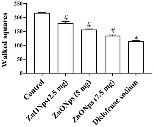
The tranquiliser efficacy of V. amygdalina was evaluated by the behaviour changes of experimental animals in open field equipment. When contrast to normal mice, the V. amygdalina (2.5 and 5 mg) induced mice does was not displayed any considerable (p < .05) modifications while the integer of squares was crossed by the V. amygdalina (7.5 mg) induced mice. But, the morphine treated mice exposed diminished behavioural alterations ().
Figure 10. The effects of ZNO-NP in the open field test. All values are illustrated as mean ± SD of six animals. The statistical significance level was calculated by one-way ANOVA followed by the Dunnet’s post hoc test; note: #p < .05 when compared with control group and *p < .05 when compared with ZnONPs administered groups.
Discussion
The chemicals, thermal, and electrical induced inflammatory pain models were extensively used in the preclinical researches [Citation36]. These models can be regarded as a way to mimic the inflammatory pain symptoms noted in the clinical in the case of tissue inflammation, where symptoms of allodynia and mechanical allodynia are observed in the patients [Citation37]. In the current study, we examined the dose-dependent anti nociceptive and anti-irritation effects of V. amygdalina in diverse animal models that explained the effect was completely contrast with the regular drugs. The anti-inflammatory exploit of V. amygdalina was well established by evaluation of leukocyte invasion in abdominal lining and also by the plane of inflammatory cytokines. The UV-Vis absorption range of ZnO nanoparticles by Nyctanthes tristis established an absorbance at 369 nm [Citation38]. Also, the FTIR series of ZnO nanoparticles exhibited the crest at 417.52 cm−1, denoted the assimilation of ZnO connection and the extensive absorption of peak at 3438 cm−1 confirmed the typical enclosure of hydroxyl group [Citation39]. As well the peak at 667.29 cm−1 indicated the expanding observance of ZnO nanoparticle which was correlated with the present outcome [Citation40]. Additionally, in XRD studies had no extra peaks that allegedly described the macromolecule amalgamation of ZnO NPs, derived elevated limpidness. The crystal-like gathering was established at 21 nm [Citation41]. The scanning Electron Microscope depicted the ZnO NPs synthesising by Trifolium pretence that had constituent dimension displayed from 100 to 190 nm [Citation42]. This correlated with our current SEM records with particles dimension arrayed from 50 to 100 nm.
Pain is the reflective response to the stimulus such as, infection, injury, etc. The nociceptive assessments were executed to evaluate the curative effect of sample agents to reduce the pain. The numerous assays were performed to such as mechanical, electrical, thermal, and chemical stimulus [Citation43]. The acetic acid-stimulated abdominal constriction is a visceral pain model, usually executed to screen the antinociceptive or anti-inflammatory effects of sample analgesic agent [Citation44]. The formalin injection to the experimental animals stimulates the biphasic pain response [Citation45]. The first phase corresponds to the neurogenic pain caused by the direct effect of formalin and the second phase is the development of an inflammatory response and the release of nociceptive mediators like serotonin, prostaglandins, bradykinin in the peripheral tissues [Citation46]. The tail immersion induced pain model is executed to examine the acute pain and it is a helpful assay to differentiate the central opioid like analgesics from peripheral analgesics [Citation43]. The open field test was performed to confirm the anti-nociceptive action of sample analgesic agents related to non-specific disturbances in the locomotor activity of the experimental animals [Citation47].
The greenery synthesis of zinc oxide nanoparticles using Moringa oleifera had rough and delicate crest from Zn and O atom and weak peaks were found form the elements like calcium and potassium [Citation48]. The study of nociceptive was executed to review the beneficiary properties of drug to lessen soreness, which carried out by geothermal, automatic or electrical incentive [Citation49]. Past research described the V. amygdalina was one of the potential therapeutic to analyse toxicity test [Citation50]. The Carrageenan provoked rat was a better model to estimate the anti-oedematous effects of herbal yield with biphasic in nature [Citation51]. The primary phase involving in the discharge of serotonin and the expressing of histamine in the second phase (after 1 h) was mediated by prostaglandins, cyclooxygenase harvest and the stability lying between two phases was offered by kinins [Citation52].
The V. amygdalina is rich in flavonoids, steroids, necessary oil and tannins, which preferably used in animal models to predict nociception [Citation53–56]. The certain stimulus of nociceptors can be reviewed by the initiation of inflammation that similar to acetic acid. These can be used to calculate writhes, which was ground to examine stomach renunciation and stretching of hind limb. The acetic acid model was performed to perceive the outcome of painkiller by the levels of prostaglandins in CNS, which usually elevated in the nociceptors [Citation57]. The current results demonstrated that V. amygdalina established repressed in the synthesis of prostaglandins E2 and prostaglandins D2 [Citation58]. Since the carrageenan induced inflammation was an important prognostic test for anti-inflammatory mediator sensitive inflammation, these outcomes suggested that the V. amygdalina was an efficient in severe inflammatory turmoil. In this study, the nanoparticles from the V. amygdalina showed the potent anti-inflammatory activity against the carrageenan induced inflammation in mice [Citation52,Citation59,Citation60].
In the present finding, the reduction in quantity of writhes was achieved by dissimilar absorption of V. amygdalina. This revealed the V. amygdalina also would inhibit the production of prostaglandins that related to sensitisation of nociceptors. Glutamate transmits the reaction by two sorts of receptors i.e. N-methyl-D-aspartate and non-NMDA receptor which activate the peripheral neurons by liberating proinflammatory cytokines [Citation61]. Correspondingly, our study of V. amygdalina pre-treated mice demonstrated glutamate-induced soreness by counting of licking number. The earlier reports about the acetone extract of V. amygdalina showed notable decline in the integer of paw licking, which induced by formalin as compared to the control [Citation50]. The two phase discharge of prostaglandins E2 stimulated nociceptive inside formalin nociception rat replica, and initiation of tenderness generated by inflammatory cytokines on centre sensation neurons [Citation62]. As correlated with this current study, the V. amygdalina drastically reduced the formalin-induced licking numbers in both neuro and inflammatory phases that explained as anti-nociceptive agent. The Naringenin restricted the carageenan provoked leukocyte invasion in intestinal lining hollow space. The increase of leukocyte production may direct the Myeloperoxidase action in animal foot edoema by reactive oxygen species produced by carrageenan [Citation63].
Spinal supportive nerve cell exude powerful pro inflammatory cytokines such as TNF-α, IL-1β and IL-6 which respond to the pain [Citation64,Citation65]. The present results showed anti tenderness of V. amygdalina and considered as space pocket model trial. The plane of inflammation promoting cytokines was down regulated in V. amygdalina treated mice that contrast to control mice. The open field examination explored the improved behavioural transformation in V. amygdalina induced mice than standard drug, which brought effective anti-nociceptive drug with minimal side effects. Therefore resistance of inflammatory cytokine production by V. amygdalina might be the motive for its anti-nociceptive possessions.
Conclusions
The present study exhibited proficient antinociceptive and anti-inflammation action of the combination of Zinc oxide nanoparticle with the extract of V. amygdalina. Further results demonstrated the beneficial effects Zinc oxide nanoparticles from the V. amygdalina through ameliorating the anti-inflammatory and antinociceptive activities. It explained the systematic base on pain improvement and managing inflammatory disarray. In dissimilar nociceptive and swelling mice models, the V. amygdalina was found as strong antinociceptive and antisoreness drug by exposing the performance of pro-inflammatory cytokines. Altogether, all the outcomes revealed the effective anti-inflammatory of zinc oxide nanoparticles from V. amygdalina in mice models.
Authors’ contribution
Xiao Wang – conceived the study design, commented on both the manuscript and figures, and approved the manuscript; Hairui Liu and Peipei Kang – analysed data; Ying Liu, Yifan An and Yanting Hu – composed this manuscript and prepared the figures; Xiyuan Jin, Xin Cao, Yunfei Qi and Thiyagarajan Ramesh – proof reading the manuscript.
Disclosure statement
No potential conflict of interest was reported by the author(s).
References
- Widyaningtyas AL, Yulizar Y, Bagus Apriandanu DO. Ag2O nanoparticles fabrication by Vernonia amygdalina Del. leaf extract: synthesis, characterization, and its photocatalytic activities. IOP Conf Ser Mater Sci Eng. 2019;509:012022.
- Vidya C, Hiremath S, Chandraprabha MN, et al. Green synthesis of ZnO nanoparticles by Calotropis gigantea. Int J Curr Eng Technol. 2013;1:118–120.
- Sabir S, Arshad M, Chaudhari SK. Zinc oxide nanoparticles for revolutionizing agriculture: synthesis and applications. Sci World J. 2014;2014:1–8.
- Khalafi T, Buazar F, Ghanemi K. Phycosynthesis and enhanced photocatalytic activity of zinc oxide nanoparticles toward organosulfur pollutants. Sci Rep. 2019;9(1):6866.
- Umar H, Kavaz D, Rizaner N. Biosynthesis of zinc oxide nanoparticles using Albizia lebbeck stem bark, and evaluation of its antimicrobial, antioxidant, and cytotoxic activities on human breast cancer cell lines. IJN 2018;14:87–100.
- Aslani F, Bagheri S, Julkapli MN, et al. Effects of engineered nanomaterials on plants growth: an overview. Sci World J. 2014;2014:1–28.
- Wiesmann N, Kluenker M, Demuth Brenner PW, et al. Zinc overload mediated by zinc oxide nanoparticles as innovative anti-tumor agent. J Trace Elem Med Biol. 2019;51:226–234.
- Elumalai K, Velmurugan S, Ravi S, et al. RETRACTED: green synthesis of zinc oxide nanoparticles using Moringa oleifera leaf extract and evaluation of its antimicrobial activity. Spectrochim Acta A Mol Biomol Spectrosc. 2015;143:158–164.
- Zhong Q, Kou H, Yang L, et al. Factors influencing variations in the thermal conductivity of polycrystalline ZnS and Cr2+:ZnS. Mater Lett. 2015;158:222–227.
- Habibi MH, Rahmati MH. The effect of operational parameters on the photocatalytic degradation of Congo red organic dye using ZnO-CdS core-shell nano-structure coated on glass by Doctor Blade method. Spectrochim Acta A Mol Biomol Spectrosc. 2015;137:160–164.
- Dimkpa CO, Singh U, Bindraban PS, et al. Zinc oxide nanoparticles alleviate drought-induced alterations in sorghum performance, nutrient acquisition, and grain fortification. Sci Total Environ. 2019;688:926–934.
- Ijeh II, Ejike CECC. Current perspectives on the medicinal potential of Vernonia amygdalina Del. J Med Plant Res. 2011;5(7):1051–1061.
- Singha SC. Medicinal plants of Nigeria. Lagos (Nigeria): Nigerian National Press; 1965.
- Masaba SC. The antimalarial activity of Vernonia amygdalina Del (Compositae). Trans R Soc Trop Med Hyg. 2000;94(6):694–695.
- Huffman MA. Animal self-medication and ethno-medicine: exploration and exploitation of the medicinal properties of plants. Proc Nutr Soc. 2003;62(2):371–381.
- Adedapo AA, Otesile AT, Soetan KO. Assessment of the anthelmintic efficacy of the aqueous crude extract of Vernonia amygdalina. Pharm Biol. 2007;45(7):564–568.
- Egedigwe CA. Effect of dietary incorporation of Vernonia amygdalina and Vernonia colorata on blood lipid profile and relative organ weights in albino rats [thesis]. Nigeria: Dep Biochem, MOUAU; 2010.
- Ferrero-Miliani L, Nielsen OH, Andersen PS, et al. Chronic inflammation: importance of NOD2 and NALP3 in interleukin-1beta generation. Clin Exp Immunol. 2007;147(2):227–235.
- Drozdova IL, Bubenchikov RA. Composition and anti-inflammatory activity of polysaccharide complexes extracted from sweet violet and low mallow. Pharm Chem J. 2005;39(4):197–200.
- Akinyemi KO, Oladapo O, Okwara CE, et al. Screening of crude extracts of six medicinal plants used in South-West Nigerian unorthodox medicine for anti-methicillin resistant Staphylococcus aureus activity. BMC Complement Altern Med. 2005;5(1):1–7.
- Prabhu KS, Lobo R, Shirwaikar AA, et al. Ocimum gratissimum: a review of its chemical, pharmacological and ethnomedicinal properties. TOALTMEDJ. 2009;1(1):1–15.,
- Ogunyemi SO, Abdallah Y, Zhang M, et al. Green synthesis of zinc oxide nanoparticles using different plant extracts and their antibacterial activity against Xanthomonas oryzae pv. oryzae. Artif Cells Nanomed Biotechnol. 2019;47(1):341–352.
- Roy S, Triparna M, Shatarupa T, et al. Biosynthesis, characterization and antifungal activity of zinc oxide nanoparticles synthesized by the fungus Aspergillus foetidus. J Nanometer Biostruc. 2013;8:197–205.
- Rad SS, Sani AM, Mohseni S. Biosynthesis, characterization and antimicrobial activities of zinc oxide nanoparticles from leaf extract of Mentha pulegium (L.). Microb Pathog. 2019;131:239–245.
- Shobha N, Nanda N, Giresha AS, et al. Synthesis and characterization of zinc oxide nanoparticles utilizing seed source of Ricinus communis and study of its antioxidant, antifungal and anticancer activity. Mater Sci Eng C Mater Biol Appl. 2019;97:842–850.
- Baliah NT, Priyatharsini SL. Biosynthesis and characterization of zinc oxide nanoparticles using onion bulb extract. IJTSRD. 2018;2(2):36–43.
- Koster R, Anderson M, De Beer EJ. Acetic acid analgesic screening. Fed Proc. 1959;18:412.
- Wakatsuki K, T-Uchimura Y, Matsubara T, et al. Peripheral nociceptive mechanisms in an experimental rat model of fibromyalgia induced by repeated cold stress. Neurosci Res. 2019;30483.
- Valek L, Auburger G, Tegeder I. Sensory neuropathy and nociception in rodent models of Parkinson’s disease. Dis Model Mech. 2019;12(6):039396.
- Hunskaar S, Fasmer OB, Hole K. Formalin test in mice, a useful technique for evaluating mild analgesics. J Neurosci Methods. 1985;14(1):69–76.
- Vinegar R, Truax JF, Selph JL. Some quantitative temporal characteristics of carrageenin-induced pleurisy in the rat. Proc Soc Exp Biol Med. 1973;143(3):711–714.
- Edwards JC, Sedgwick AD, Willoughby DA. The formation of a structure with the features of synovial lining by subcutaneous injection of air: an in vivo tissue culture system. J Pathol. 1981;134(2):147–156.
- Joe MM, Jayochitra J, Vijayapriaya M. Antimicrobial activity of some common spices against certain human pathogens. J Med Plants Res. 2009;3:1134–1136.
- Sangeetha G, Rajeshwari S, Venckatesh R. Green synthesis of zinc oxide nanoparticles by Aloe barbadeneis Miller. leaf extract: structure and optical properties. Mat Res Bull. 2011;46(12):2560–2566.
- Raut RD, Narkhede B, Gardas BB. To identify the critical success factors of sustainable supply chain management practices in the context of oil and gas industries: ISM approach. Renewable Sustainable Energy Rev. 2017;68(1):33–47.
- Walker CIB, Trevisan G, Rossato MF, et al. Antinociceptive effect of Mirabilis jalapa on acute and chronic pain models in mice. J Ethnopharmacol. 2013;149(3):685–693.
- Schlesinger N. Anti-interleukin-1 therapy in the management of gout. Curr Rheumatol Rep. 2014;16(2):398
- Jamdagni P, Poonam Khatri J, Rana S. Green synthesis of zinc oxide nanoparticles using flower extract of Nyctanthes arbor-tristis and their antifungal activity. J King Saud Univ Sci. 2016;3(8):417–432.
- Chen P, Wang H, He M, et al. Size-dependent cytotoxicity study of ZnO nanoparticles in HepG2 cells. Ecotoxicol Environ Saf. 2019;171:337–346.
- Gupta M, Tomar RS, Kaushik S, et al. Effective antimicrobial activity of green ZnO nano particles of Catharanthus roseus. Front Microbiol. 2018;9:2030.
- Zheng Y, Fu L, Han F, et al. Green biosynthesis and characterization of zinc oxide nanoparticles using Corymbia citriodora leaf extract and their photocatalytic activity. Green Chem Lett Rev. 2015;8(2):59–63.
- Dobrucka R, Długaszewska J. Biosynthesis and antibacterial activity of ZnO nanoparticles using Trifolium pratense flower extract. Saudi J Biol Sci. 2016;23(4):517–523.
- Bars DL, Gozariu M, Cadden SW. Animal models of nociception. Pharmacol Rev. 2001;53(4):597–652.
- De Souza MM, Pereira MA, Ardenghi JV, et al. Filicene obtained from Adiantum cuneatum interacts with the cholinergic, dopaminergic, glutamatergic, GABAergic, and tachykinergic systems to exert antinociceptive effect in mice. Pharmacol Biochem Behav. 2009;93(1):40–46.
- Ramirez MR, Guterres L, Odila ED, et al. Preliminary studies on the antinociceptive activity of Vaccinium ashei berry in experimental animal models. J Med Food. 2010;13(2):336–352.
- Reynoso MA, Vera N, Aristimuno ME, et al. Antinociceptive activity of fruits extracts and “arrope” of Geoffroea decorticans (chañar). J Ethnopharmacol. 2013;145(1):355–362.
- Goncalves GN, Marinho DG, Almanca CCJ, et al. Antinociceptive and anti-oedematogenic properties of the hydroethanolic extract of Sidastrum micranthum leaves in mice. Rev Bras Farmacogn. 2013;23(5):836–843.
- Mishra A, Mishra DK, Bohra NK. Synthesis and characterization of zinc oxide nanoparticles by Azadirachta indica leaves. Ann Arid Zone. 2015;54(1&2):43–49.
- Sneddon LU. Evolution of nociception and pain: evidence from fish models. Philos Trans R Soc Lond B Biol Sci. 2019;374(1785):20190290.
- Adeolu Alex A, Olujoke Janet A, Ademola Adetokunbo O. Anti-oxidant, anti-inflammatory and antinociceptive properties of the acetone leaf extract of Vernonia amygdalina in some laboratory animals. Adv Pharm Bull. 2014;4(2):591–598.
- Guo J, Zhang D, Yu C, et al. Phytochemical analysis, antioxidant and analgesic activities of Incarvillea compacta Maxim from the Tibetan plateau. Molecules. 2019;24(9):1692.
- Ondua M, Njoya EM, Abdalla MA, et al. Anti-inflammatory and antioxidant properties of leaf extracts of eleven South African medicinal plants used traditionally to treat inflammation. J Ethnopharmacol. 2019;234:27–35.
- Demsie DG, Yimer EM, Berhe AH, et al. Anti-nociceptive and anti-inflammatory activities of crude root extract and solvent fractions of Curcumis ficifolius in mice model. JPR. 2019;12:1399–1409.
- Hajhashemi V, Fahmideh F, Ghanadian M. Antinociceptive effect of methanolic extract and alkaloid fractions of Berberis integerrima root in animal models. Avecenna J Phytomed. 2018;8(3):227–236.
- Ayanniyi RO, Ojuade FI, Olumoh-Abdul H, et al. Evaluation of anti-nociceptive and anti-inflammatory activities of leaf extract of Turraea vogelli Hook. f. ex. Benth. Pak J Pharm Sci. 2019;32(1):241–245.
- Parandin R, Daroogari S. Anti-inflammatory and antinociceptive activities of the ethanolic extract of propolis in male mice and rats. Zahedan J Res Med Sci. 2019;21(2):e84150.
- Gawade SP. Acetic acid induced painful endogenous infliction in writhing test on mice. J Pharmacol Pharmacother. 2012;3(4):348.
- Wang Y, Lai L, Teng L, et al. Mechanism of the anti-inflammatory activity by a polysaccharide from Dictyophora indusiata in lipopolysaccharide-stimulated macrophages. Int J Biol Macromol. 2019;126:1158–1166.
- Ferreira SH, Moncada S, Vane JR. Some effects of inhibiting endogenous prostaglandin formation on the responses of the cat spleen. Br J Pharmacol. 1973;47(1):48–58.
- Rosa MD, Giroud JP, Willoughby DA. Studies on the mediators of the acute inflammatory response induced in rats in different sites by carrageenan and turpentine. J Pathol. 1971;104(1):15–29.
- Dobrek L, Thor P. Glutamate NMDA receptors in pathophysiology and pharmacotherapy of selected nervous system diseases. Postepy Hig Med Dosw (Online). 2011;65:338–346.
- Hassani FV, Rezaee R, Sazegara H, et al. Effects of silymarin on neuropathic pain and formalin-induced nociception in mice. Iran J Basic Med Sci. 2015;18(7):715–720.
- Ratnayake WMKM, Suresh TS, Abeysekera AM, et al. Acute anti-inflammatory and anti-nociceptive activities of crude extracts, alkaloid fraction and evolitrine from Acronychia pedunculata leaves. J Ethnopharmacol. 2019;238:111827.
- Schomberg D, Olson JK. Immune responses of microglia in the spinal cord: contribution to pain states. Exp Neurol. 2012;234(2):262–270.
- Taves S, Berta T, Chen G, et al. Microglia and spinal cord synaptic plasticity in persistent pain. Neural Plast. 2013;2013:753656.