ABSTRACT
Reverse genetic systems are mainly used to rescue recombinant viral strains in cell culture. These tools have also been used to generate, by inoculating infectious clones, viral strains directly in living animals. We previously developed the “Infectious Subgenomic Amplicons” (ISA) method, which enables the rescue of single-stranded positive sense RNA viruses in vitro by transfecting overlapping subgenomic DNA fragments. Here, we provide proof-of-concept for direct in vivo generation of infectious particles following the inoculation of subgenomic amplicons. First, we rescued a strain of tick-borne encephalitis virus in mice to transpose the ISA method in vivo. Subgenomic DNA fragments were amplified using a 3-fragment reverse genetics system and inoculated intramuscularly. Almost all animals were infected when quantities of DNA inoculated were at least 20 µg. We then optimized our procedure in order to increase the animal infection rate. This was achieved by adding an electroporation step and/or using a simplified 2- fragment reverse genetics system. Under optimal conditions, a large majority of animals were infected with doses of 20 ng of DNA. Finally, we demonstrated the versatility of this method by applying it to Japanese encephalitis and Chikungunya viruses. This method provides an efficient strategy for in vivo rescue of arboviruses. Furthermore, in the context of the development of DNA-launched live attenuated vaccines, this new approach may facilitate the generation of attenuated strains in vivo. It also enables to deliver a substance free of any vector DNA, which seems to be an important criterion for the development of human vaccines.
Introduction
Development of reverse genetics has greatly facilitated the study of RNA viruses. These tools enable viruses to be recovered and generated by transfecting nucleic acids encoding the entire viral genome into eukaryotic cells.
In vitro, numerous reverse genetics methods can be used to rescue RNA viruses from DNA, the most commonly used being the infectious clone method [Citation1]. Briefly, it consists of a single circular DNA molecule containing the complete genome of the studied virus, as well as transcriptional regulatory sequences in the case of RNA viruses. The use of this type of construct requires a bacterial amplification step that can be tedious, if not impossible, due to the toxicity of the viral sequences themselves. To overcome this drawback, numerous methods have been developed since the first “bacteria-free” reverse genetic method defined by Gritsun and Gould in 1995 [Citation2,Citation3]. Some of these have recently been developed in our laboratory [Citation4,Citation5]. The “Infectious Subgenomic Amplicons” (ISA) method is based on the division of the RNA virus genome into several overlapping subgenomic DNA fragments, flanked by transcriptional regulatory sequences. Subgenomic fragments recovered by PCR amplification are used to generate several RNA viruses by direct transfection into permissive cells. By using several DNA fragments, these methods make it easy to introduce mutations into the viral sequence, which could simplify the development of attenuated strains, potentially for use in vaccinology [Citation6,Citation7].
In vivo, the generation of viruses by the injection of nucleic acids into the cells of living organisms was first studied following the discovery of the infectious power of Tobacco mosaic virus RNA by Gierer and Schramm in 1956 [Citation8]. Subsequently, advances in genetic engineering have enabled the de novo generation of viral strains directly in animals from the inoculation of cloned viral genomes within DNA vectors or directly from corresponding RNA transcripts produced in vitro [Citation9,Citation10]. In particular, these techniques have been used to develop live attenuated vaccines (LAV) through the direct injection of DNA [Citation11]. The stability and engineering potential of DNA may make DNA-launched LAV easier to develop and distribute than conventional LAV. Progress in DNA and RNA vaccine approaches has led to the development of numerous tools to improve nucleic acid penetration into targeted cells. For example, chemical reagents such as cationic lipids and polymers are commonly used in the transfection field and physical methods such as electroporation are already employed in clinical trials on DNA vaccines [Citation12–16].
In this work, using the tick-borne encephalitis virus (TBEV, Hypr strain) as a model, we provide proof of concept for the direct in vivo application of a previously developed reverse genetics method, the ISA method, by rescuing viral particles after injection of subgenomic amplicons in mice. We also enhance the infection efficiency using an electroporation protocol or a simplified reverse genetic system. Finally, we applied this method to rescue another flavivirus, the Japanese encephalitis virus (JEV) and an alphavirus, the Chikungunya virus (CHIKV).
Methods
Cells
Baby Hamster Kidney 21 cells (BHK21; ATCC-CCL10) were grown at 37 °C with 5% CO2 in minimal essential medium (Life Technologies) supplemented with 5% heat-inactivated fetal calf serum (FCS; Life Technologies), 1% penicillin/streptomycin (PS, 5000 U.mL−1 and 5000 μg mL−1 respectively; Life Technologies), 1% L-Glutamine (Gln; 200 mmol.l−1; Life Technologies) and 1% Tryptose phosphate browth (Trp; Life Technologies).
ISA method
Reverse genetic systems
The reverse genetics systems based on the ISA method were created from the genomes of the JEV_CNS769_Laos_2009 (KC196115), TBEV Hypr (Ref-SKU: 001V-03676; EVAg) and CHIKV LR2006 OPY1 (EU224268). The complete genome of each virus is flanked by the human cytomegalovirus (pCMV) promoter at the 5’ end and by the hepatitis delta ribozyme followed by the simian virus 40 polyadenylation signal (HDR/SV40pA) at the 3’ end. For JEV and TBEV, DNA fragments were synthesized de novo (Genscript) and amplified by PCR. For CHIKV, DNA fragments were obtained from an infectious clone. The first fragment was directly amplified by PCR from an infectious clone, which had previously been linearized and digested with the restriction enzyme Dpn1 (New England Biolabs). Purification products of the first CHIKV fragment, which contained a small amount of digested infectious clones, were verified to be non-infectious when tested alone in vitro. Second and third fragments were first cloned into a bacterial vector (“StrataClone Vector Mix amp/kan,” Agilent) before being amplified by PCR, as previously described [Citation5].
Overlapping DNA fragment preparation
Each DNA fragment was amplified by PCR (Platinum™ SuperFi™ PCR Master Mix and Platinum™ PCR SuperMix High Fidelity; Invitrogen). PCR reactions were performed according to the supplier’s protocol. Briefly, 50 µl reactions were conducted with 2 µl of DNA template (1 ng/µl initial), 2.5 µl of forward and reverse primers (10 µM initial), 25 µl of 2X Mastermix and 18 µl of H2O in a thermocycler with recommended parameters. Details of primer sequences and PCR cycles are available in Table S1. DNA fragments were concentrated and purified by steric exclusion using Millipore 2 ml 100KDa columns (Amicon). Briefly, PCR products were placed in the columns and diluted in water. DNA fragments were centrifuged at 3500RPM for 30 min, resolubilized in water and centrifuged again at 3500RPM for 30 min. DNA fragments were recovered by flipping purification columns and finally centrifuged at 1000G for 1 min. Size of DNA fragments was verified using 0.8% agarose gel electrophoresis and their quantity was determined using a nanodrop device. DNA fragments are freeze at −20°C and equimolarly pooled before in vitro and in vivo experiments.
In vitro transfection and viral production
To verify the infective power of the DNA fragments, 96-well plates containing approximately 80% confluent BHK21 cells were transfected with the produced DNA fragments. Briefly, 100 ng of equimolar mixture of the produced fragments were transfected using the Lipofectamine 3000 kit (Invitrogen) following the supplier’s recommendations. Viral productions using DNA fragments were performed as previously described and determination of infectious titres was performed as described in the section “Tissue Culture Infectious Dose 50 (TCID50) assay” [Citation5].
In vivo experiments
Experiments were approved by the local ethical committee (C2EA—14) and the French “Ministère de l'Enseignement supérieur et de la Recherche” (APAFIS#9327 and #38899) and performed in accordance with the French national guidelines and the European legislation covering the use of animals for scientific purposes. All animal experiments were conducted in biosafety level (BSL) 3 laboratory.
Animal experiments
Three-weeks-old female C57BL/6J mice were provided by Charles River Laboratories (France). Animals were kept in ISOcage P - Bioexclusion System (Techniplast) with unlimited access to water/food and 14 h/10 h light/dark cycle. Animals were weighed and monitored daily to detect the appearance of any clinical signs of illness/suffering, JEV and TBEV specific signs of infection (neuromuscular disorder) and CHIKV specific signs of infection (dehydration and pale mucous membrane). Clinical signs were used to calculate a cumulative score for each animal (Table S2). Mice were euthanized before the end of the experiment if they reached a cumulative clinical score greater than or equal to 10. Virus infection, intramuscular injection and electroporation were realized under general anesthesia (Isoflurane) and organs were collected after euthanasia (cervical dislocation realized under anesthesia).
DNA fragments formulation
DNA fragments were formulated in different solutions. NaCl 0.9% was composed of sodium chloride powder and Ultrapure water (Invitrogen). Tyrode’s salt solution was realized using Tyrode’s salts powder (Sigma) and CaCl2 (Invitrogen) dissolved in Ultrapure water. Lutrol (Pluronic F-68, Invitrogen) formulation was diluted one day prior inoculation in, NaCl 0.9% or Tyrode salts solution (2X).
DNA fragment injection and viral infection
Intramuscular injections of equimolar mixture of DNA fragments were performed in posterior tibial muscles of mice (2 × 50 µl).
For electroporation, mice’s right legs were depilated under anesthesia (Isoflurane) one day prior inoculation. On the day of inoculation, 30 µl of a hyaluronidase solution (0.4U/µl) diluted in a Tyrode’s salts solution was inoculated at 3 or 4 sites of the tibial muscle. Two to three hours later, a 50 µl Tyrode salt solution containing the DNA fragments was inoculated into the same muscle. Immediately after DNA inoculation, a first 95 volts wave, composed of three 100 ms pulses separated by 1s interval, was applied on skin surface, around the inoculated muscle, and followed by a second wave applied perpendicularly to the first one. Paracetamol was added to the drinking water for 48 h after electroporation to reduce animal pain at the injection site. Electroporation was performed with the ECM 830 Square Wave electroporation system and genetrodes (5 mm, L-Shape), all provided by BTX group.
For infections using viral productions, animals were inoculated intraperitoneally with doses of 2.105 TCID50 for TBEV and 104 TCID50 for JEV and CHIKV. Viruses were diluted in a NaCl 0.9% solution.
For JEV and CHIKV, animals were injected with 1 mg of anti-IFNAR antibodies (Anti-Mouse IFNAR-1 – Purified in vivo GOLD™ Functional Grade; Leinco) the day prior and the day following inoculations with infectious material.
A negative control group, named “Mock” was systematically used during animal experiments. These groups were subjected to the same experimental protocol as the experimental groups, except that they were inoculated with a saline solution.
Organ collection
Brain of each animal was collected at the time of sacrifice as well as the liver and the spleen of the animals inoculated with DNA or viral form of CHIKV. These organs were then transferred in a 2 ml tube containing 1 ml of a solution of HBSS 20% SVF and a 3 mm tungsten bead. Organs were crushed using a Tissue Lyser machine (Retsch MM400) at 30 cycles/s for 5 min and centrifuged at 6000RPM for 10 min. The clarified supernatant was transferred into a 1.5 ml tube and centrifuged at 12000RPM for 10 min.
Realtime reverse-transcriptase PCR (RT-qPCR)
The presence of virus in the organs of sick animals was confirmed by RT-qPCR. Samples were extracted using the QIAamp 96 DNA kit and RNase-Free DNase Set on the automated QIAcube device (both from Qiagen), following the manufacturer’s instructions. Before extraction, 100 μl of each sample was inactivated in a S-Block (Qiagen) loaded with VXL lysis buffer containing RNA carrier and proteinase K. As extraction control, all samples were spiked with 10 μl of internal control (bacteriophage MS2) before acid nucleic extraction as previously described [Citation17].
Real-time RT-qPCR assays (GoTaq 1-step qRT-PCR, Promega) were performed using standard fast cycling parameters (i.e. 10 min at 50°C, 2 min at 95°C, and 40 amplification cycles at 95 °C for 3 s followed by 30 s at 60°C) and mix were composed of 3.8 μl of extracted RNA, 6.2 μl of RT-qPCR mix. RT-qPCR reactions were performed on QuantStudio 12 K Flex Real-Time PCR System (Applied Biosystems) and analysed with the QuantStudio 12 K Flex Applied Biosystems software v1.2.3. Details of detection system are presented in Table S3.
Tissue culture infectious dose 50 (TCID50) assay
Infectious viral yields in sick animal organs were assessed by cell culture titration. Infectious titres were determined by inoculating confluent 96-well plates of BHK21 cells with 10-fold serial dilutions of clarified organ supernatants. Each sample was first diluted 100-fold and then serially diluted 10-fold in a 2% FCS medium. After incubation at 37°C and 5% CO2 for 5 days, the presence of a CPE was observed and infectious titres were estimated as TCID50/ml or TCID50/g according to the method of Reed and Muench.
Virus isolation assay
The absence of infectious virus in samples from healthy animals was verified by virus isolation assay. Briefly, 96-well plates of confluent BHK21 cells were inoculated with a 5-fold dilution of organ clarified supernatants in culture medium (0% FCS; 1% PS; 1% Gln; 1% Trp) for 2 h at 37°C and 5% CO2. The inoculum was then replaced with medium containing FCS (2% FCS; 1%PS; 1% Gln; 1% Trp) and incubated 5 days. Two additional blind passages were conducted using the same protocol. The presence of a cytopathic effects (CPE) was checked at the 3rd passage and the cell supernatants were analysed by RT-qPCR. Since data from virus isolation assays were negative, results are plotted as null values in “Brain infectious titres” and “Organ infectious titres” graphics and expressed as TCID50/ml or TCID50/g ((B), 3(B), 4(B); green symbols).
Next generation sequencing (NGS) of the full-length genome
Brain supernatant extracts were analysed by NGS and viral subpopulations up to 2% were assessed. Viral RNA was retrotranscribed and amplified using the SuperScript™ III One-Step RT–PCR System with the Platinum™ Taq High Fidelity DNA Polymerase Kit and according to the supplier's recommendations. Details of the primers used and the amplification cycle are given in Table S4. The PCR products were then purified using the Monarch® PCR & DNA Cleanup Kit (5 μg) according to the supplier’s recommendations. Size of PCR products was checked by agarose gel electrophoresis before being sent to the laboratory sequencing platform.
After Qubit quantification using Qubit® dsDNA HS Assay Kit and Qubit 2.0 fluorometer (Thermo Fisher) amplicons were sonicated (Bioruptor®, Diagenode, Liège, Belgium) into 250 bp long fragments. Libraries were built by adding to fragmented DNA barcode for sample identification and primers with Ion Plus Fragment Library Kit using AB Library Builder System (Thermo Fisher). To pool equimolarly the barcoded samples, a real-time PCR quantification step was performed using Ion Library TaqMan™ Quantitation Kit (Thermo Fisher). Next steps included an emulsion PCR of the pools and loading on 520 chips performed using the automated Ion Chef instrument (Thermo Fisher), followed by sequencing using the S5 Ion torrent technology (Thermo Fisher), following manufacturer’s instructions. Using an already existing script [Citation18], consensus sequence was obtained after trimming of reads (reads with quality score <0.99, and length <100pb were removed and the 30 first and 30 last nucleotides were removed from the reads) and mapping of the reads on a reference (GenBank strain: U39292) using CLC genomics workbench software v.21.0.5 (Qiagen). Parameters for reference-based assembly consisted of match score = 1, mismatch cost = 2, length fraction = 0.5, similarity fraction = 0.8, insertion cost = 3, and deletion cost = 3. A de novo contig was also produced to ensure that the consensus sequence was not affected by the reference sequence. Quasi species with frequency over 2% were studied.
In vitro replication fitness
96-well plates of confluent BHK21 cells were infected with clarified brain supernatants at a multiplicity of infection (MOI) of 0.01. Cells were infected with a 20 µl inoculum for 2 h. The inoculum was removed, cells were rinsed with HBSS and then 150 µl of 2% FCS medium was added to each well. Replication kinetics were performed in triplicates for each sample and for a period of 3 days. Each collection was performed on independent wells. Each day, 100 µl of culture supernatant was collected and analysed by RT-qPCR as described in the section “Quantitative real-time RT–PCR (RT-qPCR).” The detection and quantification system used is available in Table S5. Absolute RNA quantities were determined using four serial dilutions of an appropriate quantified T7-generated synthetic RNA standards.
Graphical representations and statistical analysis
Graphical representations and statistical analyses were performed with Graphpad Prism 9 (Graphpad software) except the calcul of 50% lethal dose (LD50) of DNA fragment that was performed using the AAT bioquest online tool (www.aatbio.com/tools/ld50-calculator). Statistical details for each experiment are described in corresponding Supplementary tables. Survival curve comparisons were obtained by Kaplan-Meier analysis. Animal infectious titres were analysed using a two-way ANOVA test. P-values lower than 0.05 were considered statistically significant. Experimental workflow and reverse genetic systems schemes were created on biorender.com.
Results
Proof of concept of the in vivo ISA method
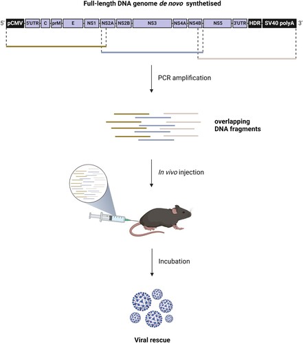
The efficacy of the in vivo application of the ISA method was evaluated by injecting overlapping subgenomic DNA fragments, encoding the full-length genome of TBEV, directly into mice (). Each subgenomic DNA fragment was de novo synthetized, cloned into a plasmid, and amplified by PCR. The full-length DNA genome is flanked by a eukaryotic transcription promoter at the 5’ end and by the hepatitis delta ribozyme followed by the simian virus 40 polyadenylation signal (HDR/SV40pA) at the 3’ end.
Figure 1. In vivo ISA method: The subgenomic DNA fragments amplified are equimolarly pooled and directly injected into animals. After an incubation period, animals show signs of viral disease that coincide with virus detection.

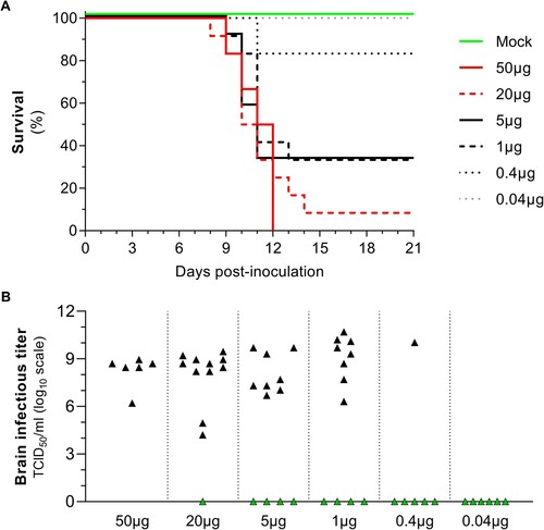
For this proof of concept, we used the Hypr strain of the TBEV (European subtype), considered highly virulent in mice [Citation19], in order to improve the detection of signs of viral disease in this model (Figure S1). Groups of 6 mice were inoculated in the posterior tibial muscle with an equimolar pool of the 3 subgenomic DNA fragments, at different doses (50-0.04 µg), formulated in NaCl 0.9% solution. The mice were monitored daily for 21 days for signs of viral disease. Survival curves, based on humane endpoints, were obtained on the basis of suffering criteria (Table S2) and brain infectious titres were determined by cell culture assays. The combined results of two independent experiments are shown in .
Figure 2. De novo generation of a highly pathogenic TBEV strain by inoculation of subgenomic DNA fragments in mice. Groups of 4 weeks-old C57BL/6J mice were inoculated intramuscularly with different amounts of DNA fragments encoding the Hypr strain of TBEV. Data represent results from two independent experiments (Table S6). Six different doses of an equimolar mix of DNA fragments formulated in NaCl 0.9% were used: 50, 0.4 and 0.04 µg (n = 6 animals) and 20, 5 and 1 µg (n = 2 × 6 animals). (A) Survival data presented as Kaplan-Meier curves. Curves of “Mock” and “5µg” groups are incremented by +2 and +1 Y-axis units, respectively, to optimized representation of the results. Statistical analysis is presented in Table S7. (B) Viral infectious titres expressed as TCID50/ml of brain clarified homogenates. Black and green symbols represent infectious titres measured in brain of sick animals and survivors, respectively.

Results showed that the in vivo ISA method is effective: inoculation of mice with DNA fragments resulted in signs of viral disease, in all groups except with the dose of 0.04 µg. At a dose greater than or equal to 1 µg, more than half the animals were infected. The rate of infection (i.e. percent of confirmed infected animals among all inoculated animals) correlated with the quantity of DNA fragments inoculated ((A)). These observations allowed us to establish a 50% lethal dose (LD50) of 0.67 µg. The presence and absence of infectious virus in the brains of sick animals (i.e. animals euthanized following the appearance of symptoms) and survivors (i.e. animals that did not show signs of illness until the end of the experiment) were confirmed by cell culture titration and viral isolation assays, respectively ((B)). The infectious titres measured in the brains of sick animals were heterogeneously distributed between 4.2 and 10.7 log10 TCID50/ml. The quantity of DNA fragment inoculated did not correlate with the quantity of viral particles in the brain of sick animals. This heterogeneity could be explained by the difference in brain collection time that ranges between 8 and 14 days post-inoculation ((A)). Moreover, viral load in brain had already been reported as dose-independent in a mouse model of peripheral TBEV infection [Citation20].
In order to optimize the rate of infection in our model, other formulations were tested during the same experiments. The use of solutions of Tyrode’s salts or Lutrol 3.5 mM, a cationic polymer, did not improve the rate of infection (Figure S2).
We previously demonstrated that the fidelity of the PCR polymerase used to produce subgenomic DNA fragments influences the genetic variability of the viral populations rescued in vitro [Citation21]. To investigate this question in vivo, we inoculated mice with 20 µg of DNA fragments amplified using a high-fidelity polymerase (Taq HiFi, 6 times fidelity of Taq polymerase) or a super high-fidelity polymerase (Taq SuperFi, over 300 times fidelity of Taq polymerase) (Figure S3). The complete genomes of the viruses present in the brains of the sick mice were sequenced, and a similar average number of mutations per animal (1.22 and 1.33) were observed in both groups (Figure S3.A-B). We also evaluated the in vitro replicative fitness of viruses collected from the brain of one animal in each group, and then compared it with the in vitro replicative fitness of viruses collected from the brain of an animal directly infected with TBEV Hypr strain (at the dose of 2.105 TCID50). Viral RNA yields were measured in cell supernatants at 24, 48, and 72 h post-infection (hpi). The viral RNA yields were 6.92, 8.41, and 7.46 log10 copies/mL at 24hpi, 10.74, 11.30, and 11.05 log10 copies/mL at 48hpi, and 11.12, 11.25, and 11.18 log10 copies/mL at 72hpi for animals inoculated with Taq Hifi DNA fragments, Taq Superfi DNA fragments and the Hypr strain, respectively (Figure S3.C). Overall, there were no major differences in genomic and fitness characteristics between viral populations generated using high-fidelity or super high-fidelity polymerase. Even so, we decided to standardized the protocol for amplifying DNA fragments by using the super high-fidelity polymerase for the following experiments in order to minimize the possible occurrence of mutations that could impact viral replication.
Optimization of the in vivo ISA method: electroporation and reduction in the number of DNA fragments
To improve the rate of infection (i.e. percent of confirmed infected animals among all inoculated animals) using the in vivo ISA method, we explored two potential optimization strategies: (1) coupling intramuscular injection with an electroporation protocol, and (2) using two overlapping subgenomic DNA fragments (Figure S4).
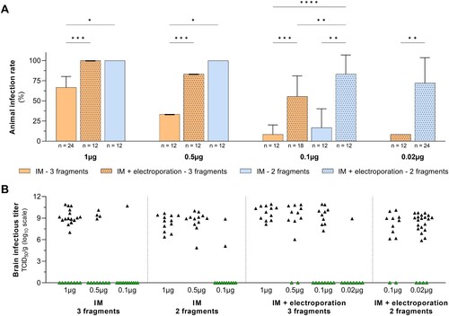
Groups of 6–18 mice were inoculated with amounts of DNA fragments ranging from 1 to 0.02 µg. For each experiment, a control group of 6 mice received 1 µg of DNA fragments intramuscularly. As previously, survival curves were obtained on the basis of suffering criteria (Table S2) and the presence of infectious virus was confirmed in the brains of sick animals, as well as the absence of virus in survivors. Animal rate of infections and brain infectious titres are presented in .
Figure 3. Optimization of the in vivo ISA method. Groups of 4 weeks-old C57BL/6J mice were inoculated intramuscularly with different amounts of DNA fragments (0.02-1 µg) encoding the Hypr strain of TBEV. Classic intramuscular injection (IM) or IM injection couples with an electroporation step were performed. TBEV genome was injected as 3 or 2 subgenomic DNA fragments. Data represents results from four independent experiments (Table S8 and Figure S5). (A) Animal infection rate. Each bar represents the rate of infection (i.e. % of confirmed sick animals among all inoculated animals) for each conditions explored. Number of mice (n) in each condition is show below the X-axis. This figure shows data presented in Figure S5 and Table S9. Statistical analysis are presented in Table S10 (Two-way ANOVA). ****, ***, ** and * symbols indicated a significant difference with a p-value ≤0.0001 and ranging between 0.0001–0.001, 0.001–0.01, and 0.01–0.05, respectively. (B) Viral infectious titres expressed as TCID50/g of brain in each condition. Black and green symbols represent infectious titres measured in brain of sick animals and survivors, respectively.

With equal amounts of DNA fragments, both electroporation protocol and the use of two DNA fragments resulted in a better rate of infection. Rates of infections were 1.5–6.7 times higher by using electroporation. This improvement was significant at all doses when using 3 (p = 0.0006) and 2 (p = 0.0021) fragments. Rates of infections were 1.5–10 times higher when using two DNA fragments instead of three. This improvement was significant at all doses when using IM injection (p = 0.024) and at 0.1 and 0.02 µg doses (p = 0.0041) by using electroporation. The infectious titres in brains of sick animals did not appear to be impacted by the viral genome fragmentation and the inoculation protocol: they were heterogeneously distributed between 4.9 and 10.9 log10 TCID50/g.
Application of the in vivo ISA method to other viruses
To evaluate the versatility of the in vivo ISA method, we used the same approach with two other arboviruses for which 3-fragmented ISA-based reverse genetic systems of wildtype (i.e pathogenic) strains were already available in the laboratory: JEV [Citation4] and CHIKV [Citation5] (Figure S6). The optimal inoculation protocol for this type of reverse genetic system, defined in the previous experiments, was used to generate these strains in mice.
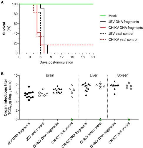
One day after receiving anti-IFNAR antibodies, groups of 12 animals were inoculated with 1 µg of DNA fragments by intramuscular route coupled with electroporation and control groups of 6 mice were inoculated with a dose of 104 TCID50 of JEV or CHIKV produced in vitro by the ISA method. As previously, survival curves were obtained on the basis of suffering criteria (Table S2) and the presence of infectious virus was confirmed in the organs of sick animals, as well as the absence of virus in survivors, using cell culture assays ().
Figure 4. De novo generation of JEV and CHIKV pathogenic strains using the in vivo ISA method. Groups of 4 weeks-old C57Bl/6J mice were inoculated with subgenomic DNA fragments encoding for a CHIKV strain or a JEV strain. Data represents results from a single experiment (Table S11). Animals were inoculated with 1 µg of DNA fragments by intramuscular route coupled with an electroporation step. As viral controls, groups of animals were inoculated with viral particles (104 TCID50) generated using same DNA fragments in vitro. (A) Survival data presented as Kaplan-Meier curves. Results from animals inoculated with viral particles (n = 6) and DNA fragments (n = 12) are presented as dotted and solid line, respectively. (B) Viral infectious titres expressed as TCID50/g of brain, liver and spleen. Black and white symbols represent infectious titres measured in organs of sick animals. Green symbols represent infectious titres measured in organs of survivors.

Results showed that the in vivo ISA method is fully applicable to other arboviruses: inoculation of DNA fragments resulted in signs of disease in all groups. Injection of the JEV strain produced in vitro caused infection in all animals, while only one survived in the group inoculated with the CHIKV strain (A). In addition, the nature of the material (ie. DNA or infectious viral particles) used to infect animals did not appear to impact the viral replication in organs. Brains were recovered from all animals. Spleens and livers were also harvested from CHIKV-infected animals. Mean infectious titres in organs of sick mice were comparable between groups: 5.6 and 5.9 log10 TCID50/g in brains of “JEV DNA fragments” and “JEV viral control” groups respectively; 6.6 log10 TCID50/g in brain of both “CHIKV” groups; 7.4 and 7.6 log10 TCID50/g in liver of “CHIKV DNA fragments” and “CHIKV viral control” groups, respectively; 7.6 log10 TCID50/g in spleen of both “CHIKV” groups, respectively ((B)).
Discussion
The ISA method is a simple procedure for rescuing viruses in vitro within days. It involves the direct transfection of susceptible cells with overlapping subgenomic DNA fragments covering a complete viral genome. In this study, we adapted this method to produce the Hypr strain of the TBEV directly in vivo and demonstrated its effectiveness. Fragments of subgenomic DNA, identical to those used in vitro, were directly inoculated intramuscularly into mice. This method was effective to generate de novo infectious viruses in mice with doses ranging from 50 to 0.4 µg formulated in a 0.9% NaCl solution. We also varied the parameters of this protocol to conclude that the use of an electroporation protocol and a lower number of fragments (2 instead of 3) increased the rate of infection of the animals and achieved 100% efficacy with 1 µg doses with 3-fragment systems. By combining the most effective parameters, we were able to rescue two flaviviruses and one alphavirus: TBEV, JEV and CHIKV.
Numerous RNA and DNA viruses have already been produced in vivo following inoculation of nucleic acid into animals. However, it generally involves inoculation of the viral genome as a single molecule, an infectious clone in most cases. To our knowledge, two other studies have reported the inoculation of fragmented genetic material to rescue viruses. In the 90’s, Sprengel et al. observed, in a duck model, the generation of infectious duck hepatitis B virus particles, through recombination of DNA fragments [Citation17]. In a more recent work, Willems et al. observed a similar phenomenon with the bovine leukaemia virus through inoculation of clones that were individually non-infectious but capable of producing viable viruses when injected simultaneously [Citation22].
The de novo generation of recombinant strains directly in vivo enables recombinant strains to bypass the positive and negative selection events that can occur during replication in cell culture and potentially impact the phenotype of the virus before inoculation into animals. Nevertheless, the main current application of DNA-launched in vivo infection is the development of new strategy to deliver LAV. By combining the ease of preparation, the genetic and thermal stability of traditional DNA vaccines with the high immunogenicity of conventional LAV, DNA-launched LAV could be a strategy for improving the biosafety and distribution of existing LAV [Citation23]. Several studies have shown that DNA coding for live attenuated viral strains of several arboviruses, such as yellow fever virus and Venezuelan equine encephalitis virus, can induce a protective immune response in mice [Citation24–36]. Some of these viruses were also generate in vivo using an electroporation protocol following intramuscular inoculation of DNA. However, these studies use an infectious clone as genetic material for transfection, which has certain disadvantages for their production within a bacterial vector.
The ISA method has the capacity to generate viral strains quickly and easily in vitro. The use of several subgenomic DNA fragments makes it possible to introduce mutations into the genome of the virus under study, thus producing attenuated viral strains. We believe that the in vivo transposition of this technique, presented in this study, will provide the same advantages. As well as the ability to directly assess the immunogenicity of these attenuated viral strains, making this a new tool for the development of LAV.
With the exception of transcriptional regulatory sequences, the genetic material used in the in vivo ISA method corresponds solely to the complete genome of the virus of interest while genomic sequences from other prokaryotes and eukaryotes were also administrated when using an infectious clone. Indeed, this is part of the current debate on the injection of foreign genetic material into humans. Nevertheless, this particularity does not prevent this method from being carefully evaluated with a view to its safe use in humans.
Author contributions
MC, XdL and AN designed the paper. MC, GM, JSD and GP performed and analysed experiments. MC wrote the paper. AN and JSD reviewed the paper. MC, XdL and AN designed and supervised experimental work. All authors have read and approved the submission of the manuscript.
Acknowledgements
We thank Rayane Amaral and Noémie Courtin (UVE; Marseille) for them technical contribution.
Supplemental Material
Download PDF (554.5 KB)Supplemental Material
Download MS Excel (3.7 MB)Disclosure statement
No potential conflict of interest was reported by the author(s).
Data availability statement
Authors can confirm that all other relevant data are included in the paper and/or its Supplementary information files.
Correction Statement
This article has been corrected with minor changes. These changes do not impact the academic content of the article.
Additional information
Funding
References
- Aubry F, Nougairède A, Gould EA, et al. Flavivirus reverse genetic systems, construction techniques and applications: a historical perspective. Antiviral Res. 2015;114:67–85. doi:10.1016/j.antiviral.2014.12.007
- Gritsun TS, Gould EA. Infectious transcripts of tick-borne encephalitis virus, generated in days by RT-PCR. Virology. 1995;214(2):611–618. doi:10.1006/viro.1995.0072
- Lindenbach BD. Reinventing positive-strand RNA virus reverse genetics. Adv Virus Res. 2022;112:1–29. doi:10.1016/bs.aivir.2022.03.001
- Aubry F, Nougairède A, de Fabritus L, et al. Single-stranded positive-sense RNA viruses generated in days using infectious subgenomic amplicons. J Gen Virol. 2014;95(Pt 11):2462–2467. doi:10.1099/vir.0.068023-0
- Driouich JS, Ali SM, Amroun A, et al. SuPReMe: a rapid reverse genetics method to generate clonal populations of recombinant RNA viruses. Emerg Microbes Infect. 2018;7(1):40. doi:10.1038/s41426-018-0040-2
- Klitting R, Riziki T, Moureau G, et al. Exploratory re-encoding of yellow fever virus genome: new insights for the design of live-attenuated viruses. Virus Evol. 2018;4(2):vey021. doi:10.1093/ve/vey021
- de Fabritus L, Nougairède A, Aubry F, et al. Utilisation of ISA reverse genetics and large-scale random codon Re-encoding to produce attenuated strains of tick-borne encephalitis virus within days. PLoS One. 2016;11(8):e0159564. doi:10.1371/journal.pone.0159564
- Gierer A, Schramm G. Infectivity of ribonucleic acid from tobacco mosaic virus. Nature. 1956;177(4511):702–703. doi:10.1038/177702a0
- Will H, Cattaneo R, Koch H-G, et al. Cloned HBV DNA causes hepatitis in chimpanzees. Nature. 1982;299(5885):740–742. doi:10.1038/299740a0
- Emerson SU, Lewis M, Govindarajan S, et al. cDNA clone of hepatitis A virus encoding a virulent virus: induction of viral hepatitis by direct nucleic acid transfection of marmosets. J Virol. 1992;66(11):6649–6654. doi:10.1128/jvi.66.11.6649-6654.1992
- Pushko P, Lukashevich IS, Weaver SC, et al. DNA-launched live-attenuated vaccines for biodefense applications. Expert Rev Vaccines. 2016;15(9):1223–1234. doi:10.1080/14760584.2016.1175943
- Gigante A, Li M, Junghänel S, et al. Non-viral transfection vectors: are hybrid materials the way forward? Medchemcomm. 2019;10(10):1692–1718. doi:10.1039/C9MD00275H
- Atianand MK, Fitzgerald KA. Molecular basis of DNA recognition in the immune system. J Immunol. 2013;190(5):1911–1918. doi:10.4049/jimmunol.1203162
- Hannaman D, Dupuy LC, Ellefsen B, et al. A phase 1 clinical trial of a DNA vaccine for Venezuelan equine encephalitis delivered by intramuscular or intradermal electroporation. Vaccine. 2016;34(31):3607–3612. doi:10.1016/j.vaccine.2016.04.077
- Vasan S, Hurley A, Schlesinger SJ, et al. In vivo electroporation enhances the immunogenicity of an HIV-1 DNA vaccine candidate in healthy volunteers. PLoS One. 2011;6(5):e19252. doi:10.1371/journal.pone.0019252
- Villemejane J, Mir LM. Physical methods of nucleic acid transfer: general concepts and applications. Br J Pharmacol. 2009;157(2):207–219. doi:10.1111/j.1476-5381.2009.00032.x
- Ninove L, Nougairede A, Gazin C, et al. RNA and DNA bacteriophages as molecular diagnosis controls in clinical virology: a comprehensive study of more than 45,000 routine PCR tests. PLoS One. 2011;6(2):e16142. doi:10.1371/journal.pone.0016142
- Driouich JS, Cochin M, Lingas G, et al. Favipiravir antiviral efficacy against SARS-CoV-2 in a hamster model. Nat Commun. 2021;12(1):1735. doi:10.1038/s41467-021-21992-w
- Asghar N, Lee Y-P, Nilsson E, et al. The role of the poly(A) tract in the replication and virulence of tick-borne encephalitis virus. Sci Rep. 2016;6:39265. doi:10.1038/srep39265
- Hayasaka D, Nagata N, Fujii Y, et al. Mortality following peripheral infection with tick-borne encephalitis virus results from a combination of central nervous system pathology, systemic inflammatory and stress responses. Virology. 2009;390(1):139–150. doi:10.1016/j.virol.2009.04.026
- Driouich JS, Moureau G, de Lamballerie X, et al. Reverse genetics of RNA viruses: ISA-based approach to control viral population diversity without modifying virus phenotype. Viruses. 2019;11(7). doi:10.3390/v11070666
- Willems L, Kettmann R, Dequiedt F, et al. In vivo infection of sheep by bovine leukemia virus mutants. J Virol. 1993;67(7):4078–4085. doi:10.1128/jvi.67.7.4078-4085.1993
- Pushko P, Lukashevich IS, Johnson DM, et al. Single-Dose immunogenic DNA vaccines coding for live-attenuated alpha- and flaviviruses. Viruses. 2024;16(3). doi:10.3390/v16030428
- Tretyakova I, Lukashevich IS, Glass P, et al. Novel vaccine against Venezuelan equine encephalitis combines advantages of DNA immunization and a live attenuated vaccine. Vaccine. 2013;31(7):1019–1025. doi:10.1016/j.vaccine.2012.12.050
- Tretyakova I, Nickols B, Hidajat R, et al. Plasmid DNA initiates replication of yellow fever vaccine in vitro and elicits virus-specific immune response in mice. Virology. 2014;468-470:28–35. doi:10.1016/j.virol.2014.07.050
- Tretyakova I, Hearn J, Wang E, et al. DNA vaccine initiates replication of live attenuated chikungunya virus in vitro and elicits protective immune response in mice. J Infect Dis. 2014;209(12):1882–1890. doi:10.1093/infdis/jiu114
- Nickols B, Tretyakova I, Tibbens A, et al. Plasmid DNA launches live-attenuated Japanese encephalitis virus and elicits virus-neutralizing antibodies in BALB/c mice. Virology. 2017;512:66–73. doi:10.1016/j.virol.2017.09.005
- Tretyakova I, Tibbens A, Jokinen JD, et al. Novel DNA-launched Venezuelan equine encephalitis virus vaccine with rearranged genome. Vaccine. 2019;37(25):3317–3325. doi:10.1016/j.vaccine.2019.04.072
- Jiang X, Dalebout TJ, Lukashevich IS, et al. Molecular and immunological characterization of a DNA-launched yellow fever virus 17D infectious clone. J Gen Virol. 2015;96(Pt 4):804–814. doi:10.1099/jgv.0.000026
- Zou J, Xie X, Luo H, et al. A single-dose plasmid-launched live-attenuated Zika vaccine induces protective immunity. EBioMedicine. 2018;36:92–102. doi:10.1016/j.ebiom.2018.08.056
- Chin WX, Lee RCH, Kaur P, et al. A single-dose live attenuated chimeric vaccine candidate against Zika virus. NPJ Vaccines. 2021;6(1):20. doi:10.1038/s41541-021-00282-y
- Avila-Perez G, Nogales A, Park J-G, et al. In vivo rescue of recombinant Zika virus from an infectious cDNA clone and its implications in vaccine development. Sci Rep. 2020;10(1):512. doi:10.1038/s41598-020-57545-2
- Kum DB, Mishra N, Vrancken B, et al. Limited evolution of the yellow fever virus 17d in a mouse infection model. Emerg Microbes Infect. 2019;8(1):1734–1746. doi:10.1080/22221751.2019.1694394
- Yamshchikov V, Manuvakhova M, Rodriguez E, et al. Development of a human live attenuated West Nile infectious DNA vaccine: identification of a minimal mutation set conferring the attenuation level acceptable for a human vaccine. Virology. 2017;500:122–129. doi:10.1016/j.virol.2016.10.012
- Yamshchikov V, Manuvakhova M, Rodriguez E. Development of a human live attenuated West Nile infectious DNA vaccine: suitability of attenuating mutations found in SA14-14-2 for WN vaccine design. Virology. 2016;487:198–206. doi:10.1016/j.virol.2015.10.015
- Hall RA, Nisbet DJ, Pham KB, et al. DNA vaccine coding for the full-length infectious Kunjin virus RNA protects mice against the New York strain of West Nile virus. Proc Natl Acad Sci U S A. 2003;100(18):10460–4. doi:10.1073/pnas.1834270100
