
Abstract
Human adenoviruses (HAdV) are a common cause of clinical infections in South Africa. However, there is a lack of information regarding the prevalence and molecular identification of this virus in the environment. The objective of this study was to investigate the recovery and molecular identification of HAdV in sewage and mussel samples. All samples were subjected to transmission electron microscopy, viral DNA extraction and nested PCR amplification using adenovirus-specific primers targeting a conserved region of the hexon gene. Amplicons were cloned and sequenced and BLAST analysis revealed a closest matched sequence (98% identity) belonging to HAdV-D17.
Introduction
Viral gastroenteritis is a global health issue resulting in high mortality rates annually. The Global Burden of Disease Study (GBD) 2013 Mortality and Causes of Death Collaborators listed gastroenteritis as the fourth cause of mortality worldwide, primarily in developing countries.Citation1 Although rotavirus is the primary cause of viral gastroenteritis globally, other viral pathogens including adenovirus, norovirus and astrovirus are also recognised as significant causes of the disease.Citation2
Adenoviruses are non-enveloped, icosahedral viruses belonging to the family Adenoviridae and genus Mastadenovirus. The viral particle contains a linear double-stranded DNA genome ranging from 30-36 kilobases with a virus capsid ~70 to 100 nm in diameter; comprising of 252 capsomers (240 hexons and 12 pentons) and 12 fiber proteins. Over sixty human adenovirus (HAdV) types have been identified and these are divided into seven known species (A to G) based on a variety of biochemical, immunological and genetic parameters. The various types are associated with multiple diseases including gastroenteritis, respiratory diseases and conjunctivitis.Citation3,4
Adenoviruses have been detected in river, raw and treated drinking water in South Africa.Citation5 Recent studies have reported a prevalence of HAdV in the Buffalo River,Citation6 Tyume River,Citation7 and final effluents of wastewater treatment plants in the Eastern Cape, South Africa.Citation8 These studies screened for adenovirus-associated infections in humans, including gastroenteritis (HAdV-F40 and HAdV-F41), respiratory tract infections (HAdV-C1, HAdV-C2, HAdV-C5, HAdV-C6, HAdV-B21 and HAdV-E4), urinary tract infections (HAdV-B21) and eye infections (HAdV-B3 and HAdV-B7). In addition, Pauly et al reports on the high prevalence and detection of HAdV-D in four Sub-Saharan countries and emphasises the need for further investigations in order to identify the pathological potential of these viruses.Citation9
Aside from adenoviruses being prevalent in stool samples,Citation9–11 several studies have shown that cases of human enteric viruses causing gastroenteritis were associated with the consumption of shellfish.Citation12,13 Bivalves/shellfish can filter massive quantities of seawater as part of their feeding activities, and can accumulate and concentrate viruses and other pathogens from human faecal pollution.Citation12 Furthermore, bivalve molluscs in coastal waters (such as mussels) are occasionally exposed to urban wastewater and are well-recognised vehicles in the transmission of bacterial and viral enteric diseases.Citation12,13
The overall aim of this study was to investigate the recovery and identification of HAdV in raw sewage and mussel samples using transmission electron microscopy and molecular techniques.
Methodology
Samples
Raw sewage was collected by swabbing separation grids at the Makana Wastewater Treatment Plant in Grahamstown, Eastern Cape, South Africa. Approximately 120 mussels were obtained from the Swartkops River in Port Elizabeth, Eastern Cape, South Africa (coordinates: 33°51′36.7″ S, 25°37’12.0” E) and stored at −20 °C. All samples were collected in early May. Groups of five swab samples and 5-6 mussels were pooled separately in order to enable greater virus recovery prior to viral DNA extraction, PCR amplification and cloning. Ethical approval was not required since all research was performed on invertebrates. The mussel collection was conducted under the licence of C McQuaid (Department of Zoology and Entomology, Rhodes University), field permit RES2014/12, issued by the Department of Agriculture, Forestry and Fisheries of South Africa.
Virus recovery
Raw sewage swabs and homogenised mussel samples were pooled and diluted into glycine buffer solution (0.05 M glycine in NaCl, pH 9.0) and treated with 1% NP-40 (Sigma-Aldrich, USA) and incubated for 1 hour with shaking at room temperature. The volumes of glycine buffer used were 1 ml for swab samples and 3 ml for mussel homogenate. Samples were centrifuged at 4000 x g for 20 minutes to remove the debris. Thereafter, the supernatant was poured through cheesecloth, filtered using 0.45 μM syringe filters (Lasec, South Africa) and clarified at 4000 x g for 20 minutes. Samples were stored at −20 °C until use.
Transmission Electron Microscopy (TEM)
TEM was carried out according to the protocol described by Hayek.Citation14 Approximately 10 μl of filtered raw sewage and mussel sample was placed on individual carbon coated grids for 30 seconds and excess sample drained off using filter paper. The carbon grids were negatively stained with 5 μl of 1% uranyl acetate applied for 30 seconds and allowed to dry overnight. Grids were viewed on a Zeiss Libra 120 transmission electron microscope (Germany) and all images captured using Mega view (G2) Olympus analysis software.
Viral DNA extraction and nested PCR
Viral DNA was extracted using the ZR Viral DNA Kit™ (Zymo Research, USA) according to the manufacturer’s instructions and stored at −80 °C until further use. A nested PCR amplification was performed using degenerate primers ADHEX1F (20 380–20 400), ADHEX2R (20 632–20 652), ADHEX2F (20 485–20 503) and ADHEX1R (20 836–20 854) designed by Avellón et al.Citation15 The PCR reaction mixture for the first and second rounds (total volume 25 μl) contained 12.5 μl of KAPA Taq Ready-mix (Kapa Biosystems (Pty) Ltd, Cape Town, South Africa), 1.25 μl of each primer (10 μM stock) and about 50 ng of DNA template. The PCR cycling parameters used were described by Van Heerden et al.Citation5
DNA cloning and sequencing
Amplicons were gel-purified using the GeneJET gel extraction kit (Thermo Scientific, USA) according to the manufacturer’s guidelines and ligated into pJET1/blunt (CloneJET PCR Cloning Kit, Thermo Scientific, USA). Ligations were transformed into competent DH5α E. coli cells, according to standard protocols. Plasmids were extracted using the QIAprep Spin Miniprep Kit (Qiagen, USA) according to the manufacturer’s protocol, restricted with XbaI and XhoI endonucleases, and digests analysed by 1% agarose gel electrophoresis in Tris-acetate EDTA buffer [(TAE, 20 mm Tris, 1% v/v glacial acetic acid and 5 mM EDTA (pH 8)] with ethidium bromide.
Sequence analysis and phylogenetics
Plasmids containing inserts of interest were subjected to bi-directional sequencing (Inqaba Biotechnical Industries (Pty) Ltd., Pretoria, South Africa). Sequences were submitted to BLAST analysis to identify their most closely related matches in the NCBI GenBank database. Bioinformatics analysis was performed as follows: the raw forward and reverse ABI files were aligned and assembled into a single consensus sequence using MEGA software (version 7). Complete genomic sequences of human adenovirus species A-F were retrieved from the NCBI GenBank database. Our sequenced wastewater and mussel samples were used as query sequences to retrieve full-coverage BLAST hits against the acquired genomic sequences. These sequences were then translated in all six frames using the ExPASy translate tool to retrieve the longest uninterrupted reading frames as a preparation for codon-based sequence alignment. Selected nucleotide translations were verified using BLAST searches against the non-redundant (nr) protein database before proceeding to multiple sequence alignment using MUSCLE. Codon-based alignment of the nucleotide sequences was subsequently performed using the EMBOSS transalign tool with the aligned protein sequences as a guide. The phylogenetic tree was constructed from the aligned nucleotide sequences using the neighbour-joining algorithm with 1 000 bootstraps using the Kimura two-parameter nucleotide substitution model from MEGA software (version 7). NCBI GenBank accession numbers corresponding to the sequences used for phylogenetic analysis included: HAdV-A (NC_001460.1), HAdV-B (JX423386.1), HAdV-C (NC_001405.1), HAdV-D (AC_000006.1), HAdV-E (NC_003266.2) and HAdV-F (NC_001454.1). Bovine (AAF82136), equine (AAB88060) and avian (Y09598.1) adenovirus sequences were used as outgroups.
The sequenced samples and the nucleotide sequences pertaining to HAdV species D retrieved from the NCBI GenBank database were used to generate the multiple sequence alignments with ClustalW using the Geneious R9 software (Biomatters Ltd., Auckland, New Zealand).
Results
Virus particles with a size range (70–90 nm) typical of HAdV were observed by TEM in all samples analysed demonstrating the efficacy of the virus recovery technique used in the study (data not shown). Following DNA extraction from samples, a nested PCR assay was conducted using HAdV-specific oligonucleotides designed by Avellón et al.Citation15 These primers are degenerate and have been designed to detect all known adenovirus HAdV types. In addition, the nested PCR amplification increases the primer binding specificity for adenoviral sequences, thus reducing the probability of generating false positives for adenoviral sequences.Citation5 Furthermore, this approach allows for the detection of a small number of viral genomes in samples ranging from environmental to clinical samples.Citation15
Amplicons of approximately 168 base pairs (bp) were obtained following optimisation of the protocol, and these were subsequently cloned. Restriction enzyme digestion of selected plasmids revealed inserts of the expected size in one raw sewage and one mussel sample. DNA sequences were analysed by BLAST and the closest matched sequence (accession number AB330098.1; 98% nucleotide identity; e-value 3e-76) was the HAdV-D type 17 hexon gene in both samples sequenced. Classification into group D was confirmed by phylogenetic analysis. Although seven of the pooled samples were analysed, only one sample from pooled raw sewage and one sample from pooled mussel extract returned readable sequences.
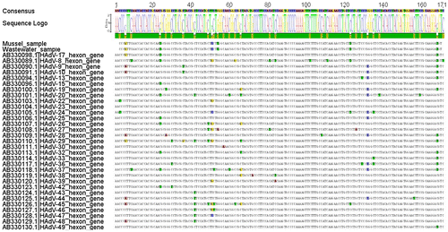
The 168 bp nucleotide sequences obtained in this study were aligned with published human adenovirus HAdV-D species nucleotide sequences (NCBI GenBank accession numbers are present in Figure ). The nucleotide multiple sequence alignment of the hexon gene sequences belonging to several HAdV-D types in comparison to the sequences found in this study is shown in Figure . A single consensus sequence was constructed for the sequences obtained from the wastewater and mussel samples as well as the serotypes in species D. The alignment revealed several single nucleotide polymorphisms (SNPs) all of which were synonymous since they did not affect the overall protein sequence (data not shown).
Figure 1: An illustration of the nucleotide multiple sequence alignment for the wastewater and mussel samples as well as the well-known adenovirus species D, with all the single-nucleotide polymorphisms highlighted according to a specific nucleotide modification.
Discussion
Recently, a virus of the species human adenovirus D was identified in diarrhoeal samples of six children in Bangladesh and was subsequently sanctioned as a novel type of HAdV-67 by the Human Adenovirus Working Group (GenBank accession number AP012302).Citation10 Moreover, Magwalivha et al provided significant insight on the incidence and prevalence of HAdV species D associated with gastroenteritis in Africa and raised awareness of the necessity for further investigations particularly in African countries.Citation11
This study describes the successful recovery and molecular identification of HAdV-D17 from raw sewage and mussel samples collected in the Eastern Cape province of South Africa. According to literature, HAdV-17 was the first species D adenovirus to be sequenced and the genome was submitted to NCBI’s GenBank in 1999.Citation16 More recently, Dehghan et al provided a corrected and newly annotated DNA sequence of the HAdV prototype 17 (accession number HQ910407)Citation16 and a comprehensive analysis of 20 HAdV-D complete genomes was subsequently described.Citation17 Importantly, HAdV-17 prototypes have been considered as a reference tool for comparative genomics of newly isolated HAdV-D adenoviruses capable of generating novel types, mainly through genome recombination events.Citation18
As yet, there appears to be minimal data available on the clinical relevance of HAdV-17, although it has been associated with keratoconjunctivitis and gastrointestinal infections.Citation4 Thus, the identification of HAdV-17 in this study is interesting and allows for more extensive screening of other biological material, including shellfish and clinical samples. Further work will involve obtaining a full genome sequence of the HAdV-17 hexon gene to determine if the virus present in these samples represents a prototypic or novel adenovirus species. Interestingly, Pauly et al suggests that the high virus diversity may also favour the emergence of recombinants with altered tropism and pathogenic characteristics.Citation9
The presence of HAdV-D17 in mussels raises concern about the ecological health of the river and the level of sewage contamination present in the Swartkops River estuary. The ecological integrity of the river is threatened by the polluting wastewater effluent originating from industrial activities and informal settlements in the region.Citation19 This observation is significant yet disturbing as not only have additional mussel samples tested positive for HAdV by nested PCR, but our studies have also detected enteric viruses such as norovirus and Aichi virus in the same tissue (data not shown). The contamination of rivers by raw sewage and other effluents is a serious threat to human health especially where it occurs in recreational areas and regions where shellfish are commercially farmed.Citation12
Conclusion and future work
To the best of our knowledge, this is the first report of HAdV-D17 being positively identified in raw sewage and mussel samples in the Eastern Cape province. The development of techniques in this study provides a platform for further studies involving more extensive sample collection as well as screening for alternative adenovirus types throughout South Africa. Ultimately this would lead to a better understanding of the prevalence of this virus in the country.
Conflict of interest
The authors declare no conflict of interest in performing this study.
Ethics statement
Ethical approval was not required for this research as only invertebrate marine molluscs (mussels) were employed.
Funding
This work was supported by SIR (Medical Research Council, South Africa) and Research Council (RC, Rhodes University) grants.
Acknowledgements
The authors gratefully acknowledge C McQuaid and J Trassierra (Department of Zoology and Entomology, Rhodes University) for their invaluable insight into this study and assistance with the mussel collection and dissection. The authors give special thanks to O Amamuddy (Research Unit in Bioinformatics, Rhodes University) for his substantial contribution and expertise pertaining to the bioinformatics analyses in this study.
References
- Naghavi M, Wang H, Lozano R, et al. GBD 2013 Mortality and Causes of Death Collaborators. Global, regional, and national age-sex specific all-cause and cause-specific mortality for 240 causes of death, 1990-2013: a systematic analysis for the Global Burden of Disease Study 2013. Lancet. 2015;385:117–71.
- Wilhelmi I, Roman E, Sanchez-Fauquier A. Viruses causing gastroenteritis. Clin Microbiol Infect. 2003;9:247–62.10.1046/j.1469-0691.2003.00560.x
- Horwitz MA. Adenoviruses. In: Fields BN, Knipe DM, Howley PM, editors. Fields virology. Philadelphia, PA: Lippincott-Raven Publishers; 1996. p. 2149–71.
- Ghebremedhin B. Human adenovirus: viral pathogen with increasing importance. Eur J Microbiol Immunol. 2014;4:26–33.10.1556/EuJMI.4.2014.1.2
- Van Heerden J, Ehlers M, Heim A, et al. Prevalence, quantification and typing of adenoviruses detected in river and treated drinking water in South Africa. J Appl Microbiol. 2005;99:234–42.10.1111/j.1365-2672.2005.02617.x
- Chigor VN, Okoh AI. Quantitative detection and characterization of human adenoviruses in the Buffalo river in the Eastern Cape Province of South Africa. Food Environ Virol. 2012;4:198–208.10.1007/s12560-012-9090-0
- Sibanda T, Okoh AI. Assessment of the incidence of enteric adenovirus species and serotypes in surface waters in the Eastern Cape province of South Africa: Tyume River as a case study. Sci World J. 2012;2012:949216.
- Osuolale O, Okoh A. Incidence of human adenoviruses and Hepatitis A virus in the final effluent of selected wastewater treatment plants in Eastern Cape Province, South Africa. Virol J. 2015;12:1.
- Pauly M, Hoppe E, Mugisha L, et al. High prevalence and diversity of species D adenoviruses (HAdV-D) in human populations of four Sub-Saharan countries. Virol J. 2014;11: 25.10.1186/1743-422X-11-25
- Matsushima Y, Shimizu H, Kano A, et al. Genome sequence of a novel virus of the species human adenovirus D associated with acute gastroenteritis. Genome Announc. 2013;1:10.
- Magwalivha M, Wolfaardt M, Kiulia NM, et al. High prevalence of species D human adenoviruses in fecal specimens from Urban Kenyan children with diarrhea. J Med Virol. 2010;82:77–84.10.1002/jmv.v82:1
- Lees D. Viruses and bivalve shellfish. Int J Food Microbiol. 2000;59:81–116.10.1016/S0168-1605(00)00248-8
- Karamoko Y, Ibenyassine K, Aitmhand R, et al. Adenovirus detection in shellfish and urban sewage in Morocco (Casablanca region) by the polymerase chain reaction. J Virol Methods. 2005;126:135–7.10.1016/j.jviromet.2005.02.003
- Hayat MA. Negative staining in electron microscopy. Biological applications. 4th ed. Cambridge: Cambridge University Press; 2000:367–99.
- Avellón A, Pérez P, Aguilar JC, et al. Rapid and sensitive diagnosis of human adenovirus infections by a generic polymerase chain reaction. J Virol Methods. 2001;92:113–20.10.1016/S0166-0934(00)00269-X
- Dehghan S, Seto J, Hudson NR, et al. Complete genome sequence of human adenovirus prototype 17. J Virol. 2011;85:11540–1.10.1128/JVI.06051-11
- Robinson CM, Singh G, Lee JY, et al. Molecular evolution of human adenoviruses. Scientific reports. 2013:3.
- Walsh MP, Chintakuntlawar A, Robinson CM, et al. Evidence of molecular evolution driven by recombination events influencing tropism in a novel human adenovirus that causes epidemic keratoconjunctivitis. PLoS One. 2009;4:e5635.10.1371/journal.pone.0005635
- Odume O, Muller W, Arimoro F, et al. The impact of water quality deterioration on macroinvertebrate communities in the Swartkops River, South Africa: a multimetric approach. Afr J Aquat Sci. 2012;37:191–200.10.2989/16085914.2012.670613