
ABSTRACT
This study aimed to estimate the levels of total serum IgE and assess the presence of eosinophils in middle ear fluid in primary and recurrent otitis media with effusion in Egyptian pediatric patients. A prospective study of 59 children with bilateral otititis media effusion (OME) were enrolled in this study. All patients had refractory OME to non-surgical lines of treatment. Full history was taken then detailed ENT examination followed by various investigations in the form of pure tone audiometry, tympanometry, serum total IgE, and eosinophilia. The incidence of allergic dermatitis, food allergy, allergic conjunctivitis, and anaphylaxis were (64.4%), (32.2%), (33.9%), (10.2%), (8.5%), and (5.1%), respectively. In this study middle ear effusion, eosinophils were positive in (61%). Total IgE was high in 40 children (67.8%). There was a strong relationship of recurrent OME with allergic manifestations, serum IgE, middle ear effusion fluid eosinophils, and blood eosinophilia suggesting intimately important role of allergy in the recurrence of OME. Allergy has a fundamental role in the pathogenesis of recurrent OME. The possibility of allergy should be considered in OME cases that did not improve with conventional lines of treatment.
Introduction
Otitis media with effusion (OME) is defined as chronic inflammation of the middle ear mucosa characterized by fluid storage behind an intact tympanic membrane (TM) within the middle ear space without signs or symptoms of an acute ear infection [Citation1]. It is one of the most common causes of childhood hearing loss that leads to surgical intervention [Citation2]. The etiology of OME has various factors. The most common pathogeneses are the Eustachian tube dysfunction and the immature function of the immune system. One of the most influencing factors is age. The peak influenced age group is between 2 and 5 years [Citation3]. Allergy also plays an important role in the pathogenesis of the OME as it causes triggered obstruction of the Eustachian tube [Citation4,Citation5].
The prevalence of OME is higher in atopic children. The associated common bilateralism and the objective hearing loss declare the allergy role in its initiation and recurrence [Citation6]. The allergy-induced cytokines act directly as key regulators of the Eustachian tube obstruction [Citation7]. Allergy may play a more significant role in recurrent than complicated OME [Citation8].
The researchers still investigate the role of IgE-mediated reactions in the pathogenesis of OME. Some studies recorded elevated total and specific IgE and other studies counteract these results [Citation9]. Thus, this point of search should be continued.
Another confusing problem is the eosinophilic otitis media. It is usually associated with bronchial asthma and is commonly resistant to conventional lines of treatment [Citation10].
The objective of this study was to estimate the levels of total serum IgE and assess the presence of eosinophils in middle ear fluid in primary and recurrent otitis media with effusion in Egyptian pediatric patients.
Patients and methods
This prospective descriptive case series study was enrolled over 59 children with OME that were admitted in the Department of Otolaryngology at Mansoura University Hospital, Mansoura, Egypt. It was conducted from April 2017 to June 2018. The diagnosis of OME was confirmed by clinical history, clinical examination, otoscopic examination, and tympanometry. Ethical approval for this work was obtained by Ethical committee board in Mansoura Faculty of Medicine, Mansoura University. The protocol of this current study was approved by the institutional review board with a proposal code number of MS/17.04.110. Consent was recruited from each of the study members.
All patients were under conservative treatment for 3 months or more planning for surgical interference under general anesthesia. The patients were selected with inclusion criteria of age less than 18 years, bilateral OME, resistance to medical treatment for 3 months. The patients with the following criteria were excluded acute otitis media, perforated tympanic membrane, cleft palate, down syndrome, and craniofacial abnormalities. The patients’ age ranged from 3 to 15 years old with mean age of 7.88 years and standard deviation ± 3.00. A complete medical history, clinical evaluation, and radiological investigations were conducted for all patients.
Blood samples were obtained from each child and collected into serum separate tubes in two aliquots. One aliquot was for eosinophils counting and expressed in cell/mm. The other was preserved for 30 min to clot then centrifuged at 2,500 rpm for 10 min. Aliquots of serum were stored at −70°C before analysis. Total IgE levels were measured by commercial ELISA kits (Diaclone, France). The sample was done in duplicate according to the manufacturer’s instructions. All values more than 100 IU/ml were considered as high total IgE. The peripheral blood eosinophils were counted on the smears prepared from 1 cc of the patients’ blood samples, transferred into oxalate, and stained with eosin using the Romanowsky method.
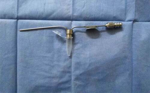
Middle ear effusions were collected from children and stored at – 40°C until used. Effusion fluid sample obtained from patients using a special suction tip during myringotomy and tympanostomy tube insertion under complete aseptic conditions by special suction tip (). Samples collected and transported within closed Eppendorf. Middle ear effusion obtained from one or both ears of the same case was considered a single sample. Samples stained by Hematoxylin and Eosin stain to be checked for the presence of eosinophils and counted.
Figure 1. Suction tip connected to Eppendorf
Statistical analysis
Data were tabulated and analyzed using the computer program SPSS (Statistical package for social science) version 23.0 to obtain: Descriptive statistics that were calculated in the form of: Mean±Standard deviation (SD) and frequency (Number-percent), Analytical statistics: in the statistical comparison between the different groups, the significance of difference was tested using inter-group comparison of categorical data by using chi-square test (χ2-value). P value <0.05 was considered statistically significant.
Results
This study was enrolled over fifty-nine children with OME. There were 30 male children (50.8%) and 29 female ones (49.2%) with mean age7.88 and SD ±3.00 years old. The allergic manifestations of the studied patients are illustrated in . Data were expressed as frequency (Number-percent). Associated allergic rhinitis was found in 38 cases (64.4%), bronchial asthma in 19 cases (32.2%), allergic dermatitis, urticaria or eczema in 20 cases (20.3%), food allergy in 6 cases (10.2%), allergic conjunctivitis in 5 cases (8.5%) and the anaphylaxis in 3 cases (5.1%).
Table 1. Frequency of allergic manifestations in the study group (59 children with OME)
Out of the studied cases, 34 children (57.6%) had otitis media with effusion for the first time (primary group) and 25 children (42.2%) with recurrent otitis media with effusion. Association between the recurrent otitis media and the allergic manifestation () reveals that there was significance between the recurrent otitis media with effusion and the bronchial asthma (P = 0.026*) and the food allergy (P = 0.03*).
Table 2. Association between recurrent OME and allergic manifestations
The total Ig E was high in 40 cases (67.8%) and low in 19 cases (32.2%). The total Ig E was high in 18 primary otitis media cases (52.9%) and 22 recurrent otitis media cases (88%) with significant P value = 0.004*. on the other hand, it was low in 16 primary cases (47.1%) and 3 recurrent cases (12%).
Eosinophilia was detected in 39 cases (66.1%) and not presented in 20 cases (33.9%). As regard the primary cases, it was positive in 18 cases (52.9%) and negative in 16 cases (47.1%). But in the recurrent otitis media, it was positive in 21 cases (84%) and negative in 4 cases (16%). There was significant association between the blood eosinophilia and the recurrent otitis media (P = 0.01*).
Eosinophils were measured in the effusion fluid in both the primary and the recurrent cases. They were positive in 16 cases (47.1%) of the primary cases and negative in 18 cases (52.9%) of the primary cases. In the recurrent otitis media with effusion, they were positive in 20 cases (80%) and negative in 5 cases (20%). There was significant association between the presence of the eosinophils and the recurrent otitis media with effusion (P = 0.01*).
Discussion
Although OME continues to be one of the most prevalent childhood diseases, a lot of controversy about its pathogenesis remains a mystery. It may start by inflammatory and immune reactions to some rhino pharyngeal infections with the production of cytokine and exudate rich inflammatory mediators [Citation11]. Further research must target the cause and prevention of OME especially those refractory to conventional treatment modalities [Citation12].
An increasingly important issue in pediatric otolaryngology is the relation between chronic otitis media with effusion and allergy [Citation13]. A connection between both disorders is recognized, but the underlying mechanism remains unclear. Allergic inflammation is the most common reason of Eustachian tube obstruction that can contribute to OME with increased susceptibility to upper airway infections or by local inflammatory reactions [Citation14].
Regarding various allergic manifestations, there was a high prevalence of allergic manifestations in the form of allergic rhinitis, bronchial asthma, allergic dermatitis, food allergy, allergic conjunctivitis, and anaphylaxis (64.4, 32.2, 33.9, 10.2, 8.5, and 5.1%), respectively. This high prevalence of allergic disorders makes the study in agreement with Chantzi et al. [Citation15] who carried out a controlled study on 88 children with OME aged 1–7 years and found a higher prevalence of allergic disorders in OME patients. On the other hand, it disagrees with Souter et al. [Citation16] who studied 89 children aged 6 or 7 years with OME concluded that the prevalence of allergic symptoms was nearly the same in both the studied group and the reference group in the same age meaning that there was a limited effect of allergy OME pathogenesis in this age group.
Symptoms of allergic rhinitis were found to affect 64.4% of the studied children. A higher frequency of allergic rhinitis symptoms in OME patients was in Alles et al. [Citation17] study who stated symptoms of AR in 89% of studied group (209 children) but this may be due to larger study group than ours. But these results agree also with Martines et al. [Citation18] who found Symptoms of allergic rhinitis in 60% of OME children. This study disagrees with Cassano et al. [Citation19] and Martines et al. [Citation18] who found that neither OME nor Eustachian tube dysfunction is significantly affected by the allergic reactions. The study results revealed that frequency of bronchial asthma was 32.2% of patients in agreement with Alles et al. [Citation17] and Passali et al. [Citation20] studies who found bronchial asthma in 36% and 37% of OME patients, respectively. Chantzi et al. [Citation15] stated that bronchial asthma was the most straightforward target for a possible intervention, the effectiveness of appropriate asthma medications in prophylaxis, and treatment of OME. As regard food allergy history, it was e positive in 10.2% of studied children and this agrees with Döner et al. [Citation21] who found a higher prevalence of food allergy in OME patients than in controls. Collection of MEE samples was collected by many methods Gomaa et al. [Citation1] and El-Sharnoby et al. [Citation22] obtained effusion fluid samples from patients by using wide bore (5 ml.) syringe in their clinical studies. But Lino et al. [Citation23], Kanazawa et al. [Citation24] and Saliba et al. [Citation25] collected MEE samples using a JuhnTym-Tap middle ear fluid aspirator/collector in their clinical studies.
Middle ear effusion samples were collected by special suction tip () under complete aseptic conditions, we choose this method as it was cheap, precise, available, and quick.
In this study Hematoxylin and Eosin stain was used for staining middle ear effusion fluid samples to examine the presence of eosinophils. This technique is direct, simple, cheap, rapid and is the most precise method in direct detection and visualization of eosinophils while immunophenotyping by flow-cytometry which is an expensive technique and used by Saliba et al. [Citation25] that used the term eosinophil-like to identify the corresponding cells of flow cytometry.
The study results showed eosinophils in middle ear effusion of 36 cases (61%) that was in agreement with both Nagamine et al. [Citation26] and Nguyen et al. [Citation27]. The total IgE was considered as high total IgE for all values more than 100 IU/ml, our results showed high IgE levels in 40 children (67.8%) It agrees with El-Sharnoby et al. [Citation22] who found that total IgE is higher in patients’ serum with OME than in the serum of the control group. Lino [Citation23] and Kanazawa et al. [Citation24] showed that total IgE levels were significantly higher in the serum samples of the patient group. These results are in agreement with the current research. In contrast with this research results, Döner et al. [Citation21] and Coulson et al. [Citation28] found serum total IgE levels showed no significance between the case (allergic) and control (not allergic) groups. In this study, 66.1% of patients show elevated blood eosinophilia. Alles et al. [Citation17] results revealed eosinophilia in only40%.
In this study recurrent OME (need second ventilation tube insertion) was found to affect 25 cases out of the 59 OME children (42.4%).
In order to assess the role of allergy in recurrent OME as there weren’t much work in the literature investigating the relation between allergy and recurrent OME, a special concern was given to describe the association between recurrence and other variables. Analysis of these 25 recurrent OME cases, revealed the association between recurrence and elevated IgE, eosinophilia, eosinophils in middle ear effusion, allergic rhinitis, bronchial asthma, allergic dermatitis, allergic conjunctivitis, food allergy and anaphylaxis as follow 88, 24, 84, 80, 76, 48, 32, 4, 20, and 4%, respectively. Bernstein et al. [Citation29] concluded that approximately one-third of patients with recurrent OME (35%) do have allergic rhinitis and two-thirds of patients with recurrent OME are not allergic.
The study results revealed high association between some allergic manifestations and OME recurrence rate in agreement with Saki et al. [Citation30] whose results showed that allergic patients were 4-fold increase than non-allergic group. In this study, the manifestations of bronchial asthma were positive in 12 recurrent cases (48%) and only positive in 7 primary cases (20.6%) with (P value equals 0.026) showing a significant association between bronchial asthma and recurrence of OME.
In this study, food allergy was positive in five recurrent cases (20%) and one primary case (2.9%) with P value = 0.03 showing a significant association between food allergy and recurrence of OME.
These results concerning high statistical significance of the association of food allergy and recurrent OME agreed with many studies (31–33).
Total serum IgE was high in 22 recurrent cases (88%) and high in 18 primary cases (52.9%) with P value equals 0.004 showing significant association between serum levels of IgE and recurrence of OME in disagreement with Döner et al. [Citation21] who found no significance between allergic and nonallergic groups. Middle ear effusion fluid samples were positive for eosinophils in 20 recurrent cases (80%) and only in 16 primary cases (47.1%) with P value = 0.01 showing significant association between middle ear fluid eosinophils and recurrence of OME. Blood eosinophilia was positive in 21 recurrent cases (84%) and 16 primary cases (52.9%) with P value = 0.01 showing a significant association between serum eosinophilia and recurrence of OME.
Conclusion
This research concluded elevated serum IgE, high serum eosinophils, and high middle ear eosinophils count in compared pediatric patients with primary and recurrent otitis media with effusion. This authorizes the role of allergy in the recurrent otitis media with effusion. Different allergy treatment modalities should be taken in consideration especially in refractory cases to treatment.
Disclosure statement
No potential conflict of interest was reported by the authors.
References
- Gomaa MA, Karim ARAA, Elsherbeny YM. Role of immunoglobulin E and gastro-esophageal reflux disease in the development of otitis media with effusion. Otolaryngologia Polska. 2014;68(3):119–123.
- Luong A, Roland PS. The link between allergic rhinitis and chronic otitis media with effusion in atopic patients. Otolaryngol Clin N Am. 2008;41:311–323.
- Elicora SS, Ozturk M, Sevinc R, et al. Risk factors for otitis media effusion in children who have adenoid hypertrophia. Int J Pediatr Otorhinolaryngol. 2015;79(3):374–377.
- Roditi RE, Veling M, Shin JJ. Age: an effect modifier of the association between allergic rhinitis and otitis media with effusion. Laryngoscope. 2015;126(7):1687–1692.
- Quaranta N, Iannuzzi L, Gelardi M. Does the type of rhinitis influence development of otitis media with effusion in children? Curr Allergy Asthma Rep. 2014;14(11):472–477.
- Quaranta N, Iannuzzi L, Gelardi M. Does the type of rhinitis influence development of otitis media with effusion in children? Curr Allergy Asthma Rep. 2014;14(11):472.
- Smirnova MG, Birchall JP, Pearson JP. The immunoregulatory and allergyassociated cytokines in the aetiology of the otitis media with effusion. Mediators Inflammation. 2004;13:75–88.
- Döner F, Yariktas M, Demirci M. The role of allergy in recurrent otitis media with effusion. J Investig Allergol Clin Immunol. 2004;14:154–158.
- Sharifian MR, Mahmoudi M, Pourmomenarabi B, et al. Correlation between allergic rhinitis and otitis media with effusion. Iran J Otorhinolaryngol. 2019 Jul;31(105):209–215.
- Kanazawa H, Yoshida N, Iino Y. New insights into eosinophilic otitis media. Curr Allergy Asthma Rep. 2015 Dec;15(12):76.
- Li J-D, Hermansson A, Ryan AF, et al. Panel 4: recent advances in otitis media in molecular biology, biochemistry, genetics, and animal models. Otolaryngol-Head Neck Surg Off J Am Acad Otolaryngol-Head neck Surg. 2013;148:E52eE63.
- Butler CC, Williams RG. The etiology, pathophysiology, and management of otitis media with effusion. Curr Infect Dis Rep. 2003;5:205–212. .
- Yang B, Brook CD. The role of allergy in otologic disease. Otolaryngol Clin North Am. 2017;50(6):1091–1101.
- Kreiner-Møller E, Chawes BLK, Caye‐Thomasen P, et al. Allergic rhinitis is associated with otitis media with effusion: a birth cohort study. Clin Exp Allergy. 2012;42(11):1615–1620.
- Chantzi FM, Kafetzis DA, Bairamis T, et al. IgE sensitization, respiratory allergy symptoms, and heritability independently increase the risk of otitis media with effusion. Allergy. 2006;61(3):332–336.
- Souter MA, Mills NA, Mahadevan M, et al. The prevalence of atopic symptoms in children with otitis media with effusion. Otolaryngology Head Neck Surg. 2009;141(1):104–107.
- Alles R, Parikh A, Hawk L, et al. The prevalence of atopic disorders in children with chronic otitis media with effusion. Pediatr Allergy Immunol. 2001;12(2):102–106.
- Martines F, Martines E, Sciacca V, et al. Otitis media with effusion with or without atopy: audiological findings on primary schoolchildren. Am J Otolaryngol. 2011;32(6):601–606.
- Cassano P, Gelardi M, Cassano M, et al. Adenoid tissue rhinopharyngeal obstruction grading based on fiberendoscopic findings: a novel approach to therapeutic management. Int J Pediatr Otorhinolaryngol. 2003;67(12):1303–1309.
- Passali D, Passali GC, Lauriello M, et al. Nasal allergy and Otitis media: a real correlation? Sultan Qaboos Univ Med J. 2014;14(1):e59.
- Döner F, Yariktas M, Demirci M. The role of allergy in recurrent otitis media with effusion. J Investig Allergol Clin Immunol. 2004;14(2):154–158.
- El-Sharnoby MKM, Ali AAAE, Omar HAH, et al. Study of the role of allergy diagnosed by immunoglobulin E in the etiology of pediatric otitis media with effusion. Menoufia Med J. 2017;30(1):151.
- Lino Y, Hara M, Hasegawa M, et al. Clinical efficacy of anti-IgE therapy for eosinophilic otitis media. Otol Neurotol. 2012;33(7):1218–1224.
- Kanazawa H, Yoshida N, Shinnabe A, et al. Antigen-specific IgE in middle ear effusion of patients with eosinophilic otitis media. Ann Allergy Asthma Immunol. 2014;113(1):88–92.
- Saliba I, Alzahrani M, Weng X, et al. Eosinophilic otitis media diagnosis using flow cytometric immunophenotyping. Acta Otolaryngol. 2018;138(2):110–115.
- Nagamine H, Iino Y, Kojima C, et al. Clinical characteristics of so called eosinophilic otitis media. Auris Nasus Larynx. 2002;29(1):19–28.
- Nguyen LH, Manoukian JJ, Sobol SE, et al. Similar allergic inflammation in the middle ear and the upper airway: evidence linking otitis media with effusion to the united airways concept. J Allergy Clin Immunol. 2004;114(5):1110–1115.
- Coulson CJ, Drake‐Lee AB, Plant T, et al. Total serum IgE and IgE antibodies specific to house dust mite found in two aged‐matched cohorts of children with and without otitis media with effusion. Clin Otolaryngol. 2006;31(2):130–133.
- Bernstein JM, Lee J, Conboy K, et al. Further observations on the role of IgE-mediated hypersensitivity in recurrent otitis media with effusion. Otolaryngology Head Neck Surg. 1985;93(5):611–615.
- Saki N, Khodadadi A, Rahim F, et al. Allergy and recurrent middle ear effusion. Apadana J Clin Res. 2012;1(1):17–20.