
Abstract
Chia seeds have a high content of polyunsaturated fatty acids (PUFAs), dietary fiber, and phenolic compounds considered to have health-promoting effects. Structural properties such as botanical integrity and particle size can affect the stability, extractability, and the availability of bioactive compounds for uptake in the gastrointestinal tract. The aim of the study was to compare the release and estimate the bioaccessibility of PUFAs and phenolic compounds during in vitro digestion of chia seeds with different particle size. The effects of temperature (23.0, 5.0, and −18.0°C) and period of storage (0, 7, 14, 21, and 28 days) of milled chia seeds were also evaluated by comparing lipid autoxidation products, but significant differences were not detected. The extractability of phenolic compounds and PUFAs were higher in chia flour with smaller particle size than in samples with larger particle size and whole chia seeds. Nevertheless, chia seeds that were included in the study serve as a richer source of omega-3 and phenolic compounds than traditional cereal crops.
PUBLIC INTEREST STATEMENT
The popularity of chia seeds is growing. This study examines the role of the access to bioactives compounds by eating chia seeds. They’re a concentrated source of fiber, high-quality protein, antioxidants and many important vitamins and minerals. They are certainly a great addition to a healthy diet. The studies indicate that the bioaccessibility of omega-3 fatty acids and phenolic compounds in chia seed is increased by milling the seeds into a flour. The best result was obtained for flour with particle size <0.73 mm. However, precautions need to be taken, e.g. by initial temperature inactivation of the enzymes in the whole seeds before milling to avoid losses during treatment and storage. Our research finding confirms that by eating the seeds of chia, preference is by ingesting the milled seeds.
Competing Interests
The authors declare no competing interests.
1. Introduction
Chia (Salvia hispanica L.) is an annual herbaceous plant that belongs to the Lamiaceae family. This plant is native to southern Mexico and northern Guatemala and has recently been marketed as a crop in South America (Ayerza & Coates, Citation2011). The use of chia may be in the form of whole seeds, flour, mucilage, or oil seed. The chia seed has been described as a good source of lipids, protein, dietary fiber, minerals, and phenolic compounds (Marineli et al., Citation2014). (1)
Phenolic compounds are basically categorized into several classes, of which phenolic acids, flavonoids, and tannins are regarded as the main dietary phenolic compounds (Martínez-Cruz & Paredes-López, Citation2014; Reyes-Caudillo, Tecante, & Valdivia-López, Citation2008). Phenolic compounds may act as antioxidants due to their capacity of transferring single electrons and/or hydrogen atoms to free radicals, and also due to their ability to bind potentially prooxidant metal ions, resulting in a stable phenoxyl radical (Espinosa, Inchingolo, Alencar, Rodriguez-Estrada, & Castro, Citation2015). The highly diverse secondary metabolites such as phenolic compounds, isolated from Salvia plants, possess excellent antimicrobial activity, as well as antioxidant capacity, and some extracts are considered to be useful for prevention of such pathological disorders as atherosclerosis, brain dysfunction, and cancer (Martínez-Cruz & Paredes-López, Citation2014).
Omega 3 fatty acids have been associated with beneficial health effects, for example, reducing cardiovascular diseases protection (Pertiwi et al., Citation2019). Atherosclerosis is an inflammatory condition associated with the genesis of several cardiovascular diseases (CVDs), including stroke and myocardial infarction, which constitute the primary cause of mortality in many countries (Espinosa et al., Citation2015).
Chia seeds are usually consumed whole or milled, and also the oil may be consumed. The bioavailability, i.e. the fraction of the ingested compounds that is available for utilization in physiological functions and storage in the body (Heaney, Citation2001), of PUFAs and phenolic compounds in different chia products is not well studied. Given the limitations of using human subjects for these kinds of investigations, in vitro models can be useful alternatives to predict bioaccessibility/bioavailability of nutrients and phytochemicals in different food matrices (Alminger et al., Citation2014; Fernández-García, Carvajal-Lérida, & Pérez-Gálvez, Citation2009; Galanakis, Citation2017; Minekus et al., Citation2014). The release of phytochemicals from foods is complex and includes several factors such as disintegration of solids, enzymatic degradation, interaction with different compounds in the food matrix, solubilization, etc. Currently, the information on the bioavailability of nutrients and phytochemicals from whole chia seed products is limited, although it can be expected to be low due to the limited digestion in the upper part of the gastrointestinal tract. Previous studies have shown that reduction of the particle size by milling may improve the extractability of phytochemicals and antioxidant properties of e.g. wheat bran (Brewer, Kubola, Siriamornpun, Herald, & Shi, Citation2014).
The objective of the study was to evaluate the impact of milling on the bioaccessibility of phenolic compounds and fatty acids during in vitro digestion of chia seed flours with three different particle sizes. Since milling can induce lipid oxidation, formation of lipid oxidation products during storage of milled chia seeds at different temperatures was also measured.
2. Materials and methods
2.1. Reagents
Methanol, chloroform, hexane, toluene, acetyl chloride, acid chloride, petroleum ether, cumene hydroperoxide (CPO), methyl tricosanoate (23:0), BHT (butylated hydroxytoluene), ammolnium thiocyanate, barium chloride, ferrous sulphate, trichloroacetic acid, thiobarbituric acid, 1,1,3,3-tetraethoxypropane (TEP), potassium persulfate, ferrous sulphate, and sodium carbonate were purchased from Sigma (United Kingdom). Reagents for in vitro digestion were purchased from Sigma–Aldrich (United Kingdom). 2,2,4-trimetylpenthane was supplied by Fisher Chemical (Loughborough, UK).
2.2. Sample preparation
Chia seeds and stabilized flour (three different batches, 200 g each) were obtained from Jasmine S.A. Brazil. For each treatment, three samples were produced separately, yielding three independent batches. Samples were kept in sealed plastic bags in a refrigerator until analysis. Upon completion of each treatment, aliquots of the samples were immediately frozen in liquid nitrogen and stored at −80°C until determination of total content and estimation of in vitro bioaccessibility. All other analyses were performed on samples on the day of preparation.
2.3. Particle size
Batch grinding was carried out with 50 g of chia seeds for 5, 30, and 60 s using a coffee mill (150 W, Model 4041, Braun, Mexico). After each grinding step, the particle size of each sample was determined using sieves with standard screen mesh (1.60, 1.25, 0.73, and 0.50 mm) stacked on each other with the smallest mesh screen at the bottom and the largest at the top. For each grinding step, the sample was placed on the top screen and the stack was shaken mechanically for 60 s using a motorized Fritsch vertical vibratory sieve shaker (model Analysett-3, Idar-Oberstein, Germany). The screen which retained particles was removed and weighed, and the mass of the individual screen increments was converted to mass fractions of the total sample.
2.4. Gastrointestinal in vitro digestion
A modified version of the standardized digestion method described by Minekus et al. (Minekus et al., Citation2014) was used for simulated gastrointestinal digestion of milled chia seeds. The method is static and includes simulation of the oral, gastric, and small intestinal phase; the fluids with enzymes and electrolytes used in the different steps are shown in Table .
Table 1. Simulated salivary (SSF), gastric (SGF) and intestinal fluids (SIF)
Oral step: Whole and milled chia seeds (3 g) were mixed with 10 mL of SSF (Simulated Salivary Fluid) in a plastic recipient. After 20 min, a sample (1 g) was removed for moisture analysis and the sample from the oral step was divided into three parts and reweighed in 50 mL screw-capped plastic tubes. The samples were mixed with 5 mL of salivary solution SSF with added alpha amylase and incubated for 2 min at 37°C on a rotary shaking plate (250 rpm) (pH 7.0).
To simulate the gastric phase, the samples were mixed with 5 mL of gastric solution SGF (Simulated Gastric Fluid) with added pepsin and the pH was adjusted to pH 3.0 ± 0.1 with 0.1 M NaHCO3 or 0.1 M HCl. Samples were incubated at 37°C and agitated on a rotary shaking plate (250 rpm). After 60 min of incubation, the pH was lowered to 3.0 with 1 M HCl and the incubation continued for 60 min. To simulate the small intestinal phase, the samples were mixed with 5 mL of SIF (Simulated Intestinal Fluid) with added bile extract and the pH was adjusted to pH 7.0 ± 0.1 with 0.1 M NaHCO3. Samples were incubated at 37°C and agitated on a rotary shaking plate (250 rpm) for 2 h and thereafter frozen at −80°C until extraction.
2.5. Storage study
The effect of milling of chia seeds on the formation of lipid oxidation products during storage was studied after storage of 50 g milled seeds (30 s using a coffee mill) at 23.0 ± 0.6°C (room temperature); 5.0 ± 0.9°C (refrigerated temperature) and −18.0 ± 1.1°C (freezing temperature). Samples were removed at day 0, 7, 14, 21, and 28. The lipid autoxidation products were quantified in chia seed samples.
2.6. Water absorption capacity (WAbC) of chia seeds
The water absorption of chia seeds milled to different particle sizes (comparison of whole and after 5, 30, and 60 s of milling) was studied during storage at room temperature (23.0 ± 0.6°C), according to the AACC method 88–04 (AACC, Citation2000). Approximate water absorption capacity was first determined by weighing 2.0 g of sample, adding water until saturation (approx. 40 mL), and centrifuging at 2000 g for 10 min using a swinging bucket centrifuge (Heraeus multifuge S-R, Kendro Laboratory Products, Germany). Excess water was discarded, the residue weighed, and the approximate water absorption capacity was calculated by dividing the increase in sample weight (g) by the quantity of water needed to complete original sample weight. Water absorption capacity (WAbC) was determined by placing samples in four tubes, adding different quantities of water to bracket the measurement (1.5 and 0.5 mL water above original weight and 1.5 and 0.5 mL water below; one in each tube), agitating vigorously in a vortex for 2 min, and centrifuging at 2000 g for 10 min. The supernatant was discarded, and the residue was weighed. Average water absorbed was calculated, and the WAbC was calculated, and expressed as g water absorbed per g of sample.
2.7. Dry matter determination
Dry matter content of the chia seeds was determined using a moisture balance (a 310 M mass balance and HA300 dryer, Precisa, Dietikon, Switzerland). A temperature of 80°C was used and initial sample weight was 0.5 ± 0.05 g.
2.8. Extraction
Samples taken at the start and after completed, gastric and intestinal digestion were subjected to a chloroform/methanol extraction (Lee, Trevino, & Chaiyawat, Citation1996). The entire sample, 4–5 mL, was vortexed with 15 mL of ice-cold chloroform/methanol (2:1, with 0.05% butyl hydroxylated toluene [BHT]) for 60 s. Five milliliters of chloroform (with 0.05% BHT) was added, and samples were vortexed for 15 s. Phase separation was obtained by adding 5 mL of 0.5% NaCl followed by 30 s of vortexing. Finally, samples were centrifuged at 5000 g for 5 min at 4°C, and aliquots from the chloroform phase were withdrawn and kept at −80°C until analysis. The chloroform and methanol/water phases were used for the determination of fat-soluble and water-soluble bioactive compounds, respectively.
2.9. Total lipids
The method for total lipid determination followed the method described by Lee et al. (Lee et al., Citation1996) with small modifications.
2.10. Analysis of fatty acids by GC-MS
Solvent (chloroform) from extracts was evaporated under a stream of nitrogen at 40°C and internal standard (fatty acid 17:0, 5 mg/mL), to which 5 mL of toluene was added, and tubes were vortexed for 30 s. After addition of 1.0 mL 10% (w/v) sodium chloride solution, the tubes were vortexed 15 s and incubated at 70°C (heat block) for 2 h, and after cooling, Milli-Q water (1 mL) was added. Methyl esters of fatty acids were extracted by mixing petroleum ether (2 mL), the vortexing for 15 s. After centrifugation (5,000 × g for 5 min), the organic phase was transferred to a new tube, and the methyl esters of fatty acids were evaporated under N2 at 40° and re-suspended in 1 mL of 2,2,4-trimethylpentane.
The extract was injected into an Agilent 7890 A GC system equipped with a J&W DB-wax column (30 m × 0.250 mm×0.25 μm) and interfaced with an Agilent 5975 C triple-axis mass spectrometric (MS) detector in electron impact mode. Injection volume was 1 μL with a 15:1 split at an inlet temperature of 27°C. The carrier gas was helium, with a fixed flow rate of 1 mL/min throughout the temperature program: 100°C for the first 4 min; 100-205°C at 4°C/min; 205-230°C at 1°C/min; and 230°C for the last 5 min. External standards of fatty acid mixtures were used for identification of the different peaks in the samples. Fatty acids were quantified against the internal standard, summed, and expressed as milligrams FA per g dry chia seeds.
2.11. Determination of total phenolic compounds
Total content of phenolic compounds (TPC) was determined according to the Folin–Ciocalteu method (Kupina, Fields, Roman, & Brunelle, Citation2018; Rufino et al., Citation2010). Extracts were mixed with a solution of Folin–Ciocalteu reagent (1:3), 20% sodium carbonate solution, and distilled water. After 1 h, absorbance at 700 nm was read in a spectrophotometer. The method was adapted to microscale using a spectrophotometer Tecan Saphire-2 microplate reader (Salzburg, Austria). Results were expressed as g gallic acid equivalents (GAE)/100 g.
2.12. Lipid autoxidation products
The same chloroform/methanol extraction ratio as described above was used for the determination of fat-soluble (Peroxide Index—PI) and water-soluble compounds (TBARS), respectively. A spectrophotometric method was employed to determine the lipid autoxidation products using a spectrophotometer Tecan Saphire-2 microplate reader (Salzburg, Austria).
2.12.1. TBARS assay
TBARS were determined in the methanol/water phase obtained after the chloroform/methanol extraction as described by Schmedes and Hølmer et al. (Schmedes & Hølmer, Citation1989). 1,1,3,3-Tetraethoxypropane (TEP) was used to prepare a standard curve for quantification. TBARS values for each sample and the results were expressed as milligrams per kilogram of malonaldehyde sample (mgMDA/Kg).
2.12.2. Peroxide index
The amount of lipid hydroperoxides in the chloroform extract obtained after the chloroform/methanol extraction was determined using the method described by Undeland et al. (Undeland, Hultin, & Richards, Citation2002). Quantification was done using a standard curve made from cumene hydroperoxide. Results were expressed as milliequivalents of peroxides per kilogram of oil (mEquiv peroxide/kg).
2.13. Statistical analysis
All treatments were repeated three times, generating three independent batches. Results are given as mean ± standard deviation (SD) of the three batches. For all analyses, variation between batches was significantly greater than variation in analysis replicates. Treatments were compared using one-way analysis of variance (ANOVA) followed by Tukey test when means were significantly different. Differences were considered significant at P < 0.05. Statistical analysis was carried out using Microsoft Excel and the PAST program.
3. Results and discussion
3.1. Particle size and moisture content of samples
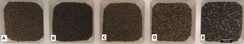
Table shows the results of the determination of particle size of chia seeds and flours, corresponding to whole seeds (CW), milled chia 5 s (Size Large—CL), 30 s (Size Medium—CM) and 60 s (Size Small—CS), and stabilized flour (CF), respectively. CF is the enzyme’s flour deactivated by low or high-temperature blanching. CS showed a higher percentage of smaller particle size (60 g/100 g) (ranging from <0.5 mm) than for CM (40 g/100 g), and CL (20 g/100 g). The CF presented the largest particle size from 0.73 to 1.25 mm (70 g/100 g), respectively. Figure presents photographs of whole bran treatments milled from the same chia seeds.
Table 2. Particle size of each sample
Figure 1. Photographs of whole bran treatments milled from the same chia seeds; scale bar represents 1.61 cm. The A treatment is described as Chia “stabilized flour”, with particle size varying (1.25-<0.5 mm). The B treatment was milled is described as Chia “Size Small”, with particle size varying (0.73-<0.5 mm). The C treatment was milled is described as Chia “Size Medium”, with particle size varying (0.73-<0.5 mm). The D treatment was milled is described as Chia “Size Big”, with particle size varying (1.25-<0.5 mm). The E treatment was milled is described as Chia “Whole”, with size varying (1.60-<0.73 mm).
The moisture values ranged from 5.10% to 5.15% for chia seeds and 9.05% to 9.13% for flour chia, which are below the limit allowed for cereals, which is 15%.
3.2. Quantification of fatty acids before and after in vitro digestion
The most abundant fatty acids in the oil obtained from the chia seeds were: palmitic acid (C16:0), stearic acid (C18:0), oleic acid (C18:1), linoleic acid (C18:2), and linolenic acid (C18:3) (Table ). Undigested samples contained 47.4–51.5 mg/Kg (w/w) of omega 3, which is in agreement with previous reports of 41.7–49.2 mg/kg (w/w) and with average values around 49 (Labanca et al., Citation2017; Taga, Miller, & Pratt, Citation1984).
Table 3. Mean values of fatty acid composition of chia seeds and flour after and before vitro digestion
The content of CL, CM, CS, and CF was measured in the digesta after centrifugation. Fatty acid content was relatively stable during the digestion process. The in vitro bioaccessibility of fatty acids (omega 3) increased significantly (p< 0.05) from 41.7 ± 1.5 to 48.9 ± 2.2% for chia seeds milled for 5 (CL) and 30 s (CM), respectively. For the other two particle sizes (milled 60 s [CS] and stabilized flour [CF]) no difference was observed. However, the whole chia seed samples did not show omega 3 or any other fatty acid, indicating that they did not suffer from the digestion process. This information is very important, since many consume the seed without milling.
Chia seeds from Brazil contained 32.1 ± 0.2 g oil/100 g, with 53.3 ± 0.6 g/100 g of linolenic acid. The total fatty acid from chia flour was 32.0 ± 0.8 g oil/100 g, with 53.7 ± 0.7 g/100 g of linolenic acid. For chia and flour, the percentage of fatty acids did not change significantly. The fatty acids (omega-3) constitute 53% of the total fatty acids in milled chia seeds. Linoleic acid constitutes 20% of total fatty acids. In addition, chia oil has the lowest amount of nutritionally undesirable-saturated fatty acids and a higher level of the desirable-monounsaturated fatty acids.
3.2.1. Total phenolic compounds (TPC)
An additional milling step did not further enhance the bioaccessibility of the phenolic compounds (by any treatment) (p < 0.05). The results were 3.66 ± 0.58; 4.68 ± 0.16; 4.33 ± 0.40; and 3.60 ± 0.04 mg GAE/g dry sample, after a second milling step with 5, 30, 60 s and flour, respectively. For whole seed chia, the total phenolic compound quantified result was close to the limit of quantification (Limited Quantified method: 20 µg/g sample) (Table ).
Table 4. Mean values of phenolic compounds, TBAR´s and PV of chia seeds and flour before and after vitro digestion
The data presented in Table verified that the samples differed with respect to the TPC content before in vitro digestion, the CM sample (4.68 mgGAE/g) being the one with the highest content of TPC and the CW sample (0.76 mgGAE/g) with the lower content. After digestion, only the CW (0.42 mgGAE/g) sample differed statistically, probably due to the reduction of the mucilage of the seed. However, the TPC content was reduced in all samples after the in vitro digestion.
The total phenolic content is known to depend on the type and polarity of the extraction solvent and other factors, such as plant cultivar, degree of seed maturation, climate, and location (Kozłowska, Gruczyńska, Ścibisz, & Rudzińska, Citation2016). AOAC recently published a single-laboratory validation of a colorimetric determination (Folin-Ciocalteau) of total phenolic content of selected dietary supplement extracts at 765 nm compared with a calibration curve generated with gallic acid standard solutions, finding a mean phenolic content of 78.03% w/w for grape seeds (Kupina et al., Citation2018).
Chia seeds are a potential source of chlorogenic acid, caffeic acid, quercetin, myricetin, and kaempferol (Romankiewicz et al., Citation2017). The total phenolic content in chia seed extract was 8.8% on dry matter basis (Reyes-Caudillo et al., Citation2008). The presence of caffeic acid, chlorogenic acid, and quercetin can be correlated with higher extents of phenolics in chia. Described that chia seed is potentially a great source of antioxidants and that the massive antioxidant potential can be utilized for better health and preservation of food lipid systems (Sargi et al., Citation2013). Ayerza and Coates (Ayerza & Coates, Citation2011) isolated polyphenolics (chlorogenic acid, caffeic acid, myricetin, quercetin, and kaempferol) from chia seed. The strong in vitro antioxidant activity of chia seed has been studied in detail (Sargi et al., Citation2013).
Total phenolic content was in agreement with previous studies. The phenolic content (mg/g of chia seed extract, expressed as gallic acid equivalents, GAE) in Jalisco seeds (0.9211 ± 0.040) and Sinaloa seeds (0.8800 ± 0.008) was not significantly different. Also, no significant difference between both hydrolyzed extracts was observed (Jalisco seeds: 0.8899 ± 0.02 and Sinaloa seeds: 0.8800 ± 0.008). The mean concentration, with this assay, was about 0.88 mg per gram of chia seed extract. Hydrolysis was chosen in order to obtain a maximum yield of polyphenolic acids as in the case of cereal grains (Kim, Tsao, Yang, & Cui, Citation2006). In terms of phenolic content, we did not find an increase of GAE as quantified by the Folin–Ciocalteau method.
The reduction in the polyphenolic compound levels studied after in vitro digestion can be justified by the chemiodiversity of the phenolic compounds, since during the digestion process, food components such as bioactive compounds are constantly exposed to different physical, chemical, and biochemical conditions, which can promote structural and chemical changes, resulting in variations in biological activity and consequently affecting the bioavailability and bioactivity of the compounds. It is important also to realize that many metabolites are formed during digestion and they are difficult to quantify since only some are available as standards; however, many of these probably have bioactive/biological effects (Celep, Charehsaz, Akyüz, Acar, & Yesilada, Citation2015).
A recent study has reported a decrease in the concentrations of all polyphenolic compounds after gastrointestinal in vitro digestion and, principally, the anthocyanin content that was severely affected in maqui berry. The gastrointestinal in vitro digestion process decreased the scavenging properties in 89.9% and 84.2% with DPPH and ABTS assays, respectively, as well as the reducing power 74.1% with respect to the non-digested sample. On the other hand, the chelating activity was increased (126.8%). At the end of the gastrointestinal in vitro digestion process, the bioaccessibility of phenolic and flavonoid compounds was 78.19% and 14.20%, respectively. The results obtained suggest that although a great amount of maqui berry polyphenolic compounds are degraded into other metabolites that also might have bioactivity during digestion process, they still have great potential as antioxidant agents (Lucas-Gonzalez et al., Citation2016).
3.3. Effects of temperature and storage time on the quality of milled chia seeds
Lipid oxidation, an inevitable phenomenon during food storage, could affect both the flavor as well as the healthiness of the product (Decker, Alamed, & Castro, Citation2010). It is crucial to analyze lipid oxidation through measurement of both primary and secondary products since it is a complex reaction. Peroxide value is a measure of the concentration of peroxides and hydroperoxides formed in the initial stages of lipid oxidation. Determination of the peroxide index is commonly used to evaluate the level of primary oxidation products in oils and fats. A PI assay was performed to investigate the change in lipid hydroperoxide concentration in the storage process, which is a primary product in the lipid oxidation reaction. The results from the PI test and MDA are shown in Table .
Table 5. Changes in the quality of milled chia seeds (CM) stored at three different temperatures
Malondialdehyde (MDA) is a degradation product generated from lipid peroxidation. MDA has been extensively used as an index for lipid peroxidation and as a marker for oxidation. The reaction of MDA with TBA has been widely adopted as a sensitive assay method for lipid peroxidation. The TBA value is considered an indicator of the amount of malonaldehyde, which is the most predominant secondary oxidation product of food lipids; hence, it is considered a good chemical test for measuring the extent of the secondary oxidation of edible lipids. The values of MDA of milled chia seeds and flour (9.43 and 9.52 mg MDA/kg, respectively) was similar to those reported by Marineli et al. (Marineli et al., Citation2014) in chia (9.58 mg MDA/kg) and oil (17.46 mg MDA/kg).
Water absorption capacity is indicative of a structure’s aptitude to spontaneously absorb water when placed in contact with a constantly moist surface or when immersed in water, and water adsorption capacity is the ability of a structure to spontaneously adsorb water when exposed to an atmosphere of constant relative humidity. It is initially a surface phenomenon but at higher hydration levels absorption can occur inside the structure, leading to swelling and eventual solubilization. The milled chia had a water absorption capacity of 5.27 g water/g sample, which is almost the same as reported for chia seeds from Argentina (6.45 g water/g sample) (Capitani, Spotorno, Nolasco, & Tomás, Citation2012) but lower compared to chia seeds from Mexico measured in the study by Alfredo et al. (Alfredo, Gabriel, Luis, & David, Citation2009) (11.73 g water/g sample).
For the milled chia seeds (CM) the PV increased from day 0–14, and then decreased from day 14–28. Peroxide value of chia seeds increased during storage. The mean PI was 3.8 meq O2/kg oil at T1 (room temperature) and 2.5 meq O2/kg oil at T2 (refrigerated temperature) and T3 (freezing temperature) (Table ). The peroxide index showed little difference (Table ), as only seeds from room temperature were significantly (P < 0.05) higher. Chia seeds PI usually ranges from 0.6 meq O2/kg to 3.9 meq O2/kg in oils extracted with different extraction methods (Ayerza, Citation2010; Dąbrowski, Konopka, Czaplicki, & Tańska, Citation2017; Labanca et al., Citation2017).
Amato et al. (Amato et al., Citation2015) analyzed commercial chia seeds from Peru and found a PV of 11.05 meq O2/kg, and Ayerza and Coates (Ayerza & Coates, Citation2004) found no differences among chia seeds grown in six tropical and subtropical ecosystems. The higher P.V. of seeds analyzed in the present work (6.3 meq O2/kg) could be at the highest temperature in 14 days. After decreased and in 28 days, the valor was the same as on day 0 (2.5 meq O2/kg oil). An important finding was to observe that none of the samples exceeded the upper limit of peroxide index value (10.0 mEq O2/kg oil) established by the Codex Alimentarius (Alimentarius, Citation1999).
For the MDA, no significant differences (p< 0.05) within each sampling could be seen over time. A recent study has reported that chia oil peroxide values of 10 mequiv/kg were observed for oils stored at 4°C while values greater than 10 mequiv/kg were observed between 60 and 120 days when stored at 20°C (Ixtaina, Nolasco, & Tomás, Citation2012). The value of chia could be one of the causes of these lower MDA values since they are known to have an antioxidant effect due to their tocopherol content (Ixtaina et al., Citation2012; Reyes-Caudillo et al., Citation2008).
TBARS of the oil chia seeds stored at 23 ± 1°C showed levels of 10.1 mg MDA/kg. Values higher than 11.1 mg MDA/kg were observed after 28 days for oil stored at 5 ± 1°C. Regarding the secondary lipid autoxidation product, the MDA value of Chilean chia oil (17.46 mg MDA/kg) was higher than those reported by Martysiak-Zurowska and Stoyhwo (Martysiak-żurowska & Stołyhwo, Citation2007) in soybean oil (9.17 mg MDA/kg) and in rapeseed oil (11.12 mg MDA/kg). However, mean absorbance of chia oil at 535 nm (0.044) was markedly lower than that of canola oil (0.53) found by Hawrysh, Shand, Tokarska, and Lin (Hawrysh, Shand, Tokarska, & Lin, Citation1988). Furthermore, the MDA value (9.58 mg MDA/kg) and mean absorbance (0.052) found in Chilean chia seeds were lower than those mentioned above.
Although the TBA test is one of the most commonly used chemical assays to determine secondary oxidation products, this method has limitations, such as the lack of specificity and sensitivity. In addition to MDA, some other substances may react with the TBA reagent and contribute to absorption, causing an overestimation of the intensity of color complex (Marineli et al., Citation2014).
4. Conclusion
The fatty acid composition was relatively stable and did not change significantly during the in vitro digestion. The studies indicate that the bioaccessibility of omega-3 fatty acids and phenolic compounds in chia seed is increased by milling the seeds into a flour, and a flour with small particle size may contribute to an increased uptake and utilization in the body. However, precautions need to be taken, e.g. by initial temperature inactivation of the enzymes in the whole seeds before milling to avoid losses during treatment and storage. Fatty acid composition, specifically linoleic acid, did not change significantly during the in vitro digestion study, and the milled chia seeds were stable against oxidation during storage for 4 weeks. It can be concluded that the chia seeds should preferably be consumed milled in order to take advantage of the fatty acids and phenolic compounds.
Highlights
Milling of chia seeds contributed to increase in vitro availability of omega 3 fatty acids and phenolic compounds.
The PUFA profile did not change during in vitro digestion of chia oil.
The extractability of phenolic compounds and PUFAs was higher in chia flour with smaller particle size.
Storage of milled seed did not show any significant changes in lipid auto-oxidation products.
Acknowledgements
The authors would like to acknowledge the financial support from the Brazilian Agencies Coordination for the Improvement of Higher Education Personnel (CAPES) and Federal University of Minas Gerais (UFMG).
Additional information
Funding
Notes on contributors
Renata Adriana Labanca
Dr Renata Adriana Labanca has graduation at Pharmacy, master’s and doctorate at Science and Technology of the Food of Brazil, postdoctoral degree in Food Sciences from the Chalmers University in Sweden. Has experience in Pharmacy, acting on the following subjects: functional food, vitro digestion and quality control.
Cecilia Svelander
Dr Cecilia Svelander work with bioactive substances in fruits and vegetables, and how they change from raw material to a finished product. Food processing can lead to loss of certain nutrients, but can also be more readily absorbed into the body or transformed into forms with higher biological activity.
Marie Alminger
Dr Marie Alminger research is focused on bioactive compounds in foods and involve characterization of plant raw materials, and studies of relationships between different processing techniques, food matrix, physical-chemical reactions and health. The research includes valorization of residues from the agricultural food chain for sustainable use of raw materials and bilateral research projects aiming to enhance food safety in low-income countries in Eastern Africa.
References
- AACC. (2000). Approved methods of the american association of cereal chemist: The Association.St. Paul, Minnesota.
- Alfredo, V. O., Gabriel, R. R., Luis, C. G., & David, B. A. (2009). Physicochemical properties of a fibrous fraction from chia (Salvia hispanica L.). LWT - Food Science and Technology, 42, 168–173. doi:10.1016/j.lwt.2008.05.012
- Alimentarius, C. (1999). STANDARD FOR NAMED VEGETABLE OILS CODEX STAN 210-1999 Adopted. Journal of Chemical Information and Modeling. Rome.
- Alminger, M., Aura, A.-M., Bohn, T., Dufour, C., El, S. N., Gomes, A., … Santos, C. N. (2014). In vitro models for studying secondary plant metabolite digestion and bioaccessibility. Comprehensive Reviews in Food Science and Food Safety, 13, 413–436. doi:10.1111/1541-4337.12081
- Amato, M., Caruso, M. C., Guzzo, F., Galgano, F., Commisso, M., Bochicchio, R., … Favati, F. (2015). Nutritional quality of seeds and leaf metabolites of Chia (Salvia hispanica L.) from Southern Italy. European Food Research and Technology, 241, 615–625. doi:10.1007/s00217-015-2488-9
- Ayerza, R. (2010). Effects of seed color and growing locations on fatty acid content and composition of two chia (Salvia hispanica L.) genotypes. JAOCS, Journal of the American Oil Chemists’ Society, 87, 1161–1165. doi:10.1007/s11746-010-1597-7
- Ayerza, R., & Coates, W. (2004). Composition of chia (Salvia hispanica) grown in six tropical and subtropical ecosystems of South America. Tropical Science, 44, 131–13. doi:10.1002/(ISSN)1556-9179
- Ayerza,, & Coates, W. (2011). Protein content, oil content and fatty acid profiles as potential criteria to determine the origin of commercially grown chia (Salvia hispanica L.). Industrial Crops and Products, 34(2), 1366–1371. doi: 10.1016/J.Indcrop.2010.12.007
- Brewer, L. R., Kubola, J., Siriamornpun, S., Herald, T. J., & Shi, Y. C. (2014). Wheat bran particle size influence on phytochemical extractability and antioxidant properties. In Brewer, L. R., J. Kubola, S. Siriamornpun, T. J. Herald, & Y. C. Shi, (Eds), Food chemistry, 152,483–90. doi: 10.1016/j.foodchem.2013.11.128
- Capitani, M. I., Spotorno, V., Nolasco, S. M., & Tomás, M. C. (2012). Physicochemical and functional characterization of by-products from chia (Salvia hispanica L.) seeds of Argentina. LWT - Food Science and Technology, 45, 94–102. doi:10.1016/j.lwt.2011.07.012
- Celep, E., Charehsaz, M., Akyüz, S., Acar, E. T., & Yesilada, E. (2015). Effect of in vitro gastrointestinal digestion on the bioavailability of phenolic components and the antioxidant potentials of some Turkish fruit wines. Food Research International, 78, 209–215. doi:10.1016/j.foodres.2015.10.009
- Dąbrowski, G., Konopka, I., Czaplicki, S., & Tańska, M. (2017). Composition and oxidative stability of oil from Salvia hispanica L. seeds in relation to extraction method. European Journal of Lipid Science and Technology, 119, 1600209. doi:10.1002/ejlt.v119.5
- Decker, E. A., Alamed, J., & Castro, I. A. (2010). Interaction between polar components and the degree of unsaturation of fatty acids on the oxidative stability of emulsions. JAOCS, Journal of the American Oil Chemists’ Society, 87, 771–780. doi:10.1007/s11746-010-1556-3
- Espinosa, R. R., Inchingolo, R., Alencar, S. M., Rodriguez-Estrada, M. T., & Castro, I. A. (2015). Antioxidant activity of phenolic compounds added to a functional emulsion containing omega-3 fatty acids and plant sterol esters. Food Chemistry, 182, 95–104. doi:10.1016/j.foodchem.2015.02.130
- Fernández-García, E., Carvajal-Lérida, I., & Pérez-Gálvez, A. (2009). In vitro bioaccessibility assessment as a prediction tool of nutritional efficiency. Nutrition Research, 29, 751–760. doi:10.1016/j.nutres.2009.09.016
- Galanakis, C. M. (2017). Introduction. Nutraceutical and Functional Food Components: Effects of Innovative Processing Techniques.
- Hawrysh, Z. J., Shand, P. J., Tokarska, B., & Lin, C. (1988). Effects of tertiary butylhydroquinone on the stability of canola oil. I. Accelerated storage. Canadian Institute of Food Science and Technology Journal, 21, 549–554. doi:10.1016/S0315-5463(88)71037-8
- Heaney, R. P. (2001). Factors influencing the measurement of bioavailability, taking calcium as a model. The Journal of Nutrition, 131, 1344S-1348S. doi:10.1093/jn/131.4.1344S
- Ixtaina, V. Y., Nolasco, S. M., & Tomás, M. C. (2012). Oxidative stability of chia (Salvia hispanica L.) seed oil: Effect of antioxidants and storage conditions. JAOCS, Journal of the American Oil Chemists’ Society, 89, 1077–1090. doi:10.1007/s11746-011-1990-x
- Kim, K. H., Tsao, R., Yang, R., & Cui, S. W. (2006). Phenolic acid profiles and antioxidant activities of wheat bran extracts and the effect of hydrolysis conditions. Food Chemistry, 95(3), 466–473.
- Kozłowska, M., Gruczyńska, E., Ścibisz, I., & Rudzińska, M. (2016). Fatty acids and sterols composition, and antioxidant activity of oils extracted from plant seeds. Food Chemistry, 213, 450–456. doi:10.1016/j.foodchem.2016.06.102
- Kupina, S., Fields, C., Roman, M. C., & Brunelle, S. L. (2018). Determination of total phenolic content using the folin-C assay: Single-Laboratory Validation, First Action 2017.13. Journal of AOAC International, 101, 1466–1472. doi:10.5740/jaoacint.18-0031
- Labanca, R. A., Svelander, C., Eliasson, L., Araújo, R. L. B., Ahrné, L., & Alminger, M. (2017). Supercritical carbon dioxide extraction and conventional extraction of chia seed oils: Chemical composition and lipid oxidation. International Journal of Research, 4(10), 563–572.
- Lee, C. M., Trevino, B., & Chaiyawat, M. (1996). A simple and rapid solvent extraction method for determining total lipids in fish tissue. Journal of AOAC International, 79(2), 487–492.
- Lucas-Gonzalez, R., Navarro-Coves, S., Pérez-Álvarez, J. A., Fernández-López, J., Muñoz, L. A., & Viuda-Martos, M. (2016). Assessment of polyphenolic profile stability and changes in the antioxidant potential of maqui berry (Aristotelia chilensis (Molina) Stuntz) during in vitro gastrointestinal digestion. Industrial Crops and Products, 94, 774–782. doi:10.1016/j.indcrop.2016.09.057
- Marineli, R. D. S., Moraes, É. A., Lenquiste, S. A., Godoy, A. T., Eberlin, M. N., & Maróstica, M. R. (2014). Chemical characterization and antioxidant potential of Chilean chia seeds and oil (Salvia hispanica L.). LWT - Food Science and Technology, 59, 1304–1310. doi:10.1016/j.lwt.2014.04.014
- Martínez-Cruz, O., & Paredes-López, O. (2014). Phytochemical profile and nutraceutical potential of chia seeds (Salvia hispanica L.) by ultra high performance liquid chromatography. Journal of Chromatography A, 1346, 43–48. doi:10.1016/j.chroma.2014.04.007
- Martysiak-żurowska, D., & Stołyhwo, A. (2007). Content of furosine in infant formulae and follow-on formulae. Polish Journal of Food and Nutrition Sciences, 57,185–190.
- Minekus, M., Alminger, M., Alvito, P., Ballance, S., Bohn, T., Bourlieu, C., … Brodkorb, A. (2014). A standardised static in vitro digestion method suitable for food-an international consensus, 5(6), 1113–24. doi: 10.1039/c3fo60702j
- Pertiwi, K., Kok, D. E., Wanders, A. J., de, G. J., Zock, P. L., & Geleijnse, J. M. (2019). Circulating n-3 fatty acids and linoleic acid as indicators of dietary fatty acid intake in post-myocardial infarction patients, nutrition, metabolism and cardiovascular diseases, 29(4), 343–350.
- Reyes-Caudillo, E., Tecante, A., & Valdivia-López, M. A. (2008). Dietary fibre content and antioxidant activity of phenolic compounds present in Mexican chia (Salvia hispanica L.) seeds. Food Chemistry, 107, 656–663. doi:10.1016/j.foodchem.2007.08.062
- Romankiewicz, D., Hassoon, W. H., Cacak-Pietrzak, G., Sobczyk, M. B., Wirkowska-WojdyBa, M., CegliNska, A., & Dziki, D. (2017). The effect of chia seeds (salvia hispanica L.) addition on quality and nutritional value of wheat bread. Journal of Food Quality, 2017, 1–7. doi:10.1155/2017/7352631
- Rufino, M. D. S. M., Alves, R. E., de Brito, E. S., Pérez-Jiménez, J., Saura-Calixto, F., & Mancini-Filho, J. (2010). Bioactive compounds and antioxidant capacities of 18 non-traditional tropical fruits from Brazil. Food Chemistry, 121, 996–1002. doi:10.1016/j.foodchem.2010.01.037
- Sargi, S. C., Silva, B. C., Santos, H. M. C., Montanher, P. F., Boeing, J. S., Santos Júnior, O. O., … Visentainer, J. V. (2013). Antioxidant capacity and chemical composition in seeds rich in omega-3: Chia, flax, and perilla. Food Science and Technology, 33, 541–548. doi:10.1590/S0101-20612013005000057
- Schmedes, A., & Hølmer, G. (1989). A new thiobarbituric acid (TBA) method for determining free malondialdehyde (MDA) and hydroperoxides selectively as a measure of lipid peroxidation. Journal of the American Oil Chemists Society, 66, 813–817. doi:10.1007/BF02653674
- Taga, M. S., Miller, E. E., & Pratt, D. E. (1984). Chia seeds as a source of natural lipid antioxidants. Journal of the American Oil Chemists’ Society, 61, 928–931. doi:10.1007/BF02542169
- Undeland, I., Hultin, H. O., & Richards, M. P. (2002). Added triacylglycerols do not hasten hemoglobin-mediated lipid oxidation in washed minced cod muscle. Journal of Agricultural and Food Chemistry, 50, 6847–6853. doi:10.1021/jf0201982