Abstract
The rapid and continuous progress in trace analysis of metals is associated with development of several new techniques such as spectrophotometry. Thiosemicarbazones have been used as chromogenic reagents in spectrophotometric determination of metals. In this study the synthesis and characterization of pyridoxal thiosemicarbazone (PDT) and 2-acetyl pyridine thiosemicarbazone (2-APT) using IR, MS, and 1HNMR were conducted. Zn(II) forms yellow colored complex with PDT and 2-APT in the pH range of 5–7 at 430 nm, and 8–10 at 360 nm, respectively. The Zn(II)-PDT and Zn(II)-2-APT complexes obeys Beer’s law in the concentration range of 0.26–2.62 and 0.25–2.56 μg/mL, respectively. The molar absorptivity and Sandell’s sensitivity of Zn(II)-PDT and Zn(II)-2-APT complexes are 1.8 × 10⁴ L/mol.cm and 0.0035 μg/cm2, 2.9 × 10⁴ L/mol.cm and 0.0167 μg/cm2, respectively. A first derivative spectrophotometry method is proposed for the determination of Zn(II) in the range 0.06–2.94 and 0.05–2.72 μg/mL of Zn(II) using PDT and 2-APT, respectively. The first derivative spectrum of Zn(II)-PDT and Zn(II)-2-APT complexes exhibits maximum amplitude at 400 and 385 nm, respectively The method was tested for Zn(II) determination in soil and vegetable samples. Using paired sample t-test at 0.05 level, the results were in good agreement with those obtained using flame atomic absorption spectrophotometer.
Public Interest Statement
Zinc is one of the trace elements found in nature and required in a trace amount by living organisms. So determination of its amount in various media such as soils and vegetables is so important. One of the techniques used for determination of Zn(II) is spectrophotmetry using chelating or complexing agents. For this reason, two Zn(II) complexes of pyridoxal thiosemicarbazone (PDT) and 2-acetyl pyridine thiosemicarbazone (2-APT) have been synthesized and characterized by various techniques. The reagents have been tested as an alternative chelating agent and therefore used for the determination of Zn(II) in real sample analysis (soil and vegetables).
1. Introduction
The presence of small quantities of zinc is essential for the growth of plants and animals including human beings. It acts as a co-factor in numerous enzymes, and plays an important role in protein synthesis and cell division (Reddy, Kumar, Ramachandraiah, Thriveni, & Reddy, Citation2007). Zinc is one of the more abundant metals in the human body and is vital for the growth, development, where over 300 enzymes contain zinc ions in their active sites (Wałęsa-Chorab et al., Citation2011). In addition, it plays a number of important biological roles such as in the synthesis of deoxyribonucleic acid (DNA) and ribosomal ribonucleic acid (RRNA) (Islam & Ahmed, Citation2013; Sarma, Kumar, Reddy, Thriveni, & Reddy, Citation2006). The deficiency of zinc leads to retarded growth, lower feed efficiency, impaired DNA synthesis, delayed wound healing, decrease in collagen synthesis, and inhibits the general well-being (Krishna Reddy, Rajesh Kumar, Subramanyam Sarma, & Varada Reddy, Citation2002; Sarma et al., Citation2006). Besides its biological importance, zinc has industrial significance (Sarma et al., Citation2006). It is used in the protection of steel against corrosion, dry batteries, photoengraving, and lithography (Bhalotra & Puri, Citation1999). On the other hand, zinc can be toxic when exposures exceed physiological needs. Zinc is present in many foods, soil and is also found in a number of pharmaceutical samples, causing environmental pollution (Reddy et al., Citation2007). Significant concentrations of zinc may reduce the soil microbial activity and also is a common contaminant in agricultural and food wastes (Ma et al., Citation2009). Zinc is released into the environment by chemical weathering of zinc minerals (Islam & Ahmed, Citation2013).
The separation and determination of trace amounts of zinc is indispensable and currently of great interest in many scientific fields, including environmental monitoring. Therefore, sensitive, selective, and rapid methods for the determination of zinc are of paramount importance. A number of analytical techniques such as atomic absorption spectrometry (AAS) (Ata et al., Citation2015; Uddin et al., Citation2016), inductively coupled plasma-atomic emission spectroscopy (ICP-AES) (Escudero, Cerutti, Martinez, Salonia, & Gasquez, Citation2013; Ozbek & Akman, Citation2016; Senila, Drolc, Pintar, Senila, & Levei, Citation2014), inductively coupled plasma-mass spectrometry (ICP-MS) (Wysocka & Vassileva, Citation2016), fluorescence spectrometry (Ma et al., Citation2009), and electroanalytical techniques (Chaiyo et al., Citation2016; Lo Coco, Ceccon, Ciraolo, & Novelli, Citation2003) are widely applied to the determination of zinc at trace level in various matrices. However, zinc can alternatively be determined by spectrophotometric method (Kiran, Citation2012). Spectrophotometry is essentially a trace analysis technique and is one of the most powerful tools in chemical analysis (Islam & Ahmed, Citation2013).
The derivative spectrophotometry is a spectral technique in which a slope of the spectrum, that is the rate of change in absorbance with wavelength, is measured as a function of wavelength, which at the same time shows good sensitivity and specificity. In the derivative spectrum, the ability to detect and to measure minor spectral features is considerably enhanced, resulting in increased sensitivity (Karpinska, Citation2004; Sánchez Rojas & Bosch Ojeda, Citation2009; Sedaira, Citation2000). This technique offers an alternative approach to the enhancement of sensitivity and specificity in mixture analysis (Bosch Ojeda & Sanchez Rojas, Citation2004). The derivative spectra starts and finished at zero, passes through zero at the same wavelength as maximum wavelength (λmax) of the absorbance band with first a positive and then a negative band, with the maximum and minimum at the same wavelengths as the inflection points in the absorbance band (Bosch Ojeda & Sanchez Rojas, Citation2004).
Thiosemicarbazone usually react with metallic cations giving complexes in which the thiosemicarbazones behave as chelating ligands (Casas, Garcı́a-Tasende, & Sordo, Citation2000). Thiosemicarbazone derivatives are of considerable interest due to their versatility as ligands bearing suitable donor atoms for coordination to metals with strong coordinating ability (Singh & Singh, Citation2015). They show a wide range of chemical properties depending on the parent aldehyde or ketone (Kovala-Demertzi, Gangadharmath, Demertzis, & Sanakis, Citation2005; Prathima, Subba Rao, Adinarayana Reddy, Reddy, & Varada Reddy, Citation2010). Zn(II) forms chelate complex with many chromogenic reagents such as thiosemicarbazones. Some representative examples are benzildithiosemicarbazone (Krishna Reddy et al., Citation2002), di-2-pyridyl ketone salicyloylhydrazone (Gaubeur, da CunhaAreias, Ávila Terra, & Suárez-Iha, Citation2002), 2,4-dihydroxybenzaldehyde isonicotinoyl hydrazone (Sivaramaiah & Raveendra Reddy, Citation2005), pyridoxal-4-phenyl-3-thiosemicarbazone (Sarma et al., Citation2006), N-ethyl-3-carbazolecarboxaldehyde-3-thiosemicarbazone (Reddy et al., Citation2007), di-2-pyridyl ketone benzoylhydrazone (Vieira et al., Citation2008), bis-[2,6-(2′-hydroxy-4′-sulpho-1′-napthylazo)]pyridine disodium salt (Barman & Barua, Citation2009), 2-benzoylpyridine thiosemicarbazone (Reddy, Reddy, & Reddy, Citation2011), and 5,7-dibromo-8-hydroxyquinoline (Islam & Ahmed, Citation2013). The aim of this work was to develop a sensitive and efficient spectrophotometric method for Zn(II) determination using chromogenic reagents containing a Schiff base. The conditions for the spectrophotometric determination of Zn(II) with PDT and 2-APT were described. Various factors influencing the sensitivity of the proposed method such as the pH and ranges of applicability of Beer’s law on the determination of Zn(II) were also studied. The method was applied to soil and vegetable samples as well and compared to standard method flame atomic absorption spectrometry (F-AAS).
2. Experimental
2.1. Chemicals and instrumentation
All the chemicals were used without purification. The chemicals used were 99% N,N-dimethylformamide (DMF), 99.9% dimethyl sulfoxide (DMSO), 98% 2-acetylpridine, 98% pyridoxal hydrochloride, 99% thiosemicarbazide, 37% hydrochloric acid, 98% sulfuric acid, and 99% zinc sulfate heptahydrate (ZnSO4·7H2O). All the glassware were cleaned with 5% HNO3. The absorption spectra of the reagents were recorded using UV 2,450 single beam spectrophotometer (Shimadzu, Japan) while the pH was measured with digital pH meter (Model Li-120, ELICO, India). The synthesized reagents were characterized by spectrum 100 IR spectrophotometer with KBr discs (Perkin-Elmer, UK), 1HNMR JEOL GSX-400 high resolution spectrometer (JEOL, USA), and Micro Mass VG-7,070 H Mass spectrometer (UK). The zinc concentration was determined using Varian AA 240FS atomic absorption spectrometer (Varian, USA).
2.2. Preparation of working solutions
A stock solution of Zn(II) (1 mg/mL) was prepared by dissolving 4.398 g of ZnSO4·7H2O in distilled water containing a few drops of conc. H2SO4 and standardized by 8-hydroxyquinoline (Vogel, Citation1989). The buffer solutions were prepared by mixing suitable portions of HCl and sodium acetate (pH 1–3), acetic acid and sodium acetate (pH 3.2–7.0), and ammonium hydroxide and ammonium chloride (pH 8.0–12.0). The PDT was prepared according to a modified procedure described in the literature (Floquet et al., Citation2009; Manikandan, Anitha, Prakash, Vijayan, & Viswanathamurthi, Citation2014; Manikandan,Vijayan, et al., Citation2014; Manikandan et al., Citation2015; Sarma et al., Citation2006; Yemeli Tido, Vertelman, Meetsma, & van Koningsbruggen, Citation2007). Equimolar amount of thiosemicarbazide and pyridoxal hydrochloride (in 10 mL ethanol) were mixed, refluxed for 5 h and cooled to room temperature. The solid material was filtered, washed, and dried. The 2-APT was prepared according to the procedure reported by Admasu, Reddy, and Mekonnen (Citation2016).
2.3. Absorption spectra of reagent solutions and metal complexes
In a 25 mL volumetric flask, 1 mL of 1 × 10⁻² M reagent solution and 10 mL of buffer solution (acetic acid and sodium acetate for PDT system, and ammonium hydroxide and ammonium chloride for 2-APT system) were added and diluted to the mark with distilled water. The absorbance of the reagent solution was measured against water blank. For measuring the absorption spectrum of complex, in a 25 mL volumetric flask 10 mL of buffer (acetic acid and sodium acetate for PDT system, and ammonium hydroxide and ammonium chloride for 2-APT system) and suitable concentration of metal ion and reagent solutions (10-fold molar excess to metal ion) were added. The contents were diluted to the mark with distilled water and the absorbance was measured against the reagent blank. To see the effects of pH, in a set of 25 mL volumetric flasks 10 mL of series of buffer solution (pH 3.0–10.0), constant amount of metal ion and reagent (usually 10-fold molar excess to metal ion) solution were added and made to the mark with distilled water. For finding optimum amount of reagent required known aliquot of reagent solution was taken in a set of 25 mL volumetric flasks, each containing 10 mL of buffer solutions (acetic acid and sodium acetate for PDT system, and ammonium hydroxide and ammonium chloride for 2-APT system) and fixed amount of metal ion and diluted to the mark with distilled water. The absorbance of each solution was measured at λmax against corresponding reagent blank. Similarly, the absorbance of the colored complex solution was measured at λmax against reagent blank at different time intervals so as to determine the stability of the complex and time interval required for full color development.
2.4. Soil and vegetable sample preparation
The soil samples were collected from three sampling spots (Adi Desta, Adi Nfas, and Adi Kula) in Abraha Atsbeha village, Tigray, Ethiopia, while the vegetable samples (tomato, cabbage, and spinach) were collected from the local market in the village with plastic bags. The soil samples were kept oven-dried at 105°C for 24 h, sieved to <2 mm, milled to the fine powder, and stored in double-cup polyethylene bottles. The vegetable samples were rinsed in distilled water, air-dried, finely powdered, and stored in double-cup polyethylene bottles. About 10 g of soil (2 g of vegetable) samples were taken in a silica crucible, heated to oxidize organic matter, and ashed at 550°C, in a muffle furnace for 4–5 h. The ash was then dissolved by heating with 10 mL of 2 N HCl and filtered (Reddy et al., Citation2007). The working calibration solutions were made up from 1,000 mg/L standards. The regression values (R2) of the calibration curve was >0.999. The concentration of Zn in the digested samples was determined at a wavelength of 213.9 nm with hallow cathode lamp using an air-acetylene flame. The analyses were conducted in triplicate and the results presented as means ± standard deviation.
2.5. Determination of the stoichiometry and stability constants of the complexes
The composition of the Zn(II)-PDT and Zn(II)-2-APT complexes were determined by Job’s continuous variation (Huang, Zhou, Yang, & Chock, Citation2003; Mansour & Danielson, Citation2012) and mole ratio methods (Mansour & Danielson, Citation2012). For Job’s method, the metal and reagent solutions were mixed in different proportions in 25 mL volumetric flask and diluted to the mark with distilled water in the presence of buffer solution (acetic acid and sodium acetate for PDT system, and ammonium hydroxide and ammonium chloride for 2-APT system). The absorbance was recorded at λmax, against the corresponding reagent blank. A plot between mole fraction of the metal ion and the absorbance was made so as to get the metal-ligand ratio based on Job’s method. The stability constants of the complexes were also calculated. For mole ratio method constant amount of metal ion and known and varying aliquots of the reagent solutions were added in a series of 25 mL volumetric flasks in the presence of buffer solution (acetic acid and sodium acetate for PDT system, and ammonium hydroxide and ammonium chloride for 2-APT system) and diluted to the mark with distilled water. The absorbance was recorded at λmax, against the corresponding reagent blank so as to get the metal-ligand ratio based on molar ratio method.
2.6. Statistical analysis
All mathematical and statistical computations were made using Excel 2007 (Microsoft Office) and OriginPro 8.5.0 SR1 (OriginLab Corporation, USA). The data were reported as mean ± standard deviation. Student t-test was used for comparison of the developed method with standard method.
3. Results and discussion
3.1. Characterization of PDT and 2-ATP
The PDT and 2-ATP were characterized by IR, 1HNMR, and MS. The IR (KBr, cm−1) spectrum of PDT shows bands corresponding to υ(N–H, asym), υ(N–H, sym), υ(C–H, pyridine), υ(C=N, Schiff’s base), υ(C–C, pyridine), υ(C–H, aromatic ring), υ(N–H, primary amide), υ(C=S), and δ(C–H, aromatic ring) at 3,342 (m), 3,279 (m, br), 3,161 (s), 1,583 (s), 1,551 (s), 1,473 (s), 1,357 (w), 1,082 (m), and 865 (m) cm−1, respectively. These results were comparable with the one reported by Casas et al. (Citation1998), Floquet et al. (Citation2009), Manikandan, Anitha, et al. Citation(2014), Manikandan, ,Vijayan, et al. Citation(2014), and Manikandan et al. (Citation2015). The 1H NMR (DMSO-d6, δ, ppm) of free ligand, PDT: 12.01 (s, 1H, H–C=N), 8.64 (s, 1H=N–NH), 8.22 (s, 2H, S=C–NH2), 8.16 (s, 1H, pyridine ring protons), 4.80 (s, 2H, C=C–CH2), 2.62 (s, 3H, –CH3). The mass spectrum of PDT shows signal at 241 (M + 1) corresponding to its molecular ion peak. The molecular formula of the reagent is C9H12N4O2S (M. Wt 240) (Floquet et al., Citation2009). The IR, 1H NMR, and mass spectra of 2-APT was already reported and discussed by Admasu et al. (Citation2016).
3.2. Absorption spectra of complexes and optimum condition for complexation
The absorption spectra of the reagent solution and the complex solutions are recorded in the wavelength 250–600 nm against the buffer solution and reagent blank, respectively. The typical spectra are presented in Figures and . The Zn(II)-PDT spectra shows an absorption maximum at 430 nm, while the reagent does not show appreciable absorbance at this wavelength at pH 6.0. Hence, 430 nm is chosen for subsequent studies. Similarly, the Zn(II)-2-APT spectra shows a strong absorbance at 360 nm, where the reagent solution does not show appreciable absorbance at pH 9.0. Hence the wavelength 360 nm is chosen for subsequent studies.
3.3. Optimum condition for complexation
The optimum pH required for the full color development of the complex solutions were examined in the pH range 1.0–10.0. It was observed that the absorbance is maximum and constant in the pH range 5.0–7.0 and 8.0–10.0 condition for PDT and 2-APT, respectively. Hence pH 6.0 and 9.0 were selected as the optimal condition for further studies of PDT and 2-APT complexes, respectively (Figures and ). The absorbance of the complex solutions at λmax of 430 and 360 nm were measured at various concentrations of the reagent solutions (PDT and 2-APT), keeping Zn(II) concentration constant at the optimized pH 6.0 and 9.0, respectively. The studies revealed that a tenfold molar excess of reagent is required for the maximum color development of Zn(II)-PDT, while fivefold for Zn(II)-2-APT. Excess of the reagent solutions had no effect on the absorbance of the complex solutions and therefore further studies were carried out using at least ten- and fivefold molar excess of reagent to Zn(II) for PDT and 2-APT, respectively. The change in the order of addition of various constituents (buffer, metal ion, and reagent) has no effect on the absorbance of the Zn(II) complexes in solution. The absorbance of the solution at 430 and 360 nm was measured at different times to ascertain the time stability of the color of the complex. The yellow color development in both cases is instantaneous and remains constant for about 1 h at the optimized conditions.
3.4. Applicability of Beer’s law, molar absorptivity, Sandell’s sensitivity, and correlation coefficient
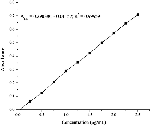
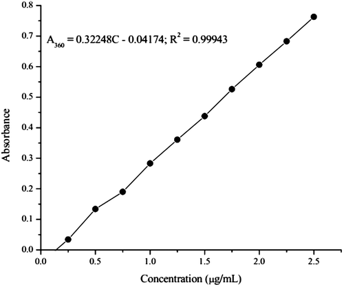
The complex systems were subjected to verification of Beer’s law to explore the possibility of trace determination of Zn(II) employing the color reaction under investigation. For the Zn(II)-PDT system, the complex system obeys Beer’s law in the concentration range of 0.26–2.62 μg/mL (Figure ). The molar absorptivity (ε) and Sandell’s sensitivity of the complex are 1.8 × 10⁴ L/mol.cm and 0.0035 μg/cm2, respectively. The specific absorptivity of the system is found to be 0.282 mL/g.cm. The replicate (n = 10) analyses of a solution containing 1.308 μg/mL of Zn(II) gave 1.322 as mean absorbance with a standard deviation of 0.0069 and a relative standard deviation (%RSD) of 0.52. The correlation coefficient value of Zn(II)-PDT system is 0.99959, indicating excellent linearity between the two variables. For the Zn(II)-2-APT system, the system obeys Beer’s law in the concentration range of 0.25–2.56 μg/mL (Figure ). The molar absorptivity and Sandell’s sensitivity of the method are 2.9 × 10⁴ L/mol.cm and 0.0167 μg/cm2, respectively. The specific absorptivity of the system is found to be 0.0098 mL/g.cm. The replicate (n = 10) analyses of a solution containing 1.0 μg/mL of Zn(II) gave 0.975 as mean absorbance with a standard deviation of 0.01131 and %RSD of 1.2. The correlation coefficient of the calibration equation of the experimental data is 0.99943. The detection limit (expressed as 3 × standard deviation of blank (n = 10) divided by the slope of the calibration line) of the Zn(II)-PDT and 2-APT methods are 0.064 and 0.084 μg/mL, respectively. Comparing the two reagents, PDT is more sensitive than 2-APT for Zn(II) determination.
3.5. Composition and stability constant of the complex
Spectrophotometric investigations of the Zn(II)-PDT and Zn(II)-2-APT complexes were made to obtain composition of the complex using the Job’s continuous variation method and molar ratio method. In order for Job’s continuous variation method to be applicable four important things should be considered: the system must conform to Beer’s law; one complex must predominate under the conditions of the experiment; the total concentration of the two binding partners must be maintained constant, and the pH and ionic strength must be maintained constant (Hill & MacCarthy, Citation1986). Job’s plot confirmed that one mole of Zn(II) reacts with one mole of the reagent showing the composition of the complex as 1:1 (M:L) for both systems. The stability constant (M−1) of Zn-PDT and Zn-2-APT are 2.4 × 10⁶ and 8.45 × 10⁵, respectively. An alternative to the Job’s continuous variation method for determining the stoichiometry of metal-ligand complexes is the mole-ratio method in which the amount of one reactant, usually the moles of metal, is held constant, while the amount of the other reactant is varied. Absorbance is monitored at a wavelength where the metal-ligand complex absorbs. Assuming the complex absorbs more than the reactants, this plot will produce an increasing absorbance up to the combining ratio. At this point, further addition of reactant will produce less increase in absorbance. Thus a break in the slope of the curve occurs at the mole ratio corresponding to the combining ratio of the complex. The molar ratio method support the composition of the complex obtained in Job’s continuous variation method is 1:1.
3.6. Determination of Zn(II) by first derivative spectrophotometry
The first derivative spectra of Zn(II)-PDT complex containing variable amounts of Zn(II) was recorded at pH 6.0 from which the analytical wavelengths were ascertained. The amplitude is maximum and proportional to the metal ion concentration. The peak zero (h) method is followed for peak height measurements and preparation of calibration plot. The maximum peak was observed at 400 nm and two zero crosses at 370 and 455 nm. Therefore, Zn(II) was determined by measuring the amplitude at 400 nm. Beer’s law is obeyed in the range 0.06–2.94 μg/mL of Zn(II). The standard deviation of the method (n = 10) of 1.32 μg/mL of Zn(II) is 0.0053 μg/mL giving RSD of 0.4%. The replicate (n = 10) analyses of a solution containing 1.308 μg/mL of Zn(II) gave 1.322 as mean absorbance with a standard deviation of 0.0069 and a relative standard deviation (%RSD) of 0.52. The slope, intercept, and correlation coefficient of the calibration equation for the experimental data are 0.7990, 0.02, and 0.99989, respectively. Similarly, the first-order derivative spectrum of Zn(II)-2-APT complex was recorded at pH 9.0. The first derivative spectrum exhibits maximum amplitude at 385 nm and two zero crosses at 358 and 419 nm. Therefore, 385 nm (peak) is chosen for further studies. Beer’s law is obeyed in the range 0.05–2.72 μg/mL of Zn(II). The standard deviation of the method (n = 10) of 1.0 μg/mL of Zn(II) is 0.0098 μg/mL giving RSD of 1.0%. The replicate (n = 10) analyses of a solution containing 1.0 μg/mL of Zn(II) gave 0.975 as mean absorbance with a standard deviation of 0.01131 and %RSD of 1.2. The slope, intercept, and correlation coefficient of the calibration equation for the experimental data are 0.05892, 0.00048, and 0.99983, respectively. A comparative analytical characteristics of the normal (zero) and first derivative spectrometric studies of both complexes is summarized in Table . Similarly, comparing the two reagents, PDT is more sensitive than 2-APT for Zn(II) determination while the first derivative is more sensitive than the normal (zero) order.
Table 1. Analytical characteristics of Zn(II)-PDT and Zn(II)-2-APT complex
3.7. Applications of the developed methods for determinations of Zn(II) in real samples
In order to confirm the applicability of the proposed method, it has been applied to the determination of Zn(II) in vegetable and soil samples. The results for this study are presented in Table . A F-AAS method was used as a standard reference method. The performance of the proposed method was compared with F-AAS method using student t-test. Using paired sample t-test at the 0.05 level, the developed methods are not significantly different with F-AAS. Therefore, the results of the developed methods are in good agreement with the standard comparative method. Moreover, comparing the results obtained from the two spectrphotometric reagents, the differences of the developed methods are not significantly different.
Table 2. Determination of Zn(II) (mean ± standard deviation, n = 3) in vegetable and soil samples
The present method was also compared with other existing spectrophotometric methods in the literature (Barman & Barua, Citation2009; Gaubeur et al., Citation2002; Islam & Ahmed, Citation2013; Krishna Reddy et al., Citation2002; Reddy et al., Citation2007, Citation2011; Sarma et al., Citation2006; Sivaramaiah & Raveendra Reddy, Citation2005; Vieira et al., Citation2008) (Table ). The developed methods are comparable with the reported methods with respect to the aforementioned analytical performances in various samples matrices (blood, water, food, biological, environmental, and pharmaceutical samples). Therefore, the proposed method could be used as an alternative method for zinc determination in various matrices.
Table 3. Comparison of characteristic performance of the developed spectrophotometric methods for the determination of Zn(II) using thiosemicarbazones with other studies
4. Conclusion
The PDT and 2-APT have been synthesized, characterized, and successfully applied as complexing agent to determine Zn(II) using spectrophotometry. The PDT is a more sensitive chelating agent than 2-APT for Zn(II) determination while the first derivative is more sensitive than the normal order in both systems. The developed methods are practical and valuable for determination of zinc since it was checked in soil and vegetable samples. Moreover, the method can be used as an alternative method for the determination of zinc in various media. The results showed good agreement with the results obtained by F-AAS methods for soil and vegetable samples.
4.1. Human and animal rights
This article does not contain any studies with human or animal subjects.
Funding
The authors received no direct funding for this research.
Acknowledgement
The Department of Chemistry, College of Natural and Computational Sciences, Mekelle University, Mekelle, Ethiopia and Ezana Mining Development Analytical Laboratory PLC, Mekelle, Ethiopia duly acknowledged for allowing the laboratory facilities and financial assistance.
Additional information
Notes on contributors
Kebede Nigussie Mekonnen
My research group is involved mainly on the analysis of pollutants (elemental pollutants and persistent organic pollutants (POPs)) in various media (aquatic environment (rivers, lakes, and dams), soils, sediments and biota such as fish and leafy vegetables) and effluents from various sources. The group is also engaged in synthesis and characterization of metal complexes as an alternative way for elemental analysis. For this purpose the group works on thiosemicarbazone as spectrophotometric chelating agents.
References
- Admasu, D., Reddy, D. N., & Mekonnen, K. N. (2016). Spectrophotometric determination of Cu(II) in soil and vegetable samples collected from Abraha Atsbeha, Tigray, Ethiopia using heterocyclic thiosemicarbazone. SpringerPlus, 5, 1169. doi:10.1186/s40064-016-2848-3
- Ata, S., Wattoo, F. H., Ahmed, M., Wattoo, M. H. S., Tirmizi, S. A., & Wadood, A. (2015). A method optimization study for atomic absorption spectrophotometric determination of total zinc in insulin using direct aspiration technique. Alexandria Journal of Medicine, 51, 19–23. doi:10.1016/j.ajme.2014.03.004
- Barman, B., & Barua, S. (2009). Spectrophotometric determination of zinc in blood serum of diabetic patients using bis-[2,6-(2′-hydroxy-4′-sulpho-1′-napthylazo)]pyridine disodium salt. Archives of Applied Science Research, 1, 74–83. http://scholarsresearchlibrary.com/AASR-first-issue/aasr-first-issue-74-83.html
- Bhalotra, A., & Puri, B. K. (1999). Trace determination of zinc in standard alloys, environmental and pharmaceutical samples by fourth derivative spectrophotometry using 1-2-(thiazolylazo)-2-naphthol as reagent and ammonium tetraphenylborate supported on naphthalene as adsorbent. Talanta, 49, 485–493. doi:10.1016/S0039-9140(99)00013-2
- Bosch Ojeda, C. B., & Sanchez Rojas, F. S. (2004). Recent developments in derivative ultraviolet/visible absorption spectrophotometry. Analytica Chimica Acta, 518, 1–24. doi:10.1016/j.aca.2004.05.036
- Casas, J. S., Rodrı́guez-Argüelles, M. C., Russo, U., Sánchez, A., Sordo, J., Vázquez-López, A., … Albertini, R. (1998). Diorganotin(IV) complexes of pyridoxal thiosemicarbazone: Synthesis, spectroscopic properties and biological activity. Journal of Inorganic Biochemistry, 69, 283–292. doi:10.1016/S0162-0134(98)00004-X
- Casas, J. S., Garcı́a-Tasende, M. S., & Sordo, J. (2000). Main group metal complexes of semicarbazones and thiosemicarbazones. A structural review. Coordination Chemistry Reviews, 209, 197–261. doi:10.1016/S0010-8545(00)00363-5
- Chaiyo, S., Mehmeti, E., Žagar, K., Siangproh, W., Chailapakul, O., & Kalcher, K. (2016). Electrochemical sensors for the simultaneous determination of zinc, cadmium and lead using a Nafion/ionic liquid/graphene composite modified screen-printed carbon electrode. Analytica Chimica Acta, 918, 26–34. doi:10.1016/j.aca.2016.03.026
- Escudero, L. A., Cerutti, S., Martinez, L. D., Salonia, J. A., & Gasquez, J. A. (2013). On-line preconcentration of zinc on ethyl vinyl acetate prior to its determination by CVG-ICP-OES. Microchemical Journal, 106, 34–40. doi:10.1016/j.microc.2012.04.013
- Floquet, S., Muñoz, M. C., Guillot, R., Rivière, E., Blain, G., Réal, J. A., & Boillot, M. L. (2009). A wide family of pyridoxal thiosemicarbazone ferric complexes: Syntheses, structures and magnetic properties. Inorganica Chimica Acta, 362, 56–64. doi:10.1016/j.ica.2008.02.057
- Gaubeur, I., da CunhaAreias, M. C., Ávila Terra, L. H. S., & Suárez-Iha, M. E. V. (2002). Spectrophotometric determination of zinc in pharmaceutical samples using di-2-pyridyl ketone salicyloylhydrazone. Spectroscopy Letters, 35, 455–465. doi:10.1081/SL-120005678
- Hill, Z. D., & MacCarthy, P. (1986). Novel approach to job's method: An undergraduate experiment. Journal of Chemical Education, 63, 162–167. doi:10.1021/ed063p162
- Huang, C. Y., Zhou, R., Yang, D. C. H., & Chock, P. B. (2003). Application of the continuous variation method to cooperative interactions: Mechanism of Fe(II)–ferrozine chelation and conditions leading to anomalous binding ratios. Biophysical Chemistry, 100, 143–149. doi:10.1016/S0301-4622(02)00275-2
- Islam, M. T., & Ahmed, M. J. (2013). Simple spectrophotometric method for the trace determination of zinc in some real, environmental, biological, pharmaceutical, milk and soil samples using 5,7-dibromo-8-hydroxyquinoline. Pakistan Journal of Analytical & Environmental Chemistry, 14(1), 1–15. http://www.ceacsu.edu.pk/Faculty/PJAEC%20Journal.html
- Karpinska, J. (2004). Derivative spectrophotometry? Recent applications and directions of developments. Talanta, 64, 801–822. doi:10.1016/j.talanta.2004.03.060
- Kiran, K. (2012). Spectrophotometric determination of zinc in water samples using 3-hydroxybenzylaminobenzoic acid. Chemical Science Transactions, 1, 669–673. doi:10.7598/cst2012.248
- Kovala-Demertzi, D., Gangadharmath, U., Demertzis, M. A., & Sanakis, Y. (2005). Crystal structure and spectral studies of a novel manganese(II) complex of 4-phenyl-2-acetylpyridine thiosemicarbazone: Extended network of [Mn(Ac4Ph)2] via hydrogen bond linkages and π–π interactions. Inorganic Chemistry Communications, 8, 619–622. doi:10.1016/j.inoche.2005.04.002
- Krishna Reddy, B., Rajesh Kumar, J., Subramanyam Sarma, L., & Varada Reddy, A. (2002). Sensitive extractive spectrophotometric determination of zinc(II) in biological and environmental samples using benzildithiosemicarbazone. Analytical Letters, 35, 1415–1427. doi:10.1081/AL-120006676
- Lo Coco, F., Ceccon, L., Ciraolo, L., & Novelli, V. (2003). Determination of cadmium(II) and zinc(II) in olive oils by derivative potentiometric stripping analysis. Food Control, 14, 55–59. doi:10.1016/S0956-7135(02)00054-3
- Ma, Q. J., Zhang, X. B., Zhao, X. H., Gong, Y. J., Tang, J., Shen, G. L., & Yu, R. Q. (2009). A ratiometric fluorescent sensor for zinc ions based on covalently immobilized derivative of benzoxazole. Spectrochimica Acta Part A: Molecular & Biomolecular Spectroscopy, 73, 687–693. doi:10.1016/j.saa.2009.03.023
- Manikandan, R., Anitha, P., Prakash, G., Vijayan, P., & Viswanathamurthi, P. (2014). Synthesis, spectral characterization and crystal structure of Ni(II) pyridoxal thiosemicarbazone complexes and their recyclable catalytic application in the nitroaldol (Henry) reaction in ionic liquid media. Polyhedron, 81, 619–627. doi:10.1016/j.poly.2014.07.018
- Manikandan, R., Anitha, P., Prakash, G., Vijayan, P., Viswanathamurthi, P., Butcher, R. J., & Malecki, J. G. (2015). Ruthenium(II) carbonyl complexes containing pyridoxal thiosemicarbazone and trans-bis(triphenylphosphine/arsine): Synthesis, structure and their recyclable catalysis of nitriles to amides and synthesis of imidazolines. Journal of Molecular Catalysis A: Chemical, 398, 312–324. doi:10.1016/j.molcata.2014.12.017
- Manikandan, R., Vijayan, P., Anitha, P., Prakash, G., Viswanathamurthi, P., Butcher, R. J., … Nandhakumar, R. (2014). Synthesis, structure and in vitro biological activity of pyridoxal N(4)-substituted thiosemicarbazone cobalt(III) complexes. Inorganica Chimica Acta, 421, 80–90. doi:10.1016/j.ica.2014.05.035
- Mansour, F. R., & Danielson, N. D. (2012). Ligand exchange spectrophotometric method for the determination of mole ratio in metal complexes. Microchemical Journal, 103, 74–78. doi:10.1016/j.microc.2012.01.008
- Ozbek, N., & Akman, S. (2016). Method development for the determination of calcium, copper, magnesium, manganese, iron, potassium, phosphorus and zinc in different types of breads by microwave induced plasma-atomic emission spectrometry. Food Chemistry, 200, 245–248. doi:10.1016/j.foodchem.2016.01.043
- Prathima, B., Subba Rao, Y. S., Adinarayana Reddy, S. A., Reddy, Y. P., & Varada Reddy, A. V. (2010). Copper(II) and nickel(II) complexes of benzyloxybenzaldehyde-4-phenyl-3-thiosemicarbazone: Synthesis, characterization and biological activity. Spectrochimica Acta Part A: Molecular & Biomolecular Spectroscopy, 77, 248–252. doi:10.1016/j.saa.2010.05.016
- Reddy, K. J., Kumar, J. R., Ramachandraiah, C., Thriveni, T., & Reddy, A. V. (2007). Spectrophotometric determination of zinc in foods using N-ethyl-3-carbazolecarboxaldehyde-3-thiosemicarbazone: Evaluation of a new analytical reagent. Food Chemistry, 101, 585–591. doi:10.1016/j.foodchem.2006.02.018
- Reddy, D. N., Reddy, K. V., & Reddy, K. H. (2011). Simple and sensitive spectrophotometric determination of Zn(II) in biological and pharmaceutical samples with 2-Benzoylpyridine thiosemicarbazone (BPT). Journal of Chemical & Pharmaceutical Research, 3, 205–213. http://jocpr.com/vol3-iss3-2011/JCPR-2011-3-3-205-213.pdf
- Sánchez Rojas, F. S., & Bosch Ojeda, C. B. (2009). Recent development in derivative ultraviolet/visible absorption spectrophotometry: 2004–2008. Analytica Chimica Acta, 635, 22–44. doi:10.1016/j.aca.2008.12.039
- Sarma, L. S., Kumar, J. R., Reddy, K. J., Thriveni, T., & Reddy, A. V. (2006). Studies of zinc(II) in pharmaceutical and biological samples by extractive spectrophotometry: Using pyridoxal-4-phenyl-3-thiosemicarbazone as chelating reagent. Journal of the Brazilian Chemical Society, 17, 463–472. doi:10.1590/S0103-50532006000300006
- Sedaira, H. (2000). Simultaneous determination of manganese and zinc in mixtures using first- and second-derivative spectrophotometry. Talanta, 51, 39–48. doi:10.1016/S0039-9140(99)00244-1
- Senila, M., Drolc, A., Pintar, A., Senila, L., & Levei, E. (2014). Validation and measurement uncertainty evaluation of the ICP-OES method for the multi-elemental determination of essential and nonessential elements from medicinal plants and their aqueous extracts. Journal of Analytical Science and Technology, 5, 391. http://www.jast-journal.com/content/5/1/3710.1186/s40543-014-0037-y
- Singh, R. K., & Singh, A. K. (2015). Synthesis, molecular structure, spectral analysis, natural bond order and intramolecular interactions of 2-acetylpyridine thiosemicarbazone: A combined DFT and AIM approach. Journal of Molecular Structure, 1094, 61–72. doi:10.1016/j.molstruc.2015.03.064
- Sivaramaiah, S., & Raveendra Reddy, P. R. (2005). Direct and derivative spectrophotometric determination of zinc with 2,4-dihydroxybenzaldehyde isonicotinoyl hydrazone in potable water and pharmaceutical samples. Journal of Analytical Chemistry, 60, 828–832. doi:10.1007/s10809-005-0190-y
- Uddin, A. H., Khalid, R. S., Alaama, M., Abdualkader, A. M., Kasmuri, A., & Abbas, S. A. (2016). Comparative study of three digestion methods for elemental analysis in traditional medicine products using atomic absorption spectrometry. Journal of Analytical Science and Technology, 7, 31. doi:10.1186/s40543-016-0085-6
- Vieira, L. E. M., Vieira, F. P., Ávila‐Terra, L. H. S., Gaubeur, I., Guekezian, M., & Suárez‐Iha, M. E. V. (2008). Spectrophotometric Determination of Zinc in Pharmaceutical and Biological Samples with Di‐2‐pyridyl Ketone Benzoylhydrazone: a Simple, Fast, Accurate and Sensitive Method. Analytical Letters, 41, 779–788. doi:10.1080/00032710801934841
- Vogel, A. I. (1989). Chapter 10. Titrimetric analysis. In G. H. Jeffery, J. Bassett, J. Mendham, & R. C. Denney (Eds.), Vogels textbook of quantitative chemical analysis (5th ed., pp. 407–408). Harlow: Longman Scientific and Technical.
- Wałęsa-Chorab, M., Stefankiewicz, A. R., Ciesielski, D., Hnatejko, Z., Kubicki, M., Kłak, J., … Patroniak, V. (2011). New mononuclear manganese(II) and zinc(II) complexes with a terpyridine ligand: Structural, magnetic and spectroscopic properties. Polyhedron, 30, 730–737. doi:10.1016/j.poly.2010.12.032
- Wysocka, I., & Vassileva, E. (2016). Determination of cadmium, copper, mercury, lead and zinc mass fractions in marine sediment by isotope dilution inductively coupled plasma mass spectrometry applied as a reference method. Microchemical Journal, 128, 198–207. doi:10.1016/j.microc.2016.05.002
- Yemeli Tido, E. W., Vertelman, E. J. M., Meetsma, A., & van Koningsbruggen, P. J. (2007). Crystal structure and magnetic behaviour of a five-coordinate iron(III) complex of pyridoxal-4-methylthiosemicarbazone. Inorganica Chimica Acta, 360, 3896–3902. doi:10.1016/j.ica.2007.03.045

![Figure 1. Absorption spectra of (A) PDT vs. water blank, (B) Zn(II)-PDT complex vs. PDT solution, [Zn(II)] = 4 × 10⁻⁵ M, [PDT] = 4 × 10⁻⁴ M, pH 6.0.](/cms/asset/4bd2b011-7172-4882-a20b-dd277fe21a77/oach_a_1249602_f0001_oc.gif)
![Figure 2. Absorption spectra of (A) 2-APT vs. water blank, (B) Zn(II)-2-APT complex vs. 2-APT solution, [Zn(II)] = 4 × 10⁻⁵ M, [2-APT] = 1 × 10⁻⁴ M, pH 9.0.](/cms/asset/0cfcee3a-6deb-4568-bbd9-488b2feff3d2/oach_a_1249602_f0002_oc.gif)
![Figure 3. Effect of pH on the absorbance of Zn(II)-PDT complex, [Zn(II)] = 4 × 10⁻⁵ M, [PDT] = 4 × 10⁻⁴ M, λmax = 430 nm.](/cms/asset/001da169-8fd1-4cb9-a2fc-2bb9e0494a29/oach_a_1249602_f0003_b.gif)
![Figure 4. Effect of pH on the absorbance of Zn(II)-2-APT complex, [Zn(II)] = 4 × 10⁻⁵ M; [2-APT] = 4 × 10⁻⁴ M, λmax = 360 nm.](/cms/asset/de04dda7-3733-49bd-b813-0b42b9c7c41f/oach_a_1249602_f0004_b.gif)

