
Abstract
Melaleuca (Melaleuca alternifolia) essential oil (MEO), commonly known as tea tree oil, is popularly used in skincare products. In the current study, we investigated the biological activity of a commercially available MEO (with terpinen-4-ol as the major active component) in pre-inflamed human dermal fibroblasts, which were designed to simulate chronic inflammation. We analyzed the levels of seventeen biomarkers that are important in inflammation and tissue remodeling. Additionally, we studied the effect of MEO on genome-wide gene expression. MEO showed a robust antiproliferative activity against the cells. It also increased the levels of monocyte chemoattractant protein 1, an inflammatory chemokine, and several tissue remodeling molecules such as epidermal growth factor receptor, matrix metalloproteinase 1, and tissue inhibitor of metalloproteinase-1 and -2. It was also noted that MEO diversely modulated global gene expression. Furthermore, Ingenuity Pathway Analysis showed that MEO affects many important signaling pathways that are closely related to metabolism, which suggests its potential modulation of metabolism. The results provide an important evidence of the biological activity of MEO in human dermal fibroblasts. They also suggest that MEO plays useful roles in tissue remodeling and metabolism; however, further research is needed to explore the mechanisms underlying these actions.
Public Interest Statement
Essential oils have become more popular globally for skincare purposes. Our study examined the effects of melaleuca essential oil (MEO, also known as tea tree oil) in a human skin disease model. The effects of MEO were determined by measuring the levels of biomarkers that are linked to inflammation, immune function, and wound healing. The effects of MEO on genome-wide gene expression were also studied. MEO showed strong anti-proliferative, tissue remodeling, and immune modulatory activities. Notably, SEO impacted critical genes and pathways that are associated with tissue remodeling and metabolism processes. The findings from this study suggest that SEO may be a good therapeutic candidate for wound care and metabolic conditions. Advanced exploration of the health benefits of MEO may lead to viable options for fighting many of these diseases. Thus, this study provides an important stepping stone for further research on MEO and its health benefits in human beings.
Competing Interests
Xuesheng Han and Tory Parker are employees at dōTERRA, where the study agent MEO was manufactured.
1. Introduction
Melaleuca (Melaleuca alternifolia) essential oil (MEO), also known as tea tree oil, is commonly used in skincare products. MEO has been shown to possess antimicrobial, antifungal, antioxidant, anti-inflammatory, immunomodulatory, and pro-wound healing properties (Pazyar, Yaghoobi, Bagherani, & Kazerouni, Citation2013). However, reports on the biological effects of MEO or its main active components (such as terpinen-4-ol, γ-terpinene, and α-terpinene) on human skin cells are scarce. Homeyer et al. (Citation2015) studied the toxicity of MEO in human fibroblasts and keratinocytes, and reported that the oil has negligible toxicity at concentrations <10%. Furthermore, several small clinical studies found that MEO seems to improve wound healing, presumably due to its antimicrobial, anti-inflammatory, and immunomodulatory activities (Chin & Cordell, Citation2013; Edmondson et al., Citation2011; Pazyar et al., Citation2013).
In the current study, we investigated the biological activity of a commercially available MEO in a human dermal fibroblast system, which was designed to simulate chronic inflammation. We analyzed the effects of MEO on 17 important protein biomarkers that are critically related to the processes of inflammation, immune response, and tissue remodeling. We then studied the effect of MEO on genome-wide gene expression. The study provides important evidence of the biological activity of MEO in human skin cells. Moreover, to the best of our knowledge, it is the first to document the impact of MEO on human genome-wide gene expression. The data provide important insights into the mechanism of action of MEO and will likely stimulate further research.
2. Materials and methods
All experiments were conducted using a Biologically Multiplexed Activity Profiling (BioMAP) system HDF3CGF (Berg et al., Citation2010; Kunkel, Dea, et al., Citation2004), which was designed to model the pathology of chronic inflammation in a robust and reproducible manner. The system comprises three components: a cell type, stimuli to create the disease environment, and a set of biomarker (protein) readouts to examine how the treatments affected the disease environment (Berg et al., Citation2010). The methodologies used in this study were essentially the same as those previously described (Han & Parker, Citation2017a, Citation2017b; Kunkel, Plavec, et al., Citation2004).
2.1. Cell culture
Primary human neonatal fibroblasts were prepared as previously described (Bergamini et al., Citation2012) and were plated under low serum conditions (0.125% fetal bovine serum) for 24 h. Then, the cell culture was stimulated with a mixture of interleukin (IL)-1β, tumor necrosis factor (TNF)-α, interferon (IFN)-γ, basic fibroblast growth factor (bFGF), epidermal growth factor (EGF), and platelet-derived growth factor (PDGF), for another 24 h. The study agent was added 1 h before stimulation and was present during the entire 24 h stimulation period. The cell culture and stimulation conditions for the HDF3CGF assays have been described in detail elsewhere and were performed in a 96-well plate (Bergamini et al., Citation2012; R Development Core Team, Citation2011).
2.2. Protein-based readouts
An enzyme-linked immunosorbent assay (ELISA) was used to measure the biomarker levels of cell-associated and cell membrane targets. Soluble factors in the supernatants were quantified using either homogeneous time-resolved fluorescence detection, bead-based multiplex immunoassay, or capture ELISA. The adverse effects of the test agents on cell proliferation and viability (cytotoxicity) were measured using the sulforhodamine B (SRB) assay. For proliferation assays, the cells were cultured and measured after 72 h, which is optimal for the HDF3CGF system, and the detailed procedure has been described in a previous study (Bergamini et al., Citation2012). Measurements were performed in triplicate wells, and a glossary of the biomarkers used in this study is provided in Supplementary Table S1.
Quantitative biomarker data are presented as the mean log10 relative expression level (compared to the respective mean vehicle control value) ± standard deviation of triplicate measurements. Differences in biomarker levels between MEO- and vehicle-treated cultures were tested for significance with the unpaired Student’s t test. A p-value <0.05, outside of the significance envelope, with an effect size of at least 10% (more than 0.05 log10 ratio units), was considered statistically significant.
2.3. RNA isolation
Total RNA was isolated from cell lysates using the Zymo Quick-RNA MiniPrep kit (Zymo Research Corp., Irvine, CA, USA) according to the manufacturer’s instructions. RNA concentration was determined using a NanoDrop ND-2000 system (Thermo Fisher Scientific, Waltham, MA, USA). RNA quality was assessed using a Bioanalyzer 2100 (Agilent Technologies, Santa Clara, CA, USA) and an Agilent RNA 6000 Nano kit. All samples had an A260/A280 ratio between 1.9 and 2.1 and a RNA integrity number score greater than 8.0.
2.4. Microarray analysis of genome-wide gene expression
The effect of 0.011% MEO on the expression of 21,224 genes was evaluated in the HDF3CGF system after a 24 h treatment. Samples for microarray analysis were processed by Asuragen, Inc. (Austin, TX, USA) according to the company’s standard operating procedures. Biotin-labeled cRNA was prepared from 200 ng of total RNA using an Illumina TotalPrep RNA Amplification kit (Thermo Fisher Scientific) and one round of amplification. The cRNA yields were quantified using ultraviolet spectrophotometry, and the distribution of the transcript sizes was assessed using the Agilent Bioanalyzer 2100. Labeled cRNA (750 ng) was used to probe Illumina human HT-12 v4 expression bead chips (Illumina, Inc., San Diego, CA, USA). Hybridization, washing, staining with streptavidin-conjugated cyanine-3, and scanning of the Illumina arrays were carried out according to the manufacturer’s instructions. The Illumina BeadScan software was used to produce the data files for each array; the raw data were extracted using Illumina BeadStudio software.
The raw data were uploaded into R (R Development Core Team, Citation2011) and analyzed for quality-control metrics using the beadarray package (Dunning, Smith, Ritchie, & Tavare, Citation2007). The data were normalized using quantile normalization (Bolstad, Irizarry, Astrand, & Speed, Citation2003), and then re-annotated and filtered to remove probes that were non-specific or mapped to intronic or intragenic regions (Barbosa-Morais et al., Citation2010). The remaining probe sets comprised the data-set for the remainder of the analysis. The fold-change expression for each set was calculated as the log2 ratio of MEO to the vehicle control. These fold-change values were uploaded onto Ingenuity Pathway Analysis (IPA, Qiagen, Redwood City, CA, USA, www.qiagen.com/ingenuity) to generate the networks and pathway analyses.
2.5. Reagents
MEO (dōTERRA Intl., Pleasant Grove, UT, USA) was diluted in dimethyl sulfoxide (DMSO) to 8 × the specified concentrations (final DMSO concentration in culture media was no more than 0.1% [v/v]). Then, 25 μL of each 8 × solution was added to the cell culture to obtain a final volume of 200 μL, and DMSO (0.1%) served as the vehicle control. The gas chromatography–mass spectrometry analysis of MEO indicated that its major chemical constitutes (i.e., >5%) were terpinen-4-ol (42%), γ-terpinene (21%), and α-terpinene (11%).
3. Results and discussion
3.1. Bioactivity profile of MEO in a human dermal fibroblast system HDF3CGF
We analyzed the activity of MEO in a dermal fibroblast system, HDF3CGF, which features the microenvironment of inflamed human skin cells with a high inflammation and immune response. Four concentrations (0.011, 0.0037, 0.0012, and 0.00041% v/v) of MEO were initially studied for their effects on cell viability. MEO was not overtly toxic to the cells at any of the concentrations tested; thus, all four concentrations were included in the further analyses. The biomarkers were designated as having key activity if their expression levels were significantly different (p < 0.05) after treatment of the cells with 0.011% v/v MEO with an effect size of at least 10% (more than 0.05 log ratio units) (Figure ).
Figure 1. Bioactivity profile of melaleuca essential oil (MEO, 0.011% v/v) in a human dermal fibroblast culture (HDF3CGF).
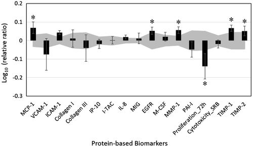
MEO showed a significant anti-proliferative activity in the human dermal fibroblasts (Figure ). In addition, it increased the levels of monocyte chemoattractant protein 1 (MCP-1), epidermal growth factor receptor (EGFR), matrix metalloproteinase 1 (MMP-1), and tissue inhibitor of metalloproteinase (TIMP)-1 and -2 in a slightly significant manner. Other biomarker readouts, including those of several inflammatory biomarkers, were not significantly affected by MEO. These results suggest that MEO may possess tissue remodeling and wound healing properties.
It has been demonstrated that MEO and its major active component terpinen-4-ol show anti-inflammatory and immunomodulatory activities (Hart et al., Citation2000; Koh, Pearce, Marshman, Finlay-Jones, & Hart, Citation2002; Low et al., Citation2015). Some small clinical studies have indicated that the anti-inflammatory, immunomodulatory, and antimicrobial activities of MEO possibly contribute to its pro-wound healing properties (Chin & Cordell, Citation2013; Edmondson et al., Citation2011; Pazyar et al., Citation2013). The results show that MEO affects molecules that are critical to the process of tissue remodeling in the skin, which suggests the involvement of an alternative mechanism in the effect of MEO on the modulation of wound healing.
3.2. Effects of MEO on genome-wide gene expression
We further explored the effects of MEO on human skin cells by studying it at a concentration of 0.011% v/v, which was the highest concentration we studied that was non-cytotoxic to the cells, on the RNA expression of 21,224 genes in the HDF3CGF system. The results showed a diverse effect of MEO on the regulation of these genes (Table S2). Among the 87 genes that were highly regulated (with a log2 [fold change ratio of expression over vehicle control] ≥ |1.5|) by MEO, a majority of them (65 genes) were significantly upregulated, whereas the rest were significantly downregulated (Table S2).
The IPA studies showed that the bioactivity of MEO significantly overlapped with many canonical signaling pathways from a literature-validated database (Figure , Table S3–S6). It was observed that many of the pathways are closely related to cellular metabolism. For instance, the phosphorylation and dephosphorylation of nicotinamide adenine dinucleotide (NAD), NAD salvage pathways, and biosynthesis of cholesterol were among these most-matched pathways. These findings indicate that MEO possibly plays a role in modulating cellular metabolism and thus, it may be a viable treatment for chronic metabolic conditions.
Figure 2. Top 20 canonical pathways matching the bioactivity profile of melaleuca essential oil (MEO, 0.011% v/v) in the HDF3CGF system produced using Ingenuity Pathway Analysis.
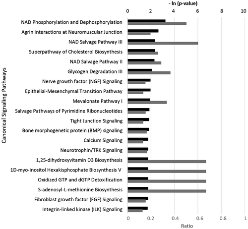
A literature search conducted by us revealed no published studies on the effects of MEO on metabolism in animal or human models. However, MEO has been found to inhibit the activities of metabolic enzymes in Candida albicans (Rajkowska, Kunicka-Styczyńska, Maroszyńska, & Dąbrowska, Citation2014). Furthermore, given that many metabolic conditions are often associated with inflammation and immune response disorders, it is reasonable to assume that MEO might be able to modulate metabolism via influencing inflammatory and immune mediators.
Collectively, the study, along with existing literature, suggests that MEO may possess tissue-remodeling and metabolism-modulating properties. However, further research into the biological and physiological mechanisms of action of MEO is recommended.
The current study has several limitations. Although the disease model was designed to simulate chronic inflammation and fibrosis, the in vitro study results cannot be directly applied to the more complex human system. In addition, the impact of MEO on gene expression was evaluated after short-term treatment of the cells with MEO. Therefore, how MEO impacts global gene expression over a longer term is unclear. Nevertheless, the protein and gene expression data are an evidence of the biological effect of MEO on human skin cells and will likely stimulate further research into the mechanism of action of MEO.
4. Conclusions
MEO significantly inhibited the proliferation of human dermal fibroblasts. Additionally, it slightly increased the levels of MCP-1, EGFR, MMP-1, and TIMP-1 and -2. Furthermore, genome-wide gene expression analysis showed that MEO modulates global gene expression. It was also observed that MEO robustly affected signaling pathways that are critical for cellular metabolism. The data obtained largely support that MEO possesses tissue-remodeling and metabolism-modulating properties.
Funding
This study was funded by dōTERRA (Pleasant Grove, UT, USA) and conducted at DiscoverX (Fremont, CA, USA).
OABI_1318476_Supplementary_Material.doc
Download MS Word (483.5 KB)Additional information
Notes on contributors
Xuesheng Han
Dr Han’s group primarily studies the health benefits of essential oils. We are specifically interested in the efficacy and safety of essential oils and their active components. Our studies of essential oils in both in vitro and clinical settings utilize a variety of experimental approaches, including analytical, biological, biochemical, and biomedical methodologies. We work closely with research institutes, hospitals, and clinics to move the study of essential oils forward. The research work discussed in this paper represents one part of a large research project, which was designed to extensively examine the impact of essential oils on human cells. This study, along with others, will further the understanding of the health benefits of essential oils for a wide research audience. Besides essential oils, we are also interested in studying the health benefits of herbal supplements and skin care products. Dr Han holds a PhD in Biological Sciences and is an elected Fellow of the American College of Nutrition. Dr Parker holds a PhD in Nutritional Sciences.
Related Research Data
References
- Barbosa-Morais, N. L., Dunning, M. J., Samarajiwa, S. A., Darot, J. F. J., Ritchie, M. E., Lynch, A. G., & Tavaré, S. (2010). A re-annotation pipeline for Illumina BeadArrays: Improving the interpretation of gene expression data. Nucleic Acids Research, 38, e17. doi:10.1093/nar/gkp942
- Berg, E. L., Yang, J., Melrose, J., Nguyen, D., Privat, S., Rosler, E., & Kunkel, E. J. (2010). Chemical target and pathway toxicity mechanisms defined in primary human cell systems. Journal of Pharmacological and Toxicological Methods, 61, 3–15. doi:10.1016/j.vascn.2009.10.001
- Bergamini, G., Bell, K., Shimamura, S., Werner, T., Cansfield, A., Müller, K., & Perrin, J. (2012). A selective inhibitor reveals PI3Kγ dependence of TH17 cell differentiation. Nature Chemical Biology, 8, 576–582. doi:10.1038/nchembio.957
- Bolstad, B. M., Irizarry, R. A., Astrand, M., & Speed, T. P. (2003). A comparison of normalization methods for high density oligonucleotide array data based on variance and bias. Bioinformatics, 19, 185–193. doi:10.1093/bioinformatics/19.2.185
- Chin, K. B., & Cordell, B. (2013). The effect of tea tree oil (Melaleuca alternifolia) on wound healing using a dressing model. The Journal of Alternative and Complementary Medicine, 19, 942–945. doi:10.1089/acm.2012.0787
- Dunning, M. J., Smith, M. L., Ritchie, M. E., & Tavare, S. (2007). beadarray: R classes and methods for Illumina bead-based data. Bioinformatics, 23, 2183–2184. doi:10.1093/bioinformatics/btm311
- Edmondson, M., Newall, N., Carville, K., Smith, J., Riley, T. V., & Carson, C. F. (2011). Uncontrolled, open-label, pilot study of tea tree (Melaleuca alternifolia) oil solution in the decolonisation of methicillin-resistant Staphylococcus aureus positive wounds and its influence on wound healing. International Wound Journal, 8, 375–384. doi:10.1111/j.1742-481X.2011.00801.x
- Han, X., & Parker, T. L. (2017a). Anti-inflammatory activity of Juniper (Juniperus communis) berry essential oil in human dermal fibroblasts. Cogent Medicine, 4, 1306200. doi:10.1080/2331205X.2017.1306200
- Han, X., & Parker, T. L. (2017b). Anti-inflammatory, tissue remodeling, immunomodulatory, and anticancer activities of oregano (Origanum vulgare) essential oil in a human skin disease model. Biochimie Open, 4, 73–77. doi:10.1016/j.biopen.2017.02.005
- Hart, P. H., Brand, C., Carson, C. F., Riley, T. V., Prager, R. H., & Finlay-Jones, J. J. (2000). Terpinen-4-ol, the main component of the essential oil of Melaleuca alternifolia (tea tree oil), suppresses inflammatory mediator production by activated human monocytes. Inflammation Research, 49, 619–626. doi:10.1007/s000110050639
- Homeyer, D. C., Sanchez, C. J., Mende, K., Beckius, M. L., Murray, C. K., Wenke, J. C., & Akers, K. S. (2015). In Vitro activity of Melaleuca alternifolia (tea tree) oil on filamentous fungi and toxicity to human cells. Medical Mycology, 53, 285–294. doi:10.1093/mmy/myu072
- Koh, K. J., Pearce, A. L., Marshman, G., Finlay-Jones, J. J., & Hart, P. H. (2002). Tea tree oil reduces histamine-induced skin inflammation. British Journal of Dermatology, 147, 1212–1217. doi:10.1046/j.1365-2133.2002.05034.x
- Kunkel, E. J., Dea, M., Ebens, A., Hytopoulos, E., Melrose, J., Nguyen, D., & Berg, E. L. (2004). An integrative biology approach for analysis of drug action in models of human vascular inflammation. FASEB Journal, 18, 1279–1281. doi:10.1096/fj.04-1538fje
- Kunkel, E. J., Plavec, I., Nguyen, D., Melrose, J., Rosler, E. S., Kao, L. T., & Wang, Y. (2004). Rapid structure-activity and selectivity analysis of kinase inhibitors by BioMAP analysis in complex human primary cell-based models. ASSAY and Drug Development Technologies, 2, 431–442. doi:10.1089/adt.2004.2.431
- Low, P., Clark, A. M., Chou, T. C., Chang, T. C., Reynolds, M., & Ralph, S. J. (2015). Immunomodulatory activity of Melaleuca alternifolia concentrate (MAC): Inhibition of LPS-induced NF-κB activation and cytokine production in myeloid cell lines. International Immunopharmacology, 26, 257–264. doi:10.1016/j.intimp.2015.03.034
- Pazyar, N., Yaghoobi, R., Bagherani, N., & Kazerouni, A. (2013). A review of applications of tea tree oil in dermatology. International Journal of Dermatology, 52, 784–790. doi:10.1111/j.1365-4632.2012.05654.x
- R Development Core Team. (2011). R: A language and environment for statistical computing. Vienna: The R Foundation for Statistical Computing. Retrieved from http://www.R-project.org/
- Rajkowska, K., Kunicka-Styczyńska, A., Maroszyńska, M., & Dąbrowska, M. (2014). The effect of thyme and tea tree oils on morphology and metabolism of Candida albicans. Acta Biochimica Polonica, 61, 305–310.