
Abbreviations
| CSF | = | cerebrospinal fluid |
| HPGD | = | hydroxyprostaglandin dehydrogenase |
| LPS | = | lipopolysaccharide |
| MRP4 | = | multidrug resistance transporter 4 |
| PGE2 | = | prostaglandin E2 |
| PGs | = | prostaglandins |
| PGT | = | prostaglandin transporter |
| SLCO2A1 | = | gene name for PGT. |
Sustained fever derives from prostaglandin E2 (PGE2) synthesized by brain capillary endothelial cells.Citation1 The “net fever signal” results from a) PGE2 synthesis, translocation, and receptor activation,Citation1 and b) PGE2 metabolic inactivation. Our understanding of the PGE2 synthesis and signaling is robust. Much less clear is how systemic PGE2 is translocated across the brain capillary endothelium into the brain parenchyma, how PGE2 synthesized in the endothelium is vectorially directed toward the parenchyma, or how the parenchyma is cleared of PGE2 when it is time to terminate the fever.
Hosotani et alCitation2 have contributed to solving these puzzles by localizing the prostaglandin transporter PGT (SLCO2A1) in the brain of rodents before and after administration of lipopolysaccharide (LPS). PGT was the first transporter discovered for which PGs are the substrate; it rapidly transports PGE2 with an affinity constant of ∼50–100 nM, depending on the species.Citation3 PGT is a lactate/prostaglandin exchanger that is poised energetically solely for PG uptake across the plasma membrane.
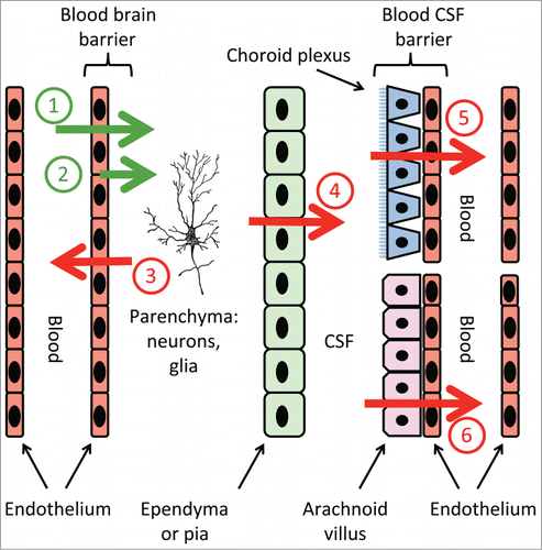
In contrast, newly-synthesized PGE2 effluxes from cells via multidrug resistance transporter 4 (MRP4).Citation4 Although MRP4 is expressed at the endothelial luminal membrane, indicating that PGE2 would be released by these cells into the bloodstream, PGT is expressed at the same membrane and, by a process of “sided reuptake” can salvage luminal PGE2 and direct it back toward the abluminal side Citation5 (Fig. 1, green arrows #1–2). In this regard, Hosotani et alCitation2 report endothelial expression of PGT in blood vessels of the subarachnoid space and choroid plexus after LPS injection. These data might indicate a role for PGT in directing PGE2 to the parenchyma.
Alternatively, or in addition, PGT in the brain might mediate PG clearance. Normally, PGs taken up by PGT are delivered to cytoplasmic oxidative enzymes, especially 15-hydroxy prostaglandin dehydrogenase (HPGD), for metabolic inactivation. Both PGT and HPGD in the same cell are required to generate the inactive metabolite 15-keto-PGE2.Citation6 Other transporters that mediate PG uptake have also identified, and some of these are also expressed in the brain (summarized in ref. Citation4). Assuming that all of these “non-MRP4” PG transporters function, like PGT, in the direction of PGE2 uptake from blood to cytoplasm, then cell types expressing these PG transporters might be expected to also express HPGD. Surprisingly, HPGD is not expressed in rat brain except to a small extent in the choroid plexus; in particular, there is no discernable HPGD expression in the parenchyma or the vasculature.Citation7 Therefore, termination of PGE2 signaling in fever likely does not occur within the brain parenchyma, but rather outside it. Since the brain parenchyma is surrounded by cells with plasma membranes, and since PGE2 does not readily penetrate plasma membranes by simple diffusion,Citation3 PGE2 must be pumped out of the brain parenchyma. How and where does this transport occur?
Figure 1 shows the putative role of PGE2 transporters in the removal and inactivation of PGE2 from the parenchyma (red arrows #3–6). PGE2 could be transported in a single step from the parenchyma across the blood brain barrier into the circulation (red arrow #3), after which it would be taken up by distant cells and enzymatically inactivated. Alternatively (or in addition), PGE2 could be cleared in 2 steps. The first would be transport (or even simple diffusion) from the parenchyma across the ependymal layer or pia into the CSF (red arrow #4). The second step would transport PGE2 into the blood across the CSF blood barrier at the choroid plexus (arrow #5) and/or the arachnoid villus (arrow #6). The novel localization of PGT in the arachnoid membrane, described by Hosotani et al,Citation2 is consistent with a role for PGT in PGE2 clearance.
Although PGT and other PG transporters have been localized to varying degrees in the brain, we have little information about the dynamic regulation of these transporters in the various phases of fever. Although Ivanov et al showed that PGT mRNA expression in the late phase of LPS-induced fever declined in liver and lung, there was no change in PGT mRNA expression in the hypothalamus in the early, mid, or late phase.Citation8 The report by Hosotani et al expands on these dynamic studies with new information on the change of PGT expression in the brain in an LPS model.Citation2 They have correlated the CSF PGE2 concentration after LPS administration with the expression levels of PGT mRNA in subarachnoid vessels and arachnoid membranes. Temporally, these correlations suggest a role for PGT in the clearance of PGE2 (arrows #3 and #6 in Fig. 1).
Much work remains, but another important piece of the puzzle has been put into place.
Figure 1. Transport pathways that might be involved when PGE2 signals fever in the brain. The green arrows show translocation of systemic PGE2 across the brain capillary endothelium, or the sided release of endothelium-derived PGE2, into the brain parenchyma. The red arrows show termination of PGE2 signaling via removal of PGE2. Numbers on arrows are discussed in the text.
Disclosure of Potential Conflict of Interest
No potential conflicts of interest were disclosed.
References
- Roth J, et al. Compr Physiol 2014; 4:1563-604; PMID:25428854; http://dx.doi.org/10.1002/cphy.c130033
- Hosotani R. et al. Temperature 2015; 2(3):425-36; http://dx.doi.org/10.1080/23328940.2015.1062953
- Schuster VL. Prostaglandins Other Lipid Mediat 2002; 68-69:633-47; PMID:12432949
- Tachikawa M, et al. Adv Pharmacol 2014; 71:337-60; PMID:25307222; http://dx.doi.org/10.1016/bs.apha.2014.06.006
- Nomura T, et al. J Biol Chem 2005; 280:28424-9; PMID:15855165; http://dx.doi.org/10.1074/jbc.M408286200
- Nomura T, et al. Mol Pharmacol 2004; 65:973-8; PMID:15044627; http://dx.doi.org/10.1124/mol.65.4.973
- Alix E, et al. Cerebrospinal Fluid Res 2008; 5:5-11; PMID:18318891; http://dx.doi.org/10.1186/1743-8454-5-5
- Ivanov AI, et al. Am J Physiol Regul Integr Comp Physiol 2003; 284:R698-706; PMID:12399253; http://dx.doi.org/10.1152/ajpregu.00570.2002