
ABSTRACT
This study examined the thermoregulatory and circulatory responses, and exercise performance of trained distance runners during exercise in the heat (31°C) at varying relative humidity (RH). In a randomized order, 11 trained male distance runners performed 5 60 min steady-state runs at a speed eliciting 70% of VO2max in RH of 23, 43, 52, 61 and 71%. This was followed immediately with an incremental exercise test to volitional exhaustion. Core (Tre) and mean skin temperature (T¯sk), cardiac output (Q), heart rate (HR), and stroke volume (SV) were recorded at regular intervals. A significant (P = 0.003) main effect was detected for RH on mean body temperature (Tb), with a significantly higher Tb detected during steady-state exercise in the 61 and 71% RH compared to that in the 23% RH. During the steady-state exercise, no differences were detected in whole body sweat loss (P = 0.183). However, a significant main effect of RH was observed for HR and SV (P = 0.001 and 0.006, respectively) but not Q (P = 0.156). The time to exhaustion of the incremental exercise test was significantly reduced at 61 and 71% RH compared with 23% RH (P = 0.045 and 0.005, respectively). Despite an increase in dry heat loss, a greater thermoregulatory and circulatory stress was evident during steady-state exercise at 61 and 71% RH. This ultimately limits the capacity to perform the subsequent incremental exercise to exhaustion. This study highlighted that in a warm environment, the range of the prescriptive zone progressively narrows as RH increases.
Introduction
It has been well established that thermoregulatory strain increases in the heat when humidity rises due to the reduced evaporative capacity of the environment (Emax).Citation1-4 This reduction in Emax limits heat dissipation from the skin surface to the environment, leading to a decline in sweating efficiency and increased area of skin wettedness.Citation5-7 The decline in sweating efficiency would, therefore, elevate skin temperature during exercise in humid conditions causing an increase in skin blood flow and greater circulatory strain.Citation8,9
The impact of relative humidity (RH) on exercise performance has been highlighted by Helou et al.Citation10 who analyzed environmental conditions and marathon performance across 6 European and American marathon races and noted that an increase in the Wet Bulb Globe Temperature (WBGT) was associated with a slowing of marathon performance. Unfortunately, Helou et al.Citation10 did not explicitly analyze the effect of RH on marathon performance, although wet bulb temperature has the greatest weighting in the calculation of WBGT.
Our current knowledge of the impact RH has on thermoregulatory responses are based on previous observations during exposure to a singleCitation1,2 or comparison of 2 (i.e., humid vs. dry heat) differing humidity conditionsCitation4,6 and 3 different relative humidity environments.Citation7 More recently, the impact of a wider range of RH on the capacity to exercise was investigated at lowCitation12 (5 RH levels) and moderateCitation13 (4 RH levels) intensity where it was concluded that exercise capacity was compromised at the higher end of the RH range when exposed to a hot (35°C),Citation12 and warmCitation13 (30°C) environment, respectively.
However, the conclusions regarding thermoregulatory and circulatory responses were inconsistent with Maughan et al.Citation13 reporting no effect of humidity on either thermoregulatory or circulatory responses as RH increased from 24 to 40%, 60 and 80%. By contrast, Moyen et al.Citation12 reported a greater heat storage and body temperatures at the higher end of the RH (55–70%) range even at a much lower exercise intensity.
The observations by Maughan et al.Citation13 appear contradictory to the widely established reports on thermoregulatory responses in humid conditions by earlier studies,Citation1-4 and therefore warrants for further investigation. In addition, the investigations by Maughan et al.Citation13 and Moyen et al.Citation12 were conducted in a condition with minimal (1 m·s−1) and no convective cooling, respectively, which presents a challenge to thermoregulation.Citation14,15 During an outdoor running event, a well trained runner will experience a substantial convective cooling as they usually run at speeds ranging from 12–20 km·h−1. The lower air speed used during laboratory testing in these earlier studiesCitation12,13 had likely overestimated the thermoregulatory capacity and makes it difficult to generalize the impact of RH on running performance. In this study, the wind speed of each individual trial was matched to running speed in order to closely simulate the effect of air resistance on a calm day outdoors.
Thus, this study was designed to examine the thermoregulatory and circulatory responses during a one hour session of moderate intensity steady-state exercise followed by a graded exercise test to volitional exhaustion under varying RH levels in a warm conditions. This experimental design allowed us to determine the compensability of the environment and separate the contribution that the thermoregulatory and circulatory systems have on the physiological and performance responses.
Therefore, the hypothesis of the current study was that increasing RH would progressively increase thermoregulatory strain and affect the stability of the circulatory system that would ultimately reduces the capacity to perform an incremental exercise test to exhaustion.
Materials and methods
Subjects
Eleven well-trained unacclimatised male runners who regularly participated in middle and long distance running events volunteered for this study (mean ± SD: age = 30 ± 4 y; height = 180 ± 6 cm; body mass = 72 ± 6 kg; lean body mass = 64 ± 6 kg; VO2max = 61 ± 6 ml·kg−1.min−1). Participants were briefed on the experimental protocol and testing procedures and provided written informed consent to participate. The University of Sydney Human Ethics Committee approved the experimental protocol for this study (Ref. No: 99/05/46), which conformed to the current Declaration of Helsinki guidelines. All experiments were conducted in a purpose-built environmental chamber during the fall and winter season in Sydney, Australia.
Preliminary testing and familiarization
Before taking part in this study, each participant visited the environmental chamber and was familiarized with the exercise protocol, equipment, and measurement procedures used in the study. This was followed by a preliminary testing session during which anthropometric measures, including body height, weight, composition and surface area were determined. Body surface area was calculated using the method of DuBois and DuBois.Citation16 Percent body fat was measured using the hydrodensitometry underwater weighing technique described in Siri.Citation17
Each participant then performed a running economy test followed by a maximal exercise test. The running economy test required each subject to run at 4 submaximal velocities of 10, 12, 14, and 16 km.h−1, respectively, for 4 min per stage. On completion of the submaximal running, participants engaged in an active recovery, where they walked for 5 min at a speed of 5 km·h−1. This was followed by a graded exercise test to determine maximal oxygen uptake (VO2max), where the participant ran at a fixed speed of 12 km·h−1 with the treadmill gradient elevated by 2% every 2 min until volitional fatigue was attained. Oxygen consumption and heart rate were taken throughout the exercise duration. Post-exercise, a linear regression line was plotted between submaximal steady state oxygen consumption and treadmill velocity to determine the participant's running speed which elicits an intensity of 70% VO2max, which was used for the individual's experimental trials for each subsequent testing session. The preliminary and familiarization sessions were conducted in a thermoneutral environment (20°C, 40% RH).
Experimental protocol
Each participant was reminded to refrain from heavy exercise and alcohol consumption the day before testing and to avoid caffeine consumption for 12 h before the test. They were also told to maintain a similar training routine throughout the duration of the study. Participants were asked to keep a 24 h food diary before testing and replicate their diet before subsequent visits to the laboratory. To ensure that participants were euhydrated at the onset of exercise, they were asked to ingest 6 ml of water per kg lean body mass at 2 h intervals (excluding when asleep) during the day before the test as well as the morning of testing. No fluid was provided to the participants during the running trials.
The five simulated humidity conditions were 23, 43, 52, 61, and 71% RH, with the ambient temperature held at constant 31.0 ± 0.2°C within each of the trials. Upon arrival at the environmental chamber on the day of testing (∼60 min before the exercise session), participants emptied their bladder, and nude body weight was determined. The participant was then instrumented and ingested 6 ml of water per kg lean body mass while sitting passively in a thermoneutral room (20°C and 40% RH) for 15 min.
For each RH trial, participants ran for 60 min at a speed eliciting an intensity of 70% VO2max. Immediately thereafter, participants continued running at the same velocity while the treadmill gradient was elevated by 2% every 2 min until volitional exhaustion, defined as the point at which participants could no longer maintain the pace of the treadmill, upon which the test was terminated. During the 60 min steady-state exercise, the treadmill was briefly stopped (≤1 min) at 30 and 60 min to allow time for the determination of the participant's body mass. The air speed set for each trial was matched to the individual running speed and thus simulated the effect of air resistance on a calm day outdoors. Each test session was separated by 5 d to minimize acclimation effects as well to provide adequate recovery.
Measurements
Rectal temperature (Tre), as an index of core temperature, was measured by a thermistor probe (YSI 400 series; Mallinckrodt Medical, St. Louis, MO) inserted 12 cm beyond the anal sphincter and data were recorded on a portable data logger (T-logger; University of Sydney, Sydney, Australia). Skin temperature was measured at 4 different sites (left shoulder, left chest, right mid-thigh, and right mid-shin) with thermistor probes (YSI 409 Series). Rectal and skin temperatures were sampled at 60 s intervals. Weighted mean skin temperature (T¯sk) was calculated according to the equation of Ramanathan.Citation18 Mean body temperature (Tb) was calculated using the equation developed by Colin et al.Citation19 Both rectal and skin thermistor probes used in the study were calibrated before and after the study in a water bath with temperature ranging from 15 to 50°C; where the calculated accuracies were ± 0.05 and ± 0.01, respectively.
Expired respiratory gas was obtained using the Douglas bag method at 10 min intervals and analyzed for fractions of O2 and CO2 concentration using gas sensors and analysers (O2 analyzer, Ametek S-3A/I and CO2 analysers, Ametek CD-3A, Applied Electochemistry Ametek Inc., Thermox Instruments Division). The gas analysers were calibrated with known calibration gases before each analysis. Cardiac output was determined during steady-state exercise at 10, 30, and 60 min by the CO2 rebreathing method of Collier.Citation20 Stroke volume was calculated using the Fick equation. Heart rate was continuously monitored telemetrically via a short-range transmitter-receiver (Polar Electro Oy, Kempele, Finland).
Whole body sweating rate (WBSR) during steady state exercise within the first and last 30 min of steady-state exercise was calculated as the difference in pre- and post-nude body weight with correction for respiratory moisture loss.Citation21 During each trial, subjects were asked to report their ratings of perceived exertion (RPE) using the Borg 15-point ratings scale.Citation22 A thermal scale developed by the American Society of Heating, Refrigeration and Air-Conditioning (1966)Citation23 was used for the subjects to provide a subjective indication of their perception or sensations of thermal stress (TS). This TS scale consisted of a 7 point range from cold (1) to hot (7).Citation23 Subjects were also asked to rate their sensation of “sweatiness” on a 5-point skin wettedness (SW) scale ranging from dry (0) to wet (4). These subjective responses were recorded at 10 min intervals during steady-state exercise.
A partitional calorimetry software program developed by Atkins and ThompsonCitation24 was used to estimate the magnitude and avenue of body heat gain and loss during the exercise trials. Partitional calorimetry calculations were based on measures taken within the last 30 min of steady state exercise in each RH condition. Evaporative heat loss (E) in W·m−2 was estimated from the classic body heat balance equation of E = M – W (Eres + Cres) – R – C – S, where M is metabolic heat production, W is external work, Eres + Cres is heat transfer via the respiratory tract, R is radiative heat loss, C is convective heat loss, and S is body heat storage. S was estimated using the following equation: ((3474 × wt × (Tb final – Tb initial)) × t−1) × AD −1 (W.m−2), where 3474 is the average specific heat of body tissue (J·kg−1 °C−1), wt is body mass (kg), Tb is mean body temperature (°C), t is exercise time (sec) and AD is body surface area (m2). Estimation of tissue heat conductance (K) was derived from the method of Davies (1979) as K = Hsk / ((Tre– T¯sk) * AD) −1 (W·m−2 °C−1), where Hsk is the heat dissipated from the skin (E + R + C), Tre is rectal temperature, T¯sk is mean skin temperature, and AD is the DuBois body surface area in m−2. Convective and radiative heat loss in W·m−2 were estimated using the equations found in Fanger Citation25 and McIntyre,Citation26, respectively.
Statistical analysis
All statistical calculations were performed using SPSS version 19 for Windows (Chicago, IL). A two-way (RH x time) repeated measure analysis of variance (ANOVA) was used to test the significance between and within treatments across the 5 RH levels. Pair-wise differences between trials were evaluated post-hoc using one-way ANOVA with a Bonferroni adjustment applied for multiple comparisons. Thermoregulatory and circulatory responses are presented as mean and group standard deviation for the purpose of improving the clarity of the figures. The level of significance was set at P < 0.05. All data are presented as means ± SD.
Results
Steady-state exercise
Participant's body mass at the start of steady state exercise did not differ significantly between any of the RH trials (P = 0.503). There were no significant differences among participants in both Tre (P = 0.305) and T¯sk (P = 0.312) at the onset of steady-state exercise, which highlighted that the participants were in a similar physiological state. There was a significant main effect of the increase in RH for Tb (P = 0.003), Tre (P = 0.006) and T¯sk (P = 0.001). A significant interaction (RH*exercise duration) was detected for all 3 thermoregulatory dependent variables, such that, Tb during steady-state exercise was significantly higher (P < 0.01) at 61 and 71% RH when compared with 23% RH ().
Figure 1. Rectal temperature (a), mean skin temperature (b) and mean body temperature (c) responses during steady-state exercise in a warm environment with varying relative humidity (RH) levels (n = 11). Data are presented as mean and group standard deviation. *** indicates significant (P < 0.05) difference between the 23% RH trial and the 52, 61, and 71% RH trials. ** indicates significant (P < 0.05) difference between the 23% RH trial and the 61% and 71% RH trials. * indicates significant (P < 0.05) difference between the 23% RH trial and 71% RH trials.
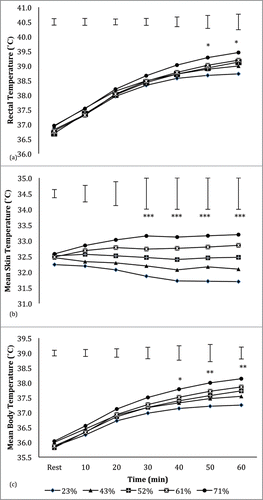
Post-hoc testing revealed that, when compared with the 23% RH, a significantly (P = 0.015) higher Tre was recorded within the last 10 min of steady-state exercise in the 71% RH (). The increased RH also resulted in a significantly (P < 0.05) greater rate of rise in Tre in the 52, 61, and 71% RH (2.4 ± 0.4°C, 2.4 ± 0.4°C, and 2.5 ± 0.5°C, respectively) compared with the 23% RH (1.9 ± 0.4°C). A significantly higher T¯sk was recorded from 30 min through to the end of steady-state exercise in the 52, 61, and 71% RH compared with the 23% RH (P = 0.001) (). Mean body temperature (Tb) during steady-state exercise was significantly higher at 61 and 71% RH when compared with 23% RH (). A significant (P < 0.05) narrowing of core to skin thermal gradient was recorded in the 52, 61 and 71% RH when compared with the 23% RH condition (6.3 ± 0.7°C, 6.3 ± 0.6°C and 6.3 ± 0.6°C vs. 7.0 ± 0.8°C).
There was no significant main effect (P = 0.141) of RH on VO2 during the steady-state exercise. At the end of steady-state exercise, S in the 43, 52, 61, and 71% RH conditions was significantly (P < 0.05) higher when compared with the 23% RH (24.1 ± 18.4 W·m−2, 36.9 ± 23.3 W·m−2, 37.16 ± 25.3 W·m−2 and 24.1 ± 18.4 W·m−2, respectively vs. 15.7 ± 22.1 W·m−2). Increased RH resulted in an enhanced dry heat loss capacity and a reduced in evaporative heat loss capacity (). A significant (P < 0.05) increase in dry heat loss capacity was recorded in the 43, 52, 61, and 71% RH levels. By contrast, E recorded a significant (P = 0.002) reduction in the 71% RH (). A significantly higher (P < 0.05) K was recorded in the 52, 61, and 71% RH trials when compared with the 23% RH trial (150.2 ± 22.2 W·m−2, 151.8 ± 15.5 W·m−2, and 155.8 ± 17.0 W·m−2, respectively vs. 140.6 ± 19.9 W·m−2; P = 0.05, 0.003, and 0.001, respectively). There was no significant main effect of a systematic rise in RH on WBSR (P = 0.183; ). WBSR remained unchanged during the last 30 min of exercise despite a steady increase in core temperature in the 61 and 71% RH conditions.
Figure 2. Evaporative and dry heat loss values at 60 min of exercise under varying levels of relative humidity (n = 11). # indicates significant (P < 0.05) difference in dry heat loss from the 23% RH trial. * indicates significant (P < 0.05) difference in evaporative heat loss from the 23% RH trial.
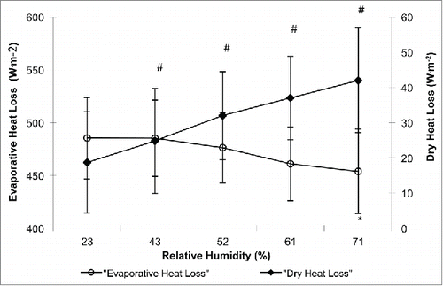
Table 1. Mean body heat storage (S) and whole body sweat rate (WBSR) during exercise across different levels of relative humidity (RH) (n = 11). Data are shown as mean ± standard deviation (SD). * indicates a significant (P < 0.05) difference from the 23% RH trial.
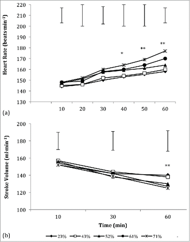
Relative humidity proved to have a significant main effect on HR and SV during the steady-state exercise phase (P = 0.001 and P = 0.006, respectively). Heart rate during steady-state exercise was significantly higher in the 61 and 71% RH trials compared with the 23% RH trial (P < 0.001) (). A systematic decline in SV during steady state exercise was recorded with rising RH and was significantly lower in the 71% RH compared with the 23% RH (). Cardiac output remained unchanged between trials and was not significantly (P = 0.156) affected with the rise in RH.
Figure 3. Heart rate (a) and stroke volume (b) responses during steady state exercise across varying relative humidity (RH) levels (n = 11). ** indicates a significant (P < 0.05) difference between the 23% RH trial and the 61% and 71% RH trials at the time interval. * indicates a significant (P < 0.05) difference between the 23% RH trial and the 71% RH trial at the time interval.
The subjective responses of RPE and TS indicated that steady-state exercise was harder to perform and more thermally stressful in the higher RH conditions of 52, 61, and 71% (). Participants further reported an increased perception of skin wettedness based on the SW rating scale at RH levels of 61 and 71% ().
Table 2. Subjective responses of rate of perceived exertion (RPE), thermal stress (TS), and skin wettedness (SW) after 60 min of exercise across different levels of relative humidity (n = 11). Data are shown as mean ± standard deviation (SD). * indicates a significant (P < 0.05) difference from the 23% RH trial.
Incremental exercise test to exhaustion
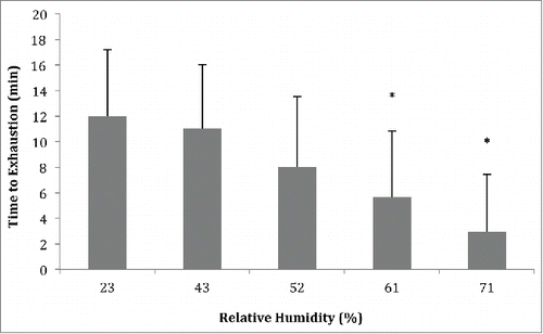
During the incremental exercise test, the time to exhaustion declined with rising RH level () and was significantly lower at 61 and 71% RH compared with 23% RH (P = 0.045 and 0.005, respectively). Mean body temperature at the time of exhaustion within the 61 and 71% RH was significantly higher than the 23% RH (38.1 ± 0.4°C and 38.3 ± 0.4°C vs 37.7 ± 0.3°C, respectively: P = 0.004 and P < 0.001, respectively). There was no significant difference (P > 0.05) in Tre between the 23% RH (39.2 ± 0.3°C) at exhaustion with the other conditions of 43% (39.3 ± 0.4°C), 52% (39.5 ± 0.2°C), and 61% (39.5 ± 0.3°C). However, Tre was significantly higher (P = 0.015) in the 71% RH trial (39.7 ± 0.3°C) as compared with the 23% RH. A was significantly higher (P < 0.001) mean skin temperature was recorded at the time of exhaustion within the 52% (32.52 ± 0.80°C), 61% (32.92 ± 0.77°C) and 71% (33.32 ± 0.81°C) when compared with the 23% RH (31.74 ± 0.93°C).
Figure 4. Time to exhaustion during a graded running exercise phase across varying relative humidity levels (n = 11). * indicates significant (P < 0.05) difference from the 23% RH trial.
There was no significant difference (P > 0.05) in HR at exhaustion across the varying levels of RH. In relative values, participants had reached ∼98% of their respective percent maximal HR at the time of exhaustion within all of the RH levels.
Discussion
The findings of this study highlight that unacclimatized well-trained male runners had experienced a greater thermoregulatory and circulatory strain during prolonged steady-state exercise in a warm environment of 30°C as RH increased to 61% and 71%. This was followed by a reduced capacity to perform incremental exercise to exhaustion within these 2 RH levels when compared with the driest RH level of 23%. These findings are consistent with more recent reports of an increased thermoregulatory and circulatory strain during prolonged steady-state exercise in a more humid conditionCitation12 and a reduced exercise capacity in a warm environment with rising RH level.Citation13
The current observation has demonstrated that the ability to regulate body temperature during running exercise at an intensity of 70% VO2max at 31°C ambient temperature was progressively compromised as RH increased, and becomes substantially and statistically so when RH exceeds 60%. This finding also highlights that the range in the prescriptive zone, previously described by LindCitation27 as a range of environmental conditions in which an individual's body temperature is independent of climatic conditions, progressively narrows as RH increases. This is reflected by the non-linear increase in core temperature and in addition, an elevated level of skin temperature during the steady-state exercise within these 2 RH levels. The increase in rectal and mean skin temperature would narrow the thermal gradient between the core and the skin, thus, resulting in a reduced capacity to transfer heat for dissipation. A recent study by Cuddy et al.Citation28 concluded that the capacity to perform aerobic exercise was progressively diminished with a reduced core to skin temperature gradient. In addition, elevated mean skin temperature seen in the present study within the rising levels of RH would coincide with a greater skin blood flow and venous compliance, which augments cardiovascular strain.Citation8,9
Prior to the study by Maughan et al.Citation13 who had examined the effect of a systematic rise in RH on exercise performance in the warm environment, many others had reported an increased thermoregulatory strain during exercise when comparison was made between a hot-humid and hot-dry condition.Citation1-3,6 Pandolf et al.Citation1 reported that during exercise at 25% VO2max, 6 unacclimatized subjects had a higher core and skin temperature in humid conditions than dry. Another later study by Sen Gupta et al.,Citation3 described a steady increase in core and skin temperature during light exercise at 3 different workloads (i.e., 65, 82 and 98 W) in hot-humid conditions compared with hot-dry and thermoneutral conditions. This consistent pattern of increasing thermoregulatory strain was observed in this experiment which is believed to be due to the reduced Emax as RH level increased which restricts the rate of sweat evaporation.Citation11 In the present study, Emax ranged from 1000.5 W.m−2 in the 23%RH to almost half of this in 71%RH (577.5 W.m−2). Interestingly, using the heat strain index (HSI) as an estimate of the thermal compensability of the environmentCitation29 only in the 71% RH trial did the steady-state exercise become uncompensable (HSI = 1.01), whereas all other trials appeared compensable (HSI = 0.98–0.99).
While a rise in core temperature during exercise in an uncompensable heat stress environment has been widely documented,Citation30,31 the present study observed a pronounced elevation in skin temperature during steady-state exercise from 30 minutes onwards within the 52, 61 and 71% RH when compared with the driest level of 23% RH. Consistent with an earlier finding by Maw et al.Citation32 a greater perception of exercise effort and thermal discomfort was recorded in the present study, when mean skin temperature remained elevated during steady-state exercise within these 3 RH levels. In comparison to the internal heat strain, a significant level of heat strain was only recorded within the last 10 minutes of the steady state exercise. It is plausible that the elevated level of skin temperature could be a limiting factor for exercise capacity in a more humid environment. Earlier publications have proposed that high skin temperature can lead to fatigue and impair endurance exercise capacity.Citation9,33 It is, however, noteworthy to highlight that while an elevated skin temperature presents a detrimental effect for a greater cutaneous blood flow, it is, however, a major requirement for maintaining heat loss from the skin surface to the environment especially in a condition where Emax is reduced such as in the higher humidity environment.Citation34 According to Kerslake,Citation34 an elevated skin temperature would raise the vapor pressure at the skin surface which is a strategy for maintaining heat transfer from the skin surface to the environment under a humid condition where Emax is reduced.
The earlier study by Maughan et al.Citation13 did not find any main effect of RH on core temperature responses, which was at odds with many earlier observations.Citation1-4,6,12 We speculated that had the participants in the study by Maughan et al.Citation13 been able to continue to exercise past the 50 min mark, within the highest RH level of 80%, they could have well observed an effect of RH on core temperature responses. This speculation was based on the current observation that the rise in core temperature during steady-state exercise reached significant level within the 50 to 60 min in the 71% RH. Extrapolating the reported rate of rise in core temperature within the 80% RH of Maughan et al.Citation13 study, core temperature could potentially be significantly higher than the 24% RH level, taken into account, the intensity of exercise was similar to the current study (70% VO2max) and their participants were also similarly categorized as well trained.
A greater magnitude of cardiovascular drift was observed during the steady-state exercise within the 61 and 71% RH trials compared to the driest condition. Others have suggested that this effect can be attributed to a greater rate of increase in skin blood flowCitation8,9 driven by an increased core temperature. Although skin blood flow was not measured directly in the present study, the increase in tissue heat conductance, previously described as an indicator of skin blood flow,Citation35 would suggest greater skin blood flow during exercise in the more humid conditions. In addition, displacement of central blood volume to the cutaneous veins has been previously shown to occur in conditions where skin temperature remained elevated,Citation36 and results in decreased stroke volume, mean arterial pressure, right arterial mean pressure, total peripheral resistance, and an increase in heart rate.Citation36
In the present study, we had also examined the interaction of heat exchange modes under varying levels of RH. The initial work by NielsenCitation37 demonstrated that humans can regulate their core temperature during moderate intensity exercise, independent of the environmental conditions over a relatively wide range of temperatures (5–30°C) by increasing their evaporative heat loss capacity. This range of environmental conditions was later referred to as the prescriptive zone by Lind.Citation27 The earlier work by NielsenCitation37 was conducted in a relatively dry (35–55% RH) conditions with increasing ambient temperature. Contrary to the finding by Nielsen,Citation36 the present study observed a greater reliance on dry heat loss as the capacity of evaporative heat loss is reduced as the RH increased as shown in . Despite the increase in dry heat loss capacity, it was not sufficient to control the rise in body temperature during exercise in the higher RH conditions. This reaffirms the notion that evaporative heat loss plays a vital role as the main avenue for heat dissipation during exercise in the a heat-stress environment.Citation11 While the importance of dry heat loss to thermoregulation in a reduced Emax environment has been previously suggested,Citation11 to our knowledge, this is the first study to systematically demonstrate the rise in dry heat loss capacity during exercise with rising RH conditions.
Another interesting observation in the present study was that despite a strong afferent input from the rising core and elevated skin temperature, within the 61 and 71% RH, the rate of whole body sweat loss remained unchanged. This observation contradicted the notion of a linear relationship between sweating rate and rectal temperature which was previously demonstrated during exercise in a drier (∼40% RH) heat stress environment.Citation38,39 The current observation on total sweat loss remaining unchanged during exercise in a humid condition has been previously reported.Citation7,40 In a more recent observation by Maughan et al.,Citation13 total sweat loss (based on changes in pre- and post-exercise body weight) had also remained unchanged during exercise across the RH levels of 24% (1.49 ± 0.46 L) to 80% (1.33 ± 0.26 L). However, when Maughan et al.Citation13 reported the amount of total sweat loss over the exercise duration (sweat rate), the sweat rate was higher in the 80% RH (a shorter exercise period of 46 ± 14 mins) when compared to the sweat rate during a longer exercise duration of 68 ± 19 mins in the 20% RH. The inability to increase the total sweat loss during the steady-state exercise had suggested that some level of sweat suppression occurs with rising RH, leading to increasing levels of skin wettedness and sweat drippage.Citation5,7 In the current study, the participants' subjective responses to the sweatiness scale had indicated that they felt an increasing area of skin wettedness during the steady state exercise within the 61% and 71% RH. The level of skin wettedness induced by sweating is positively correlated with thermal discomfort which ultimately influence exercise performance with heat stress.Citation41
After experiencing a greater thermoregulatory and circulatory strain during prolonged exercise in the higher RH levels humid of 61 and 71% RH, we observed the ability to continue the graded exercise test to exhaustion was drastically reduced. At exhaustion, the value of core temperature was lower than previously referred to the critical level of 40°C.Citation30,31 A lower core temperature at exhaustion following dynamic exercise has been previously reported in situations where evaporative heat loss is restricted.Citation42 Similarly, observation of a lower core temperature at fatigue following dynamic exercise has been previously documented in both fieldCitation43 and laboratoryCitation44,45 settings. It is interesting to note, however, heart rate at exhaustion had almost reached the maximal level. Thus, indicating that the circulatory strain was pushed to the limit during an all-out exercise in the high RH level, unlike core temperature that was lower than the proposed critical level. The current observation of a reduced exercise capacity in the higher RH condition is consistent with several earlier reports.Citation3,4,13
In conclusion, the present study has demonstrated that despite an increase in dry heat loss in the 61 and 71% RH condition, a greater thermoregulatory and circulatory stress was evident during the steady-state exercise. This combined thermoregulatory and circulatory stress ultimately limits the capacity to perform an all-out exercise to exhaustion. It is evident from this study that the range of the prescriptive zone progressively narrows as RH increases.
Abbreviations
| E | = | Evaporative heat loss |
| Emax | = | Evaporative capacity of the environment |
| HR | = | Heart rate |
| K | = | Tissue heat conductance |
| Q | = | Cardiac output |
| RH | = | Relative humidity |
| RPE | = | Rating of perceived exertion |
| SW | = | Skin wettedness |
| SV | = | Stroke volume |
| T¯sk | = | Mean skin temperature |
| Tb | = | Mean body temperature |
| Tre | = | Rectal temperature |
| TS | = | Thermal stress |
| WBSR | = | Whole body sweat rate |
Disclosure of potential conflicts of interest
No potential conflicts of interest were disclosed.
Acknowledgments
This article is dedicated to the memory of Dr. Martin Thompson, who passed away during the revision of this manuscript. The authors also gratefully acknowledge the contribution of a dedicated group of runners and the technical assistance of Ray Patton, Tim Turner and Dr. Pat Ruell. We also acknowledge suggestions and assistance of Dr. Barry Holcombe during the course of this study.
References
- Pandolf KB, Gonzalez RR, Gagge AP. Physiological strain during light exercise in hot-humid environments. Aerospace Med 1974; 45:359-65; PMID:4821728
- Kobayashi K, Horvath SM, Diaz FJ, Bransford DR, Drinkwater BL. Thermoregulation during rest and exercise in different postures in a hot humid environment. J Appl Physiol 1980; 48:999-1007; PMID:7380712
- Sen Gupta J, Swamy YV, Pichan G, Dimri GP. Physiological responses during continuous work in hot dry and hot humid environments in Indians. Int J Biometeorol 1984; 28:137-46; PMID:6735516; http://dx.doi.org/10.1007/BF02191726
- Nielsen B, Strange S, Christensen NJ, Warberg J, Saltin B. Acute and adaptive responses in humans to exercise in a warm, humid environment. Pflugers Arch 1997; 434:49-56; PMID:9094255; http://dx.doi.org/10.1007/s004240050361
- Candas V, Libert JP, Vogt, JJ. Human skin wettedness and evaporative efficiency of sweating. J Appl Physiol 1979; 46:522-8; PMID:438022
- Frye AJ, Kamon E. Sweating efficiency in acclimated men and women exercising in humid and dry heat. J Appl Physiol 1983; 54:972-7; PMID:6853304
- Alber-Wallerstrom B, Holmer I. Efficiency of sweat evaporation in unacclimatized man working in a hot humid environment. Eur J Appl Physiol 1985; 54:480-7; PMID:4085475; http://dx.doi.org/10.1007/BF00422956
- Gonzalez-Alonso J Human thermoregulation and the cardiovascular system. Exp Physiol 2012; 97:340-6; PMID:22227198; http://dx.doi.org/10.1113/expphysiol.2011.058701
- Sawka MN, Cheuvront SN, Kenefick RW. High skin temperature and hypohydration impair aerobic performance. Exp Physiol 2012; 97:327-32; PMID:22143882; http://dx.doi.org/10.1113/expphysiol.2011.061002
- Helou NE, Tafflet M, Berthelot G, Tolaini J, Marc A, Guillaume M, Hausswirth C, Toussaint J. Impact of environmental parameters on marathon running performance. PloS One 2012; 7:1-9; PMID:22649525; http://dx.doi.org/10.1371/journal.pone.0037407
- Gagge A.P, Gonzalez, R.R. Mechanisms of heat exchange: Biophysics and physiology. Compr Physiol 2011; Supplement 14: Handbook of Physiology, Environmental Physiology:45-84. First published in print 1996
- Moyen NE, Ellis CLV, Ciccone, AB, Thurston, TS, Cochrane, KC, Brown, LE, Coburn, JW, Judelson, DA. Increasing relative humidity impacts low-intensity exercise in the heat. Aviat Space Environ Med 2014; 85:112-9; PMID:24597154; http://dx.doi.org/10.3357/ASEM.3787.2014
- Maughan RJ, Otani H, Watson P. Influence of relative humidity on prolonged exercise capacity in a warm environment. Eur J Appl Physiol 2012; 112:2313-21; PMID:22012542; http://dx.doi.org/10.1007/s00421-011-2206-7
- Adams WC, Mack GW, Langhans GW, Nadel ER. Effects of varied air velocity on sweating and evaporative rates during exercise. J Appl Physiol 1992; 73:2668-74; PMID:1490985
- Saunders AG, Dugas JP, Tucker R, Lambert MI, Noakes TD. The effects of different velocities on heat storage and body temperature in humans cycling in a hot, humid environment. Acta Physiol Scand 2005; 183:241-55; PMID:15743384; http://dx.doi.org/10.1111/j.1365-201X.2004.01400.x
- DuBois D, DuBois EF. A formula to estimate the approximate surface area if height and weight be known. Arch Int Med 1916; 17:863-71; PMID:2520314; http://dx.doi.org/10.1001/archinte.1916.00080130010002
- Siri WE. Body composition from fluid space and density. In J. Brozek & A. Hanschel (Eds.), Techniques for Measuring Body Composition. National Academy of Science, Washington, DC, 223-44, 1961.
- Ramanathan NL. A new weighting system for mean surface temperature of the human body. J Appl Physiol 1964; 19:531-3; PMID:14173555
- Colin J, Timbal J, Houdas Y, Boutelier C, Guieu JD. Computation of mean body temperature from rectal and skin temperature. J Appl Physiol 1971; 31:484-9; PMID:5111868
- Collier CR. Determination of mixed venous CO2 tensions by rebreathing. J Appl Physiol 1956; 9:25-29; PMID:13357408
- Mitchell JW, Nadel ER, Stolwijk JA. Respiratory weight losses during exercise. J Appl Physiol 1972; 32:474-6; PMID:5026494
- Borg GA. Psychophysical bases of perceived exertion. Med Sci Sports Exerc 1982; 14:377-81; PMID:7154893
- American Society of Heating, Refrigeration and Air-Conditioning. Thermal comfort conditions. ASHRAE standard 1996; 55:66. New York
- Atkins K, Thompson MW. A spreadsheet for partitional calorimetry. Sportscience 2000; 4: sportsci.org/jour/0003/ka.html
- Fanger PO. Thermal Comfort. McGraw-Hill, New York, 1970.
- McIntyre DA. Indoor Climate. Applied Science Publishers, London, 1980.
- Lind AR. A physiological criterion for setting thermal environmental limits for everyday work. J Appl Physiol 1963; 18:51-6; PMID:13930723
- Cuddy JS, Hailes, WS, Ruby BC. A reduced core to skin temperature gradient, not a critical core temperature, affects aerobic capacity in the heat. J Therm Biol 2014; 43:7-12; PMID:24956952; http://dx.doi.org/10.1016/j.jtherbio.2014.04.002
- Cheung SS, McLellan TM, Tenaglia, S. The thermophysiology of uncompensable heat stress. Physiological manipulations and individual characteristics. Sports Med. 2000; 29:329-59; PMID:10840867; http://dx.doi.org/10.2165/00007256-200029050-00004
- Nielsen B, Hales J, Strange S, Christensen N, Warberg J, Saltin B. Human circulatory and thermoregulatory adaptations with heat acclimation and exercise in a hot, dry environment. J Physiol 1993; 460:467-85; PMID:8487204; http://dx.doi.org/10.1113/jphysiol.1993.sp019482
- Gonzalez-Alonso J, Teller C, Andersen S, Jensen F, Hyldig T, Nielsen, B. Influence of body temperature on the development of fatigue during prolonged exercise in the heat. J Appl Physiol 1999; 86:1032-9; PMID:10066720
- Maw GJ, Boutcher SH, Taylor NA. Ratings of perceived exertion and affect in hot and cool environments. Eur J Appl 1993; 67:174-9; PMID:8223525; http://dx.doi.org/10.1007/BF00376663
- Cheuvront SN, Kenefick RW, Montain SJ, Sawka MN. Mechanisms of aerobic performance impairment with heat stress and dehydration. J Appl Physiol 2010; 109:1989-95; PMID:20689090; http://dx.doi.org/10.1152/japplphysiol.00367.2010
- Kerslake DM. The Stress of Hot Environments. Cambridge University Press, Cambridge, 1972.
- Nielsen B. Thermoregulation in rest and exercise. Acta Physiol Scand Suppl 1969; 323:1-74; PMID:5345560; http://dx.doi.org/10.1111/j.1748-1716.1969.tb04445.x
- Rowell LB, Brengelmann GL, Murray JA, Kraning II, KK, Kusumi F. Human metabolic responses to hyperthermia during mild to maximal exercise. J Appl Physiol 1969; 26:395-402; PMID:5775323
- Nielsen M. Die regulation der korpertemperatur bei muskelarbeit. Scand Arch Physiol 1938; 79:193-230; http://dx.doi.org/10.1111/j.1748-1716.1938.tb01246.x
- Nadel ER, Bullard RW, Stolwijk JA. Importance of skin temperature in the regulation of sweating. J Appl Physiol 1971; 31:80-87; PMID:5556967
- Kondo N, Shibasaki M, Aoki K, Koga S, Inoue Y, Crandall CG. Function of human eccrine sweat glands during dynamic exercise and passive heat stress. J Appl Physiol 2001; 90:1877-1881; PMID:11299281
- Keatisuwan W, Ohnaka T, Tochihara Y. Physiological responses of men and women during exercise in hot environments with equivalent WBGT. Appl Human Sci 1996; 15:249-58; PMID:9008978; http://dx.doi.org/10.2114/jpa.15.249
- Filingeri D, Havenith G. Human skin wetness perception: psychological and neurophysiological bases. Temp 2015; 2:86-104; http://dx.doi.org/10.1080/23328940.2015.1008878
- Sawka MN, Latzka WA, Montain SJ, Cadarette BS, Kolka MA, Kraning KK, Gonzalez RR. Physiologic tolerance to uncompensable heat: intermittent exercise, field vs. laboratory. Med Sci Sports Exerc 2001; 33:422-30; PMID:11252069; http://dx.doi.org/10.1097/00005768-200103000-00014
- Byrne C, Lee JK, Chew SA, Lim CL, Tan EY. Continuous thermoregulatory response to mass-participation distance running in the heat. Med Sci Sports Exerc 2006; 38:803-10; PMID:16672830; http://dx.doi.org/10.1249/01.mss.0000218134.74238.6a
- Ely BR, Ely MR, Cheuvront SN, Kenefick RW, DeGroot DW, Montain SJ. Evidence against a 40°C core temperature threshold for fatigue in humans. J Appl Physiol 2009; 107:1519-25; PMID:19713430; http://dx.doi.org/10.1152/japplphysiol.00577.2009
- Ely BR, Cheuvront SN, Kenefick RW, Sawka MN. Aerobic performance is degraded, despite modest hyperthermia, in hot environments. Med Sci Sports Exerc 2010; 42:135-41; PMID:20010120; http://dx.doi.org/10.1249/MSS.0b013e3181adb9fb