
Abstract
Posterior-stabilized (PS) total knee arthroplasty (TKA) designs were introduced to compensate for the resected or deficient posterior cruciate ligament and to avoid paradoxical anterior sliding of the femur. Knee joint stability may differ among patients and, therefore, orthopaedic companies developed several solutions to compensate for these differences. In particular, conventional PS designs are used for conventional TKA and semi-constrained PS designs are mainly applied in revision surgery or in conventional surgery when there is a compromised soft tissue envelope. However, despite good functional long-term results, a better understanding of forces acting on the post of the polyethylene insert is needed to find an eventual correlation with the risk of post-cam failure or loosening of the tibial baseplate for the two PS solutions. To the authors’ best knowledge, no literature data is currently available to compare the two solutions. In this paper, a validated numerical model was developed to analyse and compare the posterior of the post-cam mechanism of the same TKA design with conventional and semi-constrained inserts during several motor tasks. For each solution, different motor tasks show different values of maximum post-cam force. Comparing the two solutions, a general slightly higher force (range between 3 and 5%) is observed in the semi-constrained solution. The results concerning the conventional solution are in agreement with the literature. After this analysis, we can report that a semi-constrained design shows only slightly higher posterior post-cam contact force, and thus would not be expected to significantly increase the risk of failure of the post-cam mechanism, as compared to a conventional PS design.
Introduction
The success of total knee arthroplasty (TKA) in relieving pain and improving function has led to its widespread use worldwide (Rosen et al. 202). Furthermore, the increasing size of the aging population will only further test the longevity and durability of TKA (Rosen et al. Citation2002; Kurtz et al. Citation2010).
Posterior-stabilized (PS) TKA was introduced in the mid ’70s (Insall et al. Citation1985). This concept was developed to prevent posterior subluxation of the tibia, as a replacement for a degenerative posterior cruciate ligament, and to improve the range of movement of the knee by allowing femoral rollback and increasing the moment arm of the quadriceps (Colizza et al. Citation1995). It has been widely used for more than two decades, and long-term follow-up studies have reported satisfactory results (Schai et al. Citation1998; Rodriguez et al. Citation2001; Sultan et al. Citation2003).
Recently, surgeons have sought a greater range of movement after TKA (Schai et al. Citation1999). Many patients, even those with a TKA without scar, pain and back-related problems, avoid kneeling as reported in the New Zealand orthopaedic registry (available at www.nzoa.org.nz/nz-joint-registry). However, kneeling is required for many activities of daily living and several jobs. More than 50% of patients with TKA consider kneeling as important (Matsuda et al. Citation1997). It is thought that patients who undergo a PS TKA will achieve a greater range of movement than those who undergo a cruciate retaining TKA because the post-cam mechanism in a PS TKA will promote posterior rollback of the femur (Mauerham Citation2003).
Intra-operative and post-operative benefits of a PS TKA over a cruciate retaining TKA – include easier ligament balancing, easier correction of severe deformity by eliminating a tight posterior cruciate ligament, increased predictability in restoration of knee kinematics, improved range of motion, and potentially minimized polyethylene wear – because of the option to use more congruent articular surfaces (Schai et al. Citation1998; Rodriguez et al. Citation2001; Sultan et al. Citation2003). Potential disadvantages with PS TKA include tibial post polyethylene wear, soft tissue impingement and the risk of dislocation or instability in flexion (Mestha et al. Citation2000; Mikulak et al. Citation2001; Puloski et al. Citation2001; Callaghan et al. Citation2002).
To widen the use of PS designs, semi-constrained tibial inserts have been introduced; these designs are also called constrained condylar knee implants or varus–valgus (V–V)-constrained solutions, in which, usually, only the post geometry is changed with respect to the conventional PS inserts (Scuderi Citation2001). TKA surgeons commonly use semi-constrained implants to provide greater constraint and stability through the interaction of the tibial spine in the femoral intercondylar housing. The increase in spine dimensions provides constraint by limiting V–V and internal–external (I–E) rotation (McAuley et al. Citation2003). This solution is quite important for patients in whom the soft tissue envelope is compromised even at the time of a primary implant. Simply stated, the post of a semi-constrained knee cannot be expected in isolation to provide stability in the long term. However, the post-cam provides short-term support for healing collateral structures or in association with collateral reconstruction (McBride et al. Citation2010). The trade-off is the theoretical disadvantage of increased stress transmission to the component –bone interfaces. Despite this, at least in the short term, the clinical results seem comparable to less constrained designs, however, in the longer term, the results may not be quite as encouraging (Dahlkvist et al. Citation1982; McBride et al. Citation2010).
It is known that altered post-cam forces will increase polyethylene stresses that could lead to post-cam failure and consequently the need for a revision component. So, it is important to know what level of contact force is generated at the post-cam interface. Biomechanical studies of deep flexion of the knee have been performed (Wilk et al. Citation1996; Nagura et al. Citation2002; Li, Zayontz, Most, et al. Citation2004) and some have shown that a high anteroposterior shear force is generated at the tibiofemoral joint during deep flexion (Li et al. Citation2002; Li, Zayontz, DeFrate, et al. Citation2004; Fekete et al. Citation2012). However, to date, post-cam stresses and forces have not been completely investigated, for most of the daily activities, especially for the semi-constrained designs.
For these reasons, this paper presents a numerical study on a fixed-bearing PS TKA to analyse the post-cam mechanism in its conventional and in its semi-constrained version during several motor tasks. The purpose of this study is not to analyse the behaviour and performance of this specific TKA design, but rather to determine, in general, how conventional and semi-constrained PS contact forces are depending on several daily activity movements.
Having regard to the wide use of numerical simulations in the biomechanical field (Catani et al. Citation2010; Lin et al. Citation2011; Innocenti et al. Citation2014), we chose to perform our analysis with a validated numerical model because neither clinical nor in vitro cadaver test could allow us the versatility, reproducibility and saving in time and costs to test the effect of the design on post-cam contact forces. In the literature, several studies (Todo et al. Citation2008; Pianigiani et al. Citation2012) use computational models to investigate TKA contact mechanics, but they usually investigate only the conventional TKA type, mainly during walking and not during high flexion movements.
Methods
Models
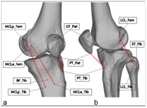
A commercially available rigid solid body kinematics numerical model (LifeMOD/KneeSIM 2008.1.0, LifeModeler Inc., San Clemente, CA) was used for this work. The numerical model was previously validated both for contact forces and kinematics outputs (Innocenti et al. Citation2011; Pianigiani et al. Citation2012). The model includes the reconstruction of a full leg, without the foot, obtained from a CT scan of a cadaveric full leg of a Caucasian male (age 82 years, height 1.88 m and weight 72.6 kg). No cartilage and menisci have been reconstructed. The physiological knee model was built assuming physiological positions of the main soft tissue insertions as described in literature (La Prade et al. Citation2003; Netter Citation2006; LaPrade et al. Citation2007) and adapted to the specific patient geometries (Figure ).
Figure 1. Bone models and position of the soft tissues insertion points considered in this study: a) medial view; b) lateral view. Tibia and fibula were considered as rigid bodies; the centroid of the insertion area of the ligaments (shown as red lines) was used as the best approximation of the position of the respective insertion point: LCL_Fib lateral collateral ligament on the fibula; LCL_Fem lateral collateral ligament on the femur; MCLa_Tib anterior attachment of the medial collateral ligament on the tibia; MCLp_Tib posterior attachment of the medial collateral ligament on the tibia; MCLa_Fem anterior attachment of the medial collateral ligament on the femur; MCLp_Fem posterior attachment of the medial collateral ligament on the femur; PT_Tib patellar tendon on the tibia; PT_Pat patellar tendon on the patella; QT_Pat quadriceps tendon on the patella; BF_Tib biceps femoris on the tibia; ST_Fib semitendinosus on the fibula.
Starting from the physiological knee model, a first model was obtained virtually by implanting a PS conventional TKA (Genesis II, Smith&Nephew, Memphis, TN) according to the surgical guidelines of the manufacturer. A second model was then obtained by substituting the conventional tibial insert (Figure (a)) with a semi-constrained for the same TKA design (Figure (b)).
Figure 2. (a) Tibial polyethylene insert for the conventional version; (b) tibial polyethylene insert for the semi-constrained version; (c) top view of the semi-constrained cam (red) and the conventional cam (blue); and (d) lateral view of the semi-constrained post (red) and the conventional post (blue).
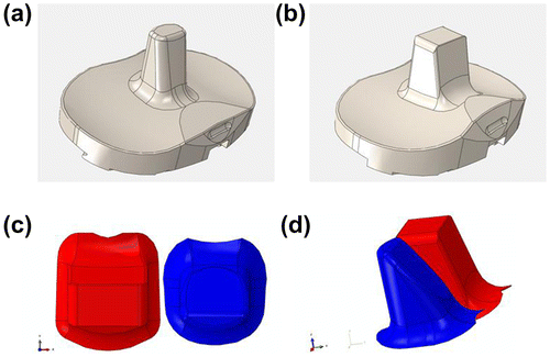
The only differences between the two tibial insert solutions are in the shape of the post: the semi-constrained design is wider than the conventional, almost as wide as the box in the femoral component (space of 1 mm between post and box border on medial and lateral side), and features less rounded edges, resulting in an almost rectangular shape (Figure (c) and (d)).
To cover a wide range of common daily life activities and their associated knee joint kinetics and kinematics, four different load-bearing motor tasks have been analysed in this study: gait, step ascent, step descent and squat. The hardware and the boundary conditions of the experimental tests have been properly simulated considering as input a controlled quadriceps load in order to obtain a constant hip load, and considering the action of the hamstrings as constant. All the other soft tissues behave consequently following each mechanical behaviour described in the model.
Thus, each task was performed in 10 s with a proper vertical hip load and vertical hip translation. Starting from 0°, the knee flexion during walking is up to 65°, during the step descent is up to 68°, during the step ascent is up to 93°and for the squat is up to 120°. Each movement follows a standard kinematics characteristic for the analysed movement as also obtained from in vivo gait lab analyses (Desloovere et al. Citation2010). To allow a following validation for contact forces outputs, these settings also match experimental tests performed in in vitro analyses on cadaver legs (Innocenti, Follador, et al. Citation2009; Innocenti, Labey, et al. Citation2009; Victor Citation2009; Victor, Wong, et al. Citation2009; Delport et al. Citation2013).
The loaded movements were reproduced numerically, simulating an existing knee kinematics rig (Maletsky Citation1999; Victor, Van Glabbeck, et al. Citation2009) in terms of geometries, constraints, inputs and outputs using a commercial musculoskeletal modelling software (McKinnon et al. Citation2005; Al-Nazer et al. Citation2008; Morra et al. Citation2008; Monaco et al. Citation2009).
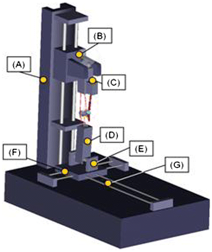
The simulated rig together with its functionality and constraints are shown in Figure .
Figure 3. The knee simulator model used in this study. It consists of: a base frame (A), a hip sled (B), a femur block (C), a tibia block (D), a tibia rotation table (E), an adduction–abduction sled (F) and an anteroposterior sled (G).
The resulting model of the knee includes tibio–femoral, patello–femoral and post-cam contacts between the TKA components, passive soft tissue structures and active muscle elements. External forces (ground reaction force and mass), muscle forces (quadriceps and hamstrings) and frictional forces are applied to the knee joint through the rig similar to a knee kinematics simulator. Mechanical properties of relevant soft tissue structures were obtained from Woo et al. (Woo et al. Citation1991; Li, Zayontz, Most, et al. Citation2004). To allow wrapping around the bone and in the trochlear groove of the femoral component, tendons were modelled as separate discrete units (more information about material models and properties in use in Innocenti et al. (Citation2011)).
For each post-cam configuration and motor task, the posterior post-cam contact force was evaluated by the numerical model during the entire movement.
Procedure to validate the numerical outputs
A dynamic knee simulator system based on the Oxford rig was also used to experimentally validate the results of the computer simulations. This electromechanical system was designed to simulate and record the motions and loads in a knee joint during squatting, and was extended used for in vitro experimental study on human knee (Innocenti, Follador, et al. Citation2009; Innocenti, Labey, et al. Citation2009; Victor Citation2009; Victor, Wong, et al. Citation2009; Delport Citation2013). Femoral components of the same TKA design used for the numerical simulations were cemented on custom designed metal fixtures, which represented femur and tibia. A conventional UHMWPE insert was used.
A pressure sensitive film (K-Scan sensor 6900/10000 psi, Tekscan® Inc., Massachusetts, USA) was covered with a 0.1 mm thin Teflon film to protect it against shear stresses and calibrated in a loading frame (858 Mini Bionix II, MTS, MN, USA). One of the four fingers of the sensor was then fixed to the posterior side of the insert post using double-sided tape. Five squats between 30° and 130° of flexion were performed on the knee simulator. The total squat time during flexion was 10 s. During the squat, the ankle loads and moments and the quadriceps load and hip position were recorded by the knee simulator. The contact force, contact area and contact pressure between insert post and the cam of the femoral component were recorded with the K-Scan sensor.
Results
Experimental results
The pressure sensor readings showed a gradual increase of the contact force as function of the flexion angle, once the cam engaged with the post. The initial contact angle was 71° flexion. The maximum contact force was 780 N, for the maximum flexion (Figure ) (Arnout et al. Citation2014).
Figure 4. Post-cam contact force during the experimental validation.
Numerical data output
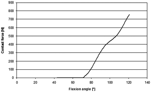
Figure shows the posterior post-cam contact force determined by the numerical simulation for the simulated squat movement. The angle at which the post engages the cam is around 50° of knee flexion.
Figure 5. Trend of the contact mechanics for the conventional design after the numerically simulated squat.
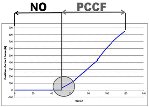
For all the analysed motor tasks, and for the two solutions, the posterior post-cam contact force shows a similar trend of the one illustrated in Figure . Generally, the contact force increases with the increase in the flexion angle, and the angle for which there is the initial PS contact is similar among the different motor tasks. However, due to the different maximum flexion angles achieved by the different motions, the maximum posterior contact force is different among the different motor tasks. An overview of the maximum contact force determined for the different analysed motions is reported in Figure . For all the designs and motions, the maximum posterior post-cam contact force always occurred close to the maximal flexion angle (walking 65°, stair descent 68°, stair ascent 93° and squat 120°).
Figure 6. Comparison of the post-cam contact force for all the motor tasks. Blue indicates the conventional design and red indicates the semi-constrained design.
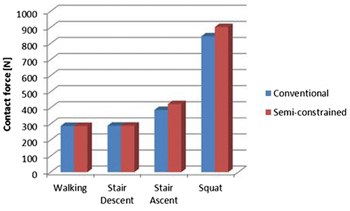
Even if different motor tasks show a different value of maximum posterior PS contact force, the values are similar for the two different tibial inserts with a slightly higher contact force (ranging from 3% during walking, up to 5% during squat) in the semi-constrained design compared to the conventional one.
As found in the literature (Arnout et al. Citation2014), designs with higher contact forces compensate for these higher forces with a larger contact area in order to reduce their contact pressure and avoid post failure. Despite this, no catastrophic failures are clinically seen for the analysed TKA design, and it has excellent long-term results without post breakage. A possible explanation for this finding is the relatively low moment on the post that possibly compensates for the high pressures.
Validation
The experimental outputs (Figure ) about the post-cam mechanism and the numerical outputs (Figure ) have been compared for the conventional design.
Numerical maximum contact forces are, in general, in agreement with literature (Fitzpatrick et al. Citation2013), but are especially in agreement with experimental results performed with the same conventional design in use for the numerical simulations (Arnout et al. Citation2014).
Discussion
The aim of our work was to estimate the maximum contact forces in the post-cam mechanism for several activities in conventional and semi-constrained PS TKA, and to check for eventual differences among the two solutions.
This work presents some limitations: only a left leg has been analysed and only one design for each solution has been analysed in this study. Moreover, a rigid body kinematics approach was considered, and therefore the eventual deformation of the polyethylene post was neglected in this study. However, despite these limitations strong agreement was found among numerical outputs and data coming from experimental activities, and from the literature. In addition, the two solutions have been tested only for daily activities and not only for tests like the pivoting one. This choice was also based on Arnout et al. paper that shows that several TKA designs present their own PS pattern during daily activity.
The modelling and simulations were performed with a validated numerical model (Innocenti et al. Citation2011; Pianigiani et al. Citation2012). The development and the definition of the numerical models were based on experimental cadaver tests. Numerical simulations achieved a similar maximum contact force to the experimental behaviour even if the post-cam engagement started earlier (Luyckx et al. Citation2009; Arnout et al. Citation2014). The reasons of these differences between experimental and numerical outputs could be explained due to the different starting points (in terms of initial flexion angle) for the two simulations (30° for the experimental tests and 0° for the numerical test). Moreover, in addition to this, also the friction coefficient in use for the two simulations is different, as the authors did not proceed with any measurements of knee lubrification and friction during the experimental activities, while the friction coefficient in use was taken from literature (Victor Citation2009) referring to metal-polyethylene coupling.
Even though the values are similar for the two insert types, different motor tasks show a different values of maximum post-cam force. A slightly higher force is observed in the semi-constrained design with respect to the corresponding conventional design. A possible explanation can be associated to the different shape of the post itself.
These results are in agreement with data shown in literature (Li et al. Citation2004; Catani et al. Citation2010; Fitzpatrick et al. Citation2013) and outcomes from experimental tests (Arnout et al. Citation2014).
Conclusions
After comparing post-cam contact forces in conventional and semi-constrained during several motor tasks, we can report that a semi-constrained design shows only a slightly higher posterior post-cam contact force, and thus would not be expected to significantly increase the risk of failure of the post-cam mechanism as compared to a conventional design, that is, assume observing only the posterior side of the PS mechanism. For this reason, a possible future development of this study could be the analysis of the lateral and medial post-cam mechanism under the same conditions and the induced knee kinematics by the two solutions.
Conflict of interest disclosure statement
No potential conflict of interest was reported by the author(s).
References
- Al Nazer R, Rantalainen T, Heinonen A, Sievanen H, Mikkola A. 2008. Flexible multibody simulation approach in the analysis of tibial strain during walking. J Biomech. 41, 1036–1043.
- Arnout N, Vanlommel L, Vanlommel J, Luyckx JP, Labey L, Innocenti B, Victor J, Bellemans J. 2014. Post-cam mechanics and tibiofemoral kinematics: a dynamic in vitro analysis of eight posterior-stabilized total knee designs. Knee Surg Sports Traumatol Arthrosc. doi:10.1007/s00167-014-3167-2.
- Callaghan JJ, O’Rourke MR, Goetz DD. 2002. Tibial post impingement in posterior-stabilized total knee arthroplasty. Clin Orthop Relat Res. 404:83–88.10.1097/00003086-200211000-00014
- Catani F, Innocenti B, Belvedere C, Labey L, Ensini A, Leardini A. 2010. The Mark Coventry Award articular: contact estimation in TKA using in vivo kinematics and finite element analysis. Clin Orthop Relat Res. 468:19–28.10.1007/s11999-009-0941-4
- Colizza WA, Insall JN, Scuderi GR. 1995. The posterior stabilized total knee prosthesis: assessment of polyethylene damage and osteolysis after a ten-year-minimum follow-up. J Bone Joint Surg [Am]. 77-A:1713–1720.
- Dahlkvist NJ, Mayo P, Seedhom BB. 1982. Forces during squatting and rising from a deep squat. Eng Med. 11:69–76.10.1243/EMED_JOUR_1982_011_019_02
- Delport HD. 2013. Collateral ligament strain of the human knee joint. Native and after total knee arthroplasty [ PhD thesis]. Leuven: KU Leuven.
- Desloovere K, Wong P, Swings L, Callewaert B, Vandenneucker H, Leardini A. 2010. Range of motion and repeatability of knee kinematics for 11 clinically relevant motor tasks. Gait Posture. 32:597–602.
- Fekete G, Csizmadia BM, Wahab MA, De Baets P, Katona G, Vanegas-Useche LV, Solanilla JA. 2012. Sliding-rolling ratio during deep squat with regard to different knee prostheses. Acta Polytech Hung. 9:5–24. IF: 0.588.
- Fitzpatrick CK, Clary CW, Cyr AJ, Maletsky LP, Rullkoetter PJ. 2013. Mechanics of post-cam engagement during simulated dynamic activity. J Orthop Res. 19:1438–1446.
- Innocenti B, Bilgen ÖF, Labey L, van Lenthe GH, Vander Sloten J, Catani F. 2014. Load sharing and ligament strains in balanced, overstuffed and understuffed UKA. A validated finite element analysis. J Arthroplasty. 29:1491–1498.
- Innocenti B, Follador M, Salerno M, Bignardi C, Wong P, Labey L. 2009. Experimental and numerical analysis of patello-femoral contact mechanics in TKA. In: Vander Sloten J, Verdonk P, Nyssen M, Haueisen J, editors. IFMBE Proceedings – 4th European Conference of the International Federation for Medical and Biological Engineering ECIFMBE 2008, 2008 November 23–27; Antwerp, Belgium. Berlin: Springer, vol. 22, 1867–1870.
- Innocenti B, Labey L, Victor J, Wong P, Bellemans J. 2009. An in vitro study of the human knee kinematics: natural vs. replaced joint. In: Vander Sloten J, Verdonk P, Nyssen M, Haueisen J, editors. IFMBE Proceedings – 4th European Conference of the International Federation for Medical and Biological Engineering ECIFMBE 2008; 2008 November 23–27; Antwerp. Berlin: Springer, vol. 22, 1789–1793.
- Innocenti B, Pianigiani S, Labey L, Victor J, Bellemans J. 2011. Contact forces in several TKA designs during squatting: a sensitivity analysis. J Biomech. 44:1573–1581.10.1016/j.jbiomech.2011.02.081
- Insall JN, Binazzi R, Soudry M, Mestriner LA. 1985. Total knee arthroplasty. Clin Orthop Relat Res. 192:13–22.
- Kurtz SM, Ong KL, Lau E, Bozic KJ, Berry D, Parvizi J. 2010. Prosthetic joint infection risk after TKA in the medicare population. Clin Orthop Relat Res. 468:52–56.
- La Prade RF, Engebretsen AH, Ly TV, Johansen S, Wentorf FA, Engebretsen L. 2007. The anatomy of the medial part of the Knee. J Bone Joint Surg Am. 89:2000–2010.10.2106/JBJS.F.01176
- La Prade RF, Ly TV, Wentorf FA, Engebretsen L. 2003. The postero lateral attachments of the knee: a qualitative and quantitative morphologic analysis of the fibular collateral ligament, popliteustendon, popliteo fibular ligament, and lateral gastrocnemius tendon. Am J Sports Med. 31:854–860.
- Li G, Most E, Otterberg E, Sabbag K, Zayontz S, Johnson T, Rubash H. 2002. Biomechanics of posterior-substituting total knee arthroplasty: an in vitro study. Clin Orthop Relat Res. 404:214–225.10.1097/00003086-200211000-00035
- Li G, Zayontz S, DeFrate LE, Most E, Suggs JF, Rubash HE. 2004. Kinematics of the knee at high flexion angles: an in vitro investigation. J Orthop Res. 22:90–95.10.1016/S0736-0266(03)00118-9
- Li G, Zayontz S, Most E, DeFrate LE, Suggs JF, Rubash HE. 2004. In situ forces of the anterior and posterior cruciate ligaments in high knee flexion, and in vitro investigation. J Orthop Res. 22:293–297.10.1016/S0736-0266(03)00179-7
- Lin KJ, Huang CH, Liu YL, Chen WC, Chang TW, Yang CT, Lai YS, Cheng CK. 2011. Influence of post-cam design of posterior stabilized knee prosthesis on tibiofemoral motion during high knee flexion. Clin Biomech. 26:847–852.10.1016/j.clinbiomech.2011.04.002
- Luyckx T, Didden K, Vandenneucker H, Labey L, Innocenti B, Bellemans J. 2009. Is there a biomechanical explanation for anterior knee pain in patients with patella alta? Influence of patellar height on patellofemoral contact force, contact area and contact pressure. J Bone Joint Surg Br. 91:344–350.10.1302/0301-620X.91B3.21592
- Maletsky, L. P. 1999. Validation of the next generation knee simulator [ PhD thesis]. West Lafayette (IN): Purdue University.
- Matsuda S, Whiteside LA, White SE, McCarthy DS. 1997. Knee kinematics of posterior cruciate ligament sacrificed total knee arthroplasty. Clin Orthop. 341:257–266.
- Mauerham DR. 2003. Fracture of the polyethylene tibial post in a posterior cruciate-substituting total knee arthroplasty mimicking patellar clunk syndrome: a report of 5 cases. J Arthroplasty. 18:942–945.10.1016/S0883-5403(03)00333-4
- McAuley JP, Engh GA. 2003. Constraint in total knee arthroplasty. When and what? J Arthroplasty. 18:51–54.
- McBride M, Ranawat CS, Rasquinha VJ. 2000. Revision total knee arthroplasty. Available from: http://www.medscape.org/viewarticle/420399
- McKinnon BW, Otto JK, McGuan S. 2005. The virtual knee. In: Bellemans J, Ries MD, Victor J, editors. Total knee arthroplasty a guide to get better performance. Heidelberg: Springer; p. 159–162.10.1007/3-540-27658-0
- Mestha P, Shenava Y, D’Arcy JC. 2000. Fracture of the polyethylene tibial post in posterior stabilized (Insall Burstein II) total knee arthroplasty. J Arthroplasty. 15:814–815.10.1054/arth.2000.6615
- Mikulak SA, Mahoney OM, dela Rosa MA, Schmalzried TP. 2001. Loosening and osteolysis with the press-fit condylar posterior-cruciate-substituting total knee replacement. J Bone Joint Surg [Am]. 83-A:398–403.
- Monaco V, Rinaldi LA, Macrì G, Micera S. 2009. During walking elders increase efforts at proximal joints and keep low kinetics at the ankle. Clin Biomech. 24:493–498.10.1016/j.clinbiomech.2009.04.004
- Morra EA, Rosca M, Greenwald JFI, Greenwald AS. 2008. The influence of contemporary knee design on high flexion: a kinematic comparison with the normal knee. J Bone Joint Surg Am. 90:195–201.10.2106/JBJS.H.00817
- Nagura T, Dyrby CO, Alexander EJ, Andriacchi TP. 2002. Mechanical loads at the knee joint during deep flexion. J Orthop Res. 20:881–886.10.1016/S0736-0266(01)00178-4
- Netter FH. 2006. Atlas of human anatomy. 4th ed. Philadelphia (PA): Saunders-Elsevier.
- Pianigiani S, Chevalier Y, Labey L, Pascale V, Innocenti B. 2012. Tibio-femoral kinematics in different total knee arthroplasty designs during a loaded squat: a numerical sensitivity study. J Biomech. 45:2315–2323.10.1016/j.jbiomech.2012.06.014
- Puloski SK, McCalden RW, MacDonald SJ, Rorabeck CH, Bourne RB. 2001. Tibial post wear in posterior stabilized total knee arthroplasty: an unrecognized source of polyethylene debris. J Bone Joint Surg [Am]. 83-A:390–397.
- Rodriguez JA, Bhende H, Ranawat CS. 2001. Total condylar knee replacement: a 20-year followup study. Clin Orthop Relat Res. 388:10–17.10.1097/00003086-200107000-00004
- Rosen S, Andrew L, Scuderi G. 2002. Patella resurfacing. Tech Knee Surg. 1:72–76.10.1097/00132588-200209000-00009
- Schai PA, Thornhill TS, Scott RD. 1998. Total knee arthroplasty with the PFC system: results at a minimum of ten years and survivorship analysis. J Bone Joint Surg [Br]. 80-B:850–858.10.1302/0301-620X.80B5.8368
- Schai PA, Scott RD, Thornhill TS. 1999. Total knee arthroplasty with posterior cruciate retention in patients with rheumatoid arthritis. Clin Orthop Relat Res. 367:195–200.
- Sultan PG, Most E, Schule S, Li G, Rubash HE. 2003. Optimizing flexion after total arthroplasty: advances in prosthetic design. Clin Orthop. 416:167–173.10.1097/01.blo.0000081937.75404.ee
- Scuderi GR. 2001. Revision total knee arthroplasty: how much constraint is enough? Clin Orthop Relat Res. 392:300–305.10.1097/00003086-200111000-00039
- Todo M, Takahashi Y, Nagamine R. 2008. Computational analysis of stress concentration and wear for tibial insert of PS type knee prosthesis under deep flexion. ICBME, Proceedings. vol. 23, p. 1559–1563.
- Victor J. 2009. A comparative study on the biomechanics of the native human knee joint and total knee arthroplasty [Doctoral dissertation]. KUL. 135–137. Available from: https://lirias.kuleuven.be/handle/123456789/233948S
- Victor J, Van Glabbeck F, Vander Sloten J, Parizel PM, Somville J, Bellemans J. 2009c. An experimental model for kinematic analysis of the knee. J Bone Joint Surg Am. 91:150–163.10.2106/JBJS.I.00498
- Victor J, Wong P, Witvrouw E, Van der Sloten J, Bellemans J. 2009. How isometric are the medial patello femoral, superficial medial collateral, and lateral collateral ligaments of the knee? Am J Sports Med. 37:2028–2036.10.1177/0363546509337407
- Wilk KE, Escamilla RF, Fleisig GS, et al. 1996. A comparison of tibiofemoral joint forces and electromyographic activity during open and closed kinetic chain exercises. Am J Sports Med. 24:518–527.10.1177/036354659602400418
- Woo SL, Hollis JM, Adams DJ, Lyon RM, Takai S. 1991. Tensile properties of the human femur anterior cruciate ligament tibia complex: The effects of specimen age and orientation. Am J Sports Med. 19:217–225.10.1177/036354659101900303