Abstract
Lacrosse is one of the oldest team sports in the United States and it has become the fastest growing sport in the country. The purpose of this study was to describe the kinematics of the overhead shot in youth male lacrosse players in attempt to gain an understanding of the movement patterns of the upper extremity. It was hypothesized that the kinematics observed with the overhead lacrosse shot would be similar to the mechanics of an overhead throw. Ten male youth lacrosse players (10.69 ± 2.06 years; 153.87 ± 12.90 cm; and 43.33 ± 9.25 kg) volunteered. Three accurate overhead shots were performed and the fastest shot was then selected for detailed analysis. The overhead lacrosse shot relies on trunk extension and rotation as the shot progresses and less on shoulder external rotation than a throw. Additionally, the lacrosse shot also appears to be less dependent on humeral elevation, compared to throwing, as 15.38 ± 12.10° of elevation was observed.
Keywords:
Introduction
Lacrosse is a fast-paced game that requires speed, agility, endurance, and physical contact (Hinton et al. Citation2005). Lacrosse is one of the oldest team sports in the United States and has become the fastest growing team sport (Hinton et al. Citation2005; McCulloch & Bach Citation2007; US Lacrosse Citation2012; Millard Citation2013). Since 2001, participation in lacrosse has almost tripled throughout the United States with 722,205 participants (US Lacrosse Citation2012) reported in 2012, and of those, 390,000 were youth (15 years and under). While youth participation in lacrosse has grown drastically, there is a lack of biomechanical research regarding the sport (Mercer & Nielson Citation2013; Millard Citation2013). Additionally, with the variety of shooting strategies implemented in lacrosse, the mechanics of the overhead lacrosse shot have not been examined.
Throughout the course of a lacrosse game, a player may utilize a number of different types of shots on goal. The shots that can be performed include an overhead, three-quarters, sidearm, and underhand. However, at the youth level, the overhead shot is the most frequently coached and performed by players. The overhead shot is the most accurate and is beneficial from a performance standpoint because the player can alter the release point of the ball. Altering the release point of the shot allows a player to shoot either high or low on the goal, making it more difficult for the goalie to defend. As youth players advance in their skill performance, they begin to learn the more advanced sidearm and underhand shots on goal but these shots are difficult to master and perform accurately.
A fundamental understanding of overhead shot mechanics in youth lacrosse players is necessary for sports performance enhancement and skill acquisition. By identifying key movement patterns in the overhead shot, future research can begin to identify potential injurious pathomechanics that may be present and also identify the kinematic factors that contribute to shot accuracy. Because youth participation in lacrosse comprises the majority of overall participation (US Lacrosse Citation2012), understanding young athletes’ shooting mechanics is critical for determining the mechanics necessary for producing a high-velocity, accurate shot on goal. Based on the lack of research in the area of lacrosse, it was the purpose of this study to describe the kinematics of the overhead shot in youth lacrosse players in attempt to better understand the role of the trunk and upper extremity. It was hypothesized that the kinematics observed with the overhead lacrosse shot would be similar to the mechanics previously reported for an overhead throw.
Methods
Participants
Ten male, right-hand dominant, youth lacrosse players (10.69 ± 2.06 years; 153.87 ± 12.90 cm; and 43.33 ± 9.25 kg), volunteered to participate. Participant selection criteria included coach recommendation and freedom from injury within the past six months (Oliver & Keeley Citation2010a; Oliver et al. Citation2010, Citation2011). Coach recommendation was sought to ensure that the participants were experienced at playing lacrosse and that they frequently take shots on goal during game situations. While freedom from injury within the past six months was one of the criteria for selection, none of the participants reported a history of injury to their upper extremity. Testing was conducted indoors in a biomechanics laboratory. The University’s Institutional Review Board approved all testing protocols. Once the participants arrived to data collection, all testing procedures were explained and parental consent and participant assent were obtained.
Procedures
The MotionMonitor™ (Innovative Sports Training, Chicago, IL) synced with an electromagnetic tracking system (Track Star, Ascension Technologies Inc., Burlington, VT) was used to collect all the data. With electromagnetic tracking systems, field distortion has been shown to be the cause of error in excess of 5° at a distance of 2 m from an extended range transmitter (Day et al. Citation2000), but increase in instrumental sensitivity have reduced this error to near 10° prior to system calibration and 2° following system calibration (Meskers et al. Citation1999; Perie et al. Citation2002; Myers Citation2005). Thus, prior to data collection, the current system was calibrated using previously established techniques (Meskers et al. Citation1998, Citation1999; Day et al. Citation2000; Perie et al. Citation2002). Following calibration, magnitude of error in determining the position and orientation of the electromagnetic sensors within the calibrated world axes system was less than 0.01 m and 3°, respectively (Oliver & Plummer Citation2011; Plummer & Oliver Citation2013, Citation2014). Participants had a series of 11 electromagnetic sensors (Track Star, Ascension Technologies Inc., Burlington, VT) attached at the following locations: [1] seventh cervical vertebra [C7] spinous process; [2] the pelvis at sacral vertebrae 1 [S1]; [3–4] deltoid tuberosity of the humerus; [5–6] bilateral wrist, dorsal aspect of the wrist between the radial and ulnar styloid processes; [7–8] bilateral shank centered between the head of the fibula and lateral malleolus; [9–10] bilateral midpoint of the lateral aspect of the femur (Myers Citation2005; Oliver Citation2013; Oliver & Keeley Citation2010b; Plummer & Oliver Citation2013); and [11] the midpoint of the third metatarsal on the stride foot. A student researcher trained in the application techniques applied sensors. Sensors were affixed to the skin using PowerFlex cohesive tape (Andover Healthcare, Inc., Salisbury, MA) to ensure that the sensors remained secure throughout the testing. An additional sensor was attached to a stylus and used to digitize the trunk and upper extremity bony landmarks, described in Table , to create a skeletal model of the participant (Wu et al. Citation2002, Citation2005; Myers Citation2005). Participants were instructed to stand in anatomical position during digitization to guarantee accurate bony landmark identification. A link segment model was developed through digitization of joint centers for the ankle, knee, hip, shoulder, thoracic vertebrae 12 [T12] to lumbar vertebrae 1 [L1], and C7 to thoracic vertebrae 1 [T1]. The spinal column was defined as the digitized space between the associated spinous processes, whereas the ankle and knee were defined as the midpoints of the digitized medial and lateral malleoli, and medial and lateral femoral condyles, respectively. The shoulder joint center was determined manually by digitizing a point anterior and posterior to the joint and then calculating the midpoint (Blackburn et al. Citation2003). The hip joint center was estimated using the rotation method, which is implemented by stabilizing the joint and then passively moving the joint in 10 positions in a small circular pattern (Huang et al. Citation2010). This method of calculating a joint center has been reported as providing accurate positional data (Veeger Citation2000; Huang et al. Citation2010). The hip joint center was calculated from rotation of the femur relative to the pelvis. The variation in the measurement of the joint center was a root mean square error less than 0.003 m in order to be accepted.
Table 1. Description of the bony landmarks digitized.
Kinematic data describing the position and orientation of the electromagnetic sensors were sampled at 100 Hz. Raw data regarding sensor orientation and position were transformed to locally based coordinate systems for each of the respective body segments following ISB standards (Wu et al. Citation2002). Raw data were independently filtered along each global axis using a fourth order Butterworth filter with a cutoff frequency of 20.0 Hz (Oliver & Keeley Citation2010a, Citation2010b; Plummer & Oliver Citation2013). All data were synchronized passively via a data acquisition board and time stamped through MotionMonitor™.
Following digitization, participants dressed in full lacrosse protective equipment (helmet, shoulder pads, elbow pads, and gloves) and were instructed to perform their pre-competition warm-up routine. Participants were allowed to perform their own warm-up routine in order to ensure that they felt they were physically ready to complete the maximal effort shots required during the testing procedure and decrease the risk of injury. While participants were allotted an unlimited time to warm-up, the average length of warm-up for the participants was 10 ± 2 min. All participants used their own lacrosse stick to complete this study. Participants shot a standard sized lacrosse ball (5.0 oz) into a regulation-sized goal (182.8 cm H × 182.8 cm W × 213.4 cm D) positioned 9.14 m away. All participants were required to perform at least three right-handed overhead shots to insure proper landing placement on the force plate. The ground surface was positioned so that the participant’s stride foot would land on top of a 40 × 60 cm Bertec force plate (Bertec Corp, Columbus, OH) that was anchored into the floor (Oliver & Keeley Citation2010a; Plummer & Oliver Citation2013). The force plate was used to event mark stride foot contact. Data were collected for three accurate overhead shots in which a goal was scored and the participant’s stride foot landed on the force plate. A JUGS radar gun (OpticsPlanet, Inc., Northbrook, IL) positioned in the direction of the shot determined speed of each shot. The shot with the fastest ball speed was then selected for detailed analysis (Sabick et al. Citation2004; Keeley et al. Citation2008).
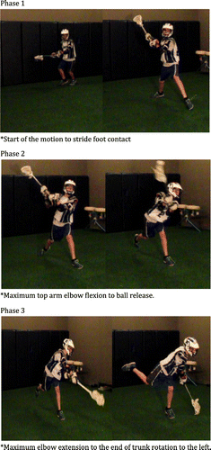
Previous research, using video analysis, has divided the lacrosse shot into six key phases (Mercer & Nielson Citation2013). The six phases are the approach, crank-back, stick acceleration, stick deceleration, follow through, and recovery phase. For the purpose of this study, we only examined the first five phases of the overhead shot because the recovery phase is when a player prepares for the next task during competition and did not apply to the controlled lab environment (Mercer & Nielson Citation2013). In addition, because we were unable to track the movement of the stick, the stick acceleration and deceleration phases were defined slightly different than previously defined phases. The approach phase began with the initiation of movement and ended with stride foot contact (Mercer & Nielson Citation2013). The crank-back phase was from foot contact to maximum elbow flexion of the top arm. The acceleration phase was defined as the period from maximum elbow flexion to ball release. Ball release was estimated as the midpoint between maximum elbow flexion and maximum elbow extension. This estimate was required because we did not have the capability to track the movement of the ball. This method to estimate ball release has been used previously in throwing literature using electromagnetic tracking, but has yet to be validated (Oliver Citation2013; Oliver & Keeley Citation2010a, Citation2010b; Plummer & Oliver Citation2014). The deceleration phase occurred from ball release to maximum elbow extension of the top arm. And the follow through was from maximum elbow extension of the top arm to the end of trunk rotation (largest value of trunk rotation) to the left (Figure ).
Results
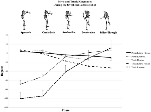
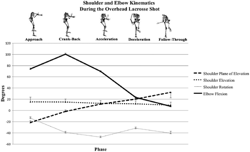
Pelvis and trunk descriptive kinematic data are presented in Figure . The pelvis and trunk began flexed to the right during the approach then during the crank-back phase there was a shift in positioning to the left, towards the goal. The pelvis and trunk segments had maximum rotation to the left (towards the goal) during the approach phase and then in the crank-back phase the rotation moved to the right. Shoulder and elbow data for the dominant arm (right) were selected for analysis. Shoulder and elbow kinematic data are presented in Figure . Shoulder elevation was 15.38° during the approach phase and decreased slightly as the motion of the shot progressed. The shoulder reached maximum external rotation (−47.51°) during the acceleration phase before progressing to internal rotation. The elbow reached 100.49° of maximum flexion during the crank-back phase and then began extending as the shot progressed.
Discussion
While lacrosse is the fastest growing team sport in the United States, there are currently no kinematic data available on shooting mechanics. To our knowledge, this is the first study to examine the kinematics of the overhead lacrosse shot in youth players. It was hypothesized that players performing the overhead lacrosse shot would exhibit similar mechanics as the overhead throw in baseball and softball. However, based on the results of this study, the kinematics of the overhead shot in youth lacrosse players is very different than overhead throwing.
One important variable that has been established in the overhead throwing motion is humeral elevation. The humerus should be elevated to 90° to reduce joint torques and maximize functional stability of the shoulder (Fortenbaugh et al. Citation2009; Matsuo et al. Citation2002). Computer simulations suggest that 90° of shoulder abduction, along with slight lateral trunk tilt, maximizes wrist and ball velocity (Matsuo et al. Citation2002), however, in the current study, maximum humeral elevation of 15.38 ± 12.10° was observed. Maximum humeral elevation during the overhead shot occurred during the approach phase and as the shooting movement progressed, humeral elevation decreased. While humeral elevation may prove to be a key variable in overhead throwing, the effects of elevation during the lacrosse shot are unknown. Lacrosse compared to other overhead sports requires the addition of an implement, the lacrosse stick, which becomes an extension of the upper extremity. The use of a lacrosse stick during the overhead shot functions as a lever that favors speed and range of motion during movement. The axis of the lever is formed from the bottom (left) hand, whereas the top (right) hand provides the force needed to accelerate the stick. With this configuration, the force is closer to the axis of rotation leading to a smaller force arm as opposed to the resistance arm. Therefore, the addition of the stick allows for a more extreme range of motion while not relying heavily on glenohumeral range of motion as seen in overhead throwing. The speed of a lacrosse game may also play a role in the shot mechanics that youth players implement. A player generally has very little time after becoming free from a defender to make an accurate shot on goal and this may decrease the amount of time that they have to elevate the stick and their arm.
As expected, the humerus progressed from a position behind the participant’s trunk and moved anteriorly as shot progressed which is similar to the overhead throwing motion. Of interest during the overhead lacrosse shot was shoulder rotation. In overhead throwing, large values of shoulder external rotation are observed during the cocking phase and then the shoulder begins to internally rotate rapidly in order to propel the ball forward to ball release. Werner et al. (Citation1993) reported that pitchers reach 185° during baseball pitching which is much greater than the maximal value observed in lacrosse. Data in the current study reveal relatively small values of external rotation during the course of the overhead shot. Maximum external rotation of the shoulder was 47.51 ± 30.73° during the acceleration phase from the progression of maximum elbow flexion to ball release. It is postulated that maximum shoulder external rotation during the lacrosse shot is likely due to the use of the lacrosse stick that increases the lever arm, and therefore, the distance in which the ball can travel. This increased distance that the ball travels may also lead to greater velocity being imparted on the ball without relying on increased external rotation at the shoulder joint. Throwing literature has reported an association between shoulder external rotation and elbow valgus stress (Werner et al. Citation1993; Miyashita et al. Citation2008). The decreased values of shoulder external rotation may also decrease not only the forces about the shoulder but also the valgus stress about the elbow during the shot.
The proximal segments of the pelvis and trunk should provide a stable base of support for movement of the upper extremity. In this study, the pelvis and the trunk rotated and laterally flexed to the left throughout the course of the overhead shot, which is similar to overhead throwing. Of interest in the present study is the pattern of trunk flexion and extension. It is typically observed in overhead throwing that the trunk follows a progression of increasing flexion throughout the motion. However, in the overhead lacrosse shot, alterations in the progression of trunk flexion were observed. Initially during the first two phases of the lacrosse shot this pattern occurred, however, beginning in the acceleration phase the trunk moved into extension and rotation. It is postulated that this alteration of trunk flexion to trunk extension could be counterproductive to energy transfer. Additionally, this extension may not only decrease the energy transferred to the shoulder during the acceleration phase but may also apply increased load on the spine.
Although this study provides valuable insight into the biomechanics of the overhead lacrosse shot, some important limitations should be noted. A homogenous sample of youth lacrosse players was chosen for analysis; however, this sample might not be representative of the true mechanics needed to perform an overhead shot correctly. Older, more experienced lacrosse players likely have more refined shooting mechanics than the participants examined in this study. Large standard errors were observed in this sample and it is believed to be the result of examining youth players with varying levels of experience. It is important to note that Millard (Citation2013) reported difficulty in identifying the discrete events of the overhead lacrosse in female players when examining video to sync the phases with the collected electromyographic data. The author reports that each participant had a somewhat unique style of overhead shot mechanics, which may have been related to the varying levels of experience between participants. Because this is the first study examining the kinematics of the overhead shot in lacrosse, it is difficult to draw definitive conclusions on the mechanics observed in this sample because no data are available for comparison. Only male participants were examined in this study, and therefore, female lacrosse players may exhibit different mechanics. The use of an electromagnetic tracking system, and the inability to collect high-speed video that was synced with the kinematic data, required the event of ball release to be estimated. Additionally, it is currently unknown if this method of estimating ball release is effective for the sport of lacrosse.
Conclusions
The results of this study provide valuable data on the kinematics of the overhead lacrosse shot in youth male players. The overhead lacrosse shot relies on trunk extension and rotation as the shot progresses and less on shoulder external rotation. Future research should examine the kinematics of the overhead shot in experienced high school and college-aged players. These data can then be used to better identify improper mechanics that potentially increase the risk of injury, in less experienced lacrosse players. Research should endeavor to examine kinematics, kinetics, and kinetic chain sequencing of the overhead lacrosse shot to gain a better understanding of injury predictors.
Conflict of interest disclosure statement
No potential conflict of interest was reported by the author(s).
References
- Blackburn JT, Riemann BL, Myers JB, Lephart SM. 2003. Kinematic analysis of the hip and trunk during bilateral stance on firm, foam, and multiaxial support surfaces. Clin Biomech. 18:655–661.10.1016/S0268-0033(03)00091-3
- Day J, Murdoch D, Dumas G. 2000. Calibration of position and angular data from a magnetic tracking device. J Biomech. 33:1039–1045.10.1016/S0021-9290(00)00044-0
- Fortenbaugh D, Fleisig GS, Andrews JR. 2009. Baseball pitching biomechanics in relation to injury risk and performance. Sports Health. 1:314–320.10.1177/1941738109338546
- Hinton RY, Lincoln AE, Almquist JL, Douoguih WA, Sharma KM. 2005. Epidemiology of lacrosse injuries in high school-aged girls and boys: a 3-year prospective study. Am J Sports Med. 33:1305–1314.10.1177/0363546504274148
- Huang Y-H, Wu T-Y, Learman KE, Tsai Y-S. 2010. A comparison of throwing kinematics between youth baseball players with and without a history of medical elbow pain. Chin J Physiol. 53:160–166.10.4077/CJP.2010.AMK026
- Keeley DW, Hackett T, Keirns M, Sabick MB, Torry MR. 2008. A biomechanical analysis of youth pitching mechanics. J Pediatr Orthop. 28:452–459.10.1097/BPO.0b013e31816d7258
- Matsuo T, Matsumoto T, Mochizuki Y, Takada Y, Saito K. 2002. Optimal shoulder abduction angles during baseball pitching from maximal wrist velocity and minimal kinetics viewpoints. J Appl Biomech. 18:306–320.
- McCulloch PC, Bach BR. 2007. Injuries in men’s lacrosse. Orthopedics. 30:29–34.
- Mercer JA, Nielson JH. 2013. Description of phases and discrete events of the lacrosse shot. Sport J. 16:1–1.
- Meskers CGM, Vermeulen HM, de Groot JH, van der Helm FCT, Rozing PM. 1998. 3D shoulder position measurements using a six-degree-of-freedom electromagnetic tracking device. Clin Biomech. 13:280–292.10.1016/S0268-0033(98)00095-3
- Meskers CGM, Fraterman H, van der Helm FCT, Vermeulen HM, Rozing PM. 1999. Calibration of the ‘Flock of Birds’ electromagnetic tracking device and its application in shoulder motion studies. J Biomech. 32:629–633.10.1016/S0021-9290(99)00011-1
- Millard B. 2013. Examination of lower extremity muscle activity during an overhand lacrosse shot in females. Las Vegas (NV): Kinesiology and Nutrition Sciences, University of Nevada.
- Miyashita K, Urabe Y, Kobayashi H, Yokoe K, Koshida S, Kawamura M, Ida K. 2008. The role of shoulder maximum external rotation during throwing for elbow injury prevention in baseball players. J Sports Sci Med. 7:223–228.
- Myers JB. 2005. Scapular position and orientation in throwing athletes. Am J Sports Med. 33:263–271.10.1177/0363546504268138
- Oliver GD. 2013. Relationship between gluteal muscle activation and upper extremity kinematics and kinetics in softball position players. Med Biol Eng Comput. 52:265–270.
- Oliver GD, Dwelly PM, Kwon YH. 2010. Kinematic motion of the windmill softball pitch in prepubescent and pubescent girls. J Strength Cond Res. 24:2400–2407.10.1519/JSC.0b013e3181dc43af
- Oliver GD, Keeley DW. 2010a. Pelvis and torso kinematics and their relationship to shoulder kinematics in high-school baseball pitchers. J Strength Cond Res. 24:3241–3246.10.1519/JSC.0b013e3181cc22de
- Oliver GD, Keeley DW. 2010b. Gluteal muscle group activation and its relationship with pelvis and torso kinematics in high-school baseball pitchers. J Strength Cond Res. 24:3015–3022.10.1519/JSC.0b013e3181c865ce
- Oliver GD, Plummer H. 2011. Ground reaction forces, kinematics, and muscle activations during the windmill softball pitch. J Sports Sci. 29:1071–1077.10.1080/02640414.2011.576692
- Oliver GD, Plummer HA, Keeley DW. 2011. Muscle activation patterns of the upper and lower extremity during the windmill softball pitch. J Strength Cond Res. 25:1653–1658.10.1519/JSC.0b013e3181db9d4f
- Perie D, Tate AJ, Cheng PL, Dumas GA. 2002. Evaluation and calibration of an electromagnetic tracking device for biomechanical analysis of lifting task. J Biomech. 35:293–297.10.1016/S0021-9290(01)00188-9
- Plummer HA, Oliver GD. 2013. Quantitative analysis of kinematics and kinetics of catchers throwing to second base. J Sports Sci. 31:1108–1116.10.1080/02640414.2013.770907
- Plummer HA, Oliver GD. 2014. The relationship between gluteal muscle activation and throwing kinematics in baseball and softball catchers. J Strength Cond Res. 28:87–96.10.1519/JSC.0b013e318295d80f
- Sabick MB, Torry MR, Young-Kyu K, Hawkins RJ. 2004. Humeral torques in professional baseball pitchers. Am J Sports Med. 32:892–898.10.1177/0363546503259354
- US Lacrosse 2012 Participation Survey. 2012.
- Veeger HE. 2000. The position of the rotation center of the glenohumeral joint. J Biomech. 33:349–355.
- Werner SL, Fleisig GS, Dillman CJ, Andrews JR. 1993. Biomechanics of the elbow during baseball pitching. J Orthop Sports Phys Ther. 17:274–278.10.2519/jospt.1993.17.6.274
- Wu G, Siegler S, Allard P, Kirtley C, Leardini A, Rosenbaum D, Whittle M, D’Lima D, Cristofolini L, Witte H, et al. 2002. ISB recommendation on definitions of joint coordinate systems of various joints for the reporting of human joint motion – part I: ankle, hip, and spine. J Biomech. 35:543–548.10.1016/S0021-9290(01)00222-6
- Wu G, van der Helm FCT, Veeger HEJ, Makhsous M, Anglin C, Nagels J, Karduna AR, McQuade K, Wang X, et al. 2005. ISB recommendation on definitions of joint coordinate systems of various joints for the reporting of human joint motion – part II: shoulder, elbow, wrist and hand. J Biomech. 38:981–992.10.1016/j.jbiomech.2004.05.042


