
 ?Mathematical formulae have been encoded as MathML and are displayed in this HTML version using MathJax in order to improve their display. Uncheck the box to turn MathJax off. This feature requires Javascript. Click on a formula to zoom.
ABSTRACT
This study developed a method to detect knee wobbling (KW) at low knee flexion. KW consists of quick uncontrollable medio-lateral knee movements without knee flexion, which may indicate a risk of ACL injury. Ten female athletes were recorded while performing slow, single-leg squats. Using motion capture data, the ratio of the frontal angular velocity to sagittal angular velocity (F/S) was calculated. An ‘F/S spike’ was defined when the F/S ratio exceeded 100%. The number of F/S spikes was counted before and after low-pass filtering at different cut-off frequencies. Intraclass correlation coefficients for KW and filtered F/S spike were analysed. KWs per squat cycle showed a median (range) of 3 (2–8) times. F/S spikes before and after low-pass filtering at 3-, 6-, 10-, and 15-Hz were 51 (12–108), 2 (0–6), 3 (1–12), 5 (2–21), and 9 (3–33) times, respectively. KWs and F/S spikes on motion capture with 6-Hz, low-pass filtering were well correlated (r = 0 .76). Median percentages of valgus and varus F/S spikes were 71% and 29%, respectively. After 6Hz, low-pass filtering, the number of F/S spikes was strongly correlated with observed KWs. An F/S spike assessment may be used to objectively detect KW, including flexion and varus/valgus angular velocity.
Introduction
Severe anterior cruciate ligament (ACL) injury is suffered by athletes in various sports; therefore, cost-effective screening and a prevention strategy are needed. ACL reconstructive surgery increased 22% from 2002 to 2014 in the U.S., reaching 74.6 cases per 100,000 person-years (Herzog et al. Citation2018). Moreover, ACL injury requires 6 months (Kvist Citation2004) to 2 years (Nagelli and Hewett Citation2017) of rehabilitation before an injured athlete can return to sports. A 2012 survey in the U.S. found that the total annual cost of ACL reconstruction had reached 2.8 USD billion (Mather et al. Citation2013). Further, athletes who return to sports after ACL reconstruction have a high risk of re-injury (Wiggins et al. Citation2016) and an increased likelihood of early onset of knee osteoarthritis (OA) (Harris et al. Citation2014). The incidence of ACL injury among female college basketball and soccer athletes is 3–4 times higher than among male athletes (Agel et al. Citation2005). Although research on ACL injury prevention has been conducted internationally (Hewett et al. Citation1999; Mandelbaum et al. Citation2005), the incidence of ACL injury in female athletes has not decreased (Agel et al. Citation2016). A screening method to predict ACL injury has yet to be developed (Bahr Citation2016), but is needed to identify athletes at high-risk.
An optimal screening method should identify the potential injury mechanism. Previous studies on the mechanism of ACL injury demonstrated that most injury occurred during deceleration activity with slight knee flexion (<25°), valgus, with internal or external rotation (Krosshaug et al. Citation2007; Koga et al. Citation2010), and during high-impact activity with one leg (Boden et al. Citation2000). Knee kinematics during ACL injury in female athletes were analysed using a model-based, image-matching technique and showed that the knee valgus angle increased by 12°, while the knee flexion angle increased by only 1° between the initial contact (IC) of the foot and 40 ms after IC (Koga et al. Citation2010). Therefore, the flexion component should be considered in order to detect high-risk athletes. A prospective cohort study demonstrated that injured athletes have a greater valgus angle at baseline (Hewett et al. Citation2005). Previously, screening tasks to select high-risk athletes included a drop vertical jump (DVJ) with both legs from a box 30 cm high (Hewett et al. Citation2005; Krosshaug et al. Citation2016) and a landing error scoring system (LESS) (Smith et al. Citation2012; Padua et al. Citation2015). Both of these methods utilized snap shots of knee positions at IC or maximal knee flexion during landing. Knee flexion angles at initial contact during DVJ ranged from 30.3° to 32.3° (Cortes et al. Citation2011; Mok et al. Citation2017), while those at the onset of ACL injury ranged from 9° to 23° (Krosshaug et al. Citation2007; Koga et al. Citation2010). This indicates that the knee flexion angle at IC of DVJ is greater than that at the instant of ACL injury. Studies utilizing DVJ also lacked dynamic analyses, such as repeated knee valgus/varus movement during the knee flexion phase or changes in flexion movement, since only a snapshot of the landing maneuver was analysed (Hewett et al. Citation2005; Smith et al. Citation2012; Padua et al. Citation2015; Krosshaug et al. Citation2016). Moreover, the double-leg task during DVJ may diminish abnormal knee movement due to the wide support base; thus, it may fail to detect the effects of trunk dysfunction. Therefore, previous screening methods utilizing DVJ probably have limited ability to reproduce knee position and movement at the moment of injury.
A more sensitive and specific screening method is needed to identify high-risk athletes. We hypothesized that ideally, such a screen should employ a single-leg task, simulating the knee position at the time of an ACL injury, and should include a parameter for dynamic knee movement. Repeated knee valgus/varus motion or knee wobbling (KW), comprising quick and uncontrollable medio-lateral movements (valgus/varus) with a reduction in the flexion angular velocity of the knee, can be detected at low knee flexion during single-leg squats. In our laboratory, repeated valgus/varus knee movements relative to flexion angle during single-leg landing were analysed at baseline among 71 athletes, and the relative risk was 1.35 [0.90, 2.03] (Koresawa et al. Citation2014). While investigating the optimal single-leg squat speed to detect KW, it was found that 2.2 and 1.2 KWs occurred during the descending phase of slow and fast single-leg squats (Koresawa Citation2013). However, that method has not been validated utilizing a three-dimensional motion capture system, which would provide higher accuracy. KW can be defined as an ‘F/S ratio’ of the valgus/varus angular velocity (frontal plane movement) divided by the flexion angular velocity (sagittal plane movement). An ‘F/S spike’ can then be defined as a sudden increase in the F/S ratio, exceeding 100%, which may indicate a sudden reduction of flexion movement and a sudden increase of valgus/varus movement. The purpose of this study was to develop a laboratory-based method of detecting KW at low knee flexion during a single-leg squat.
Our hypothesis was that an appropriate filtering frequency would be able to distinguish F/S spikes that correlate with KW, making F/S spikes an objective index of KW. Accordingly, this is a preliminary study to develop a method of detecting KW utilizing an objective parameter. Results of this study will be used in future validation and prospective cohort studies.
Methods
Participants
This was a cross-sectional study and the protocol was approved by Hiroshima International University. Participants were recruited from female basketball or volleyball teams on campus. Written informed consent was obtained from each participant prior to testing.
Inclusion criteria were as follows: (1) athlete participating in team sports involving stopping, jumping, and pivoting; (2) 20–25 years of age; (3) female; (4) healthy; and (5) demonstrating KW on visual observation during single-leg squatting without any additional weight or stress. Exclusion criteria were as follows: (1) prior surgical intervention, bone fracture, or ligament injury to lower extremities; (2) pain during exercise; (3) range of motion limitation; (4) neural symptoms; (5) a psychological disorder; or (6) any problem with communication or understanding the research content.
Recruiting
Ten basketball athletes and 20 volleyball athletes were invited to participate in this study, and 10 of these athletes (4 basketball and 6 volleyball athletes) agreed to participate. All 10 athletes proceeded to the screening session.
Sample size
All ten subjects were 20 years of age. Height was 1.61 [1.53, 1.69] m (mean [95% confidence interval (CI)]) and weight was 56.4 [50.8, 62.0] kg. All 10 subjects manifested KW during screening, and were included in the measurement session. According to a power analysis, eight subjects are sufficient for intraclass correlation coefficients (ICC). Therefore, a sample size of 10 provided an adequate spectrum of knee wobbling and was considered sufficient to develop a method for future validation studies.
(2) Screening
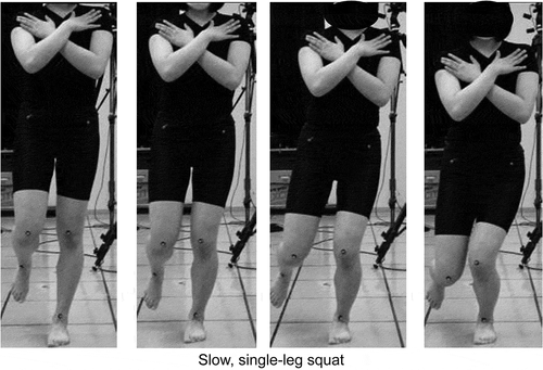
Screening to detect KW visually was performed using movies taken with 2D video cameras (Casio EX-FH25, CASIO COMPUTER Co., LTD, Japan) at 120 Hz positioned at the front and sides of the standing subject. Each camera was located 3 m from the subject. Slow, single-leg squats (SSLSs) without any additional weight or stress were chosen as the screening task (). Ten candidates were asked to perform SSLSs with each leg. After enough practice (at least 3 times, but less than 5 min), the SSLS task was performed twice to confirm that it was being performed as instructed.
Figure 1. A slow, single-leg squat was performed the same way during screening and measurement. Subjects were instructed to perform the squat with an 8-s descending phase and a 2-s ascending phase, keeping time with a metronome, and with their arms crossed in front of their chests
KW was defined as quick, uncontrollable medio-lateral movements (valgus/varus) with a reduction in the flexion angular velocity of the knee during single-leg squats. Visual observation was conducted by one of the authors while watching the screening videos. The same researcher performed all observations and rated KW during the screening session to identify candidates likely to present KW in the measurement session. All 10 candidates demonstrated at least one KW during screening and were included in further testing ().
(3) Measurement and Analysis
Measurement
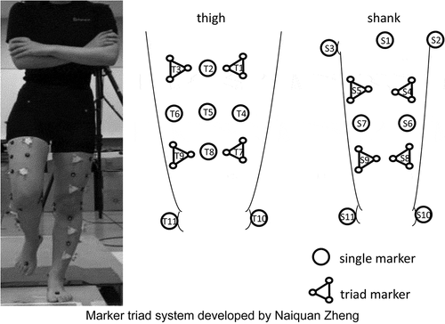
Figure 2. This system detected rotational movement of the knee in detail by distinguishing translational movement and measuring angles formed by triad markers
SSLS was chosen as the task for the measurement session. Participants were allowed two practice attempts in order to perform the movement properly. One trial of two squat cycles was performed with no rest between cycles. Each squat cycle took 10 s, 8 s for descending and 2 s for ascending, using an electro-metronome at 1 Hz. Starting from the single-leg standing position with the knee completely extended and the arms crossed in front of the chest, subjects were asked to flex the knee more than 30°. The opposite hip and knee were kept flexed with the tips of the toes ≥10 cm above the floor. The opposite hip was also slightly abducted to separate the knees by about 5 cm in order to avoid overlapping the markers. No trunk tilting in the frontal plane was allowed, but there was no restriction on trunk movement in the sagittal plane. When trunk tilting was observed, the trial was aborted and re-performed.
Analyses
Kinematic analysis was performed using a custom MATLAB 2020a (MathWorks Inc., Massachusetts USA) program developed by Gao and Zheng (Citation2008). Knee kinematics were analysed between 0° and 30° knee flexion to obtain knee flexion angular velocity (sagittal plane) and valgus/varus angular velocity (frontal plane). F/S ratio (%) was calculated using the following equation with the absolute value of the valgus/varus angular velocity:
F/S ratio >100% or <−100% was defined as an F/S spike, which was used as an index of quantitative KW. A high F/S ratio was typically observed with a low knee flexion angular velocity, while a negative F/S ratio was observed with negative knee flexion angular velocity (or knee extension motion). Knee flexion movement is also important for one ACL risk factor, such as the ability to absorb impact, as well as the direction of frontal knee movement. Especially, knee extension movement is abnormal during SSLS. On the other hand, the direction of valgus/varus action was determined using kinematic data. Knee flexion angle at the F/S spike was also determined from kinematic data.
Comparisons with visual observations
Raw kinematic data demonstrate many spikes that are not associated with knee movement, while filtered kinematic data may exclude meaningful knee movement. Therefore, the appropriate filtering frequency needed to be determined. Kinematic data were filtered using a zero-lag, fourth-order, low-pass Butterworth filter with a 3-, 6-, 10-, or 15-Hz cut-off frequency.
Identification of KWs by visual observation was based on the following criteria: (1) sudden valgus/varus movement, and (2) a reduction of flexion movement based on observation of descending knee movement. If it was difficult to determine the sagittal knee angular velocity from the frontal recording, the sagittal recording was used to determine the reduction of knee flexion movement. Two physical therapists observed the SSLS video and counted the number of knee wobbles. One observer was experienced at counting KWs and the other was not. Inter-observer reliability in detecting KW visually was tested during the measurement session.
Each filtered kinematic dataset was compared with visual observations using a graph showing time on the X-axis and F/S ratio on the Y-axis (). Visual observations were performed using animation of marker movements in the frontal plane (). The number of F/S spikes between 0° and 30° knee flexion was counted, and the timing of F/S spikes as well as directions of valgus/varus movement were determined based on kinematic data after filtering. The optimal filtering frequency was determined by comparing filtered data and visual observations by the experienced observer. When the number of F/S spikes in filtered data was correlated with the number of KWs on visual observation, the filtering frequency was considered appropriate, since kinematic data of motion capture without filtering involve both movement and noise.
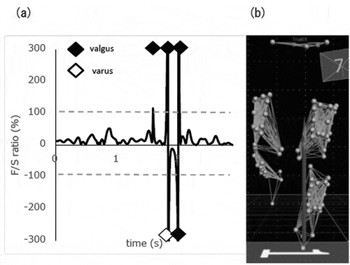
Figure 3. Detecting knee wobbling by visual observation and F/S ratio calculated as “frontal-plane angular velocity”/“sagittal-plane angular velocity” X 100. (a) The F/S ratio graph during the descending phase of one squat is shown. F/S ratio > 100 % was defined as an F/S spike, which was considered an index of quantitative KW. A positive F/S ratio indicates knee flexion angular velocity and negative angular velocity indicates knee extension angular velocity. Using absolute frontal angular velocity, a valgus F/S spike was marked with a black circle and a varus F/S spike was marked with a white circle. (b) Animation of marker movements in the frontal plane. Validation of consistency between the F/S spike and KW by visual inspection was conducted by plotting data sets as shown in this figure
The median and range of knee flexion angles at F/S spikes, the direction of valgus/varus action was obtained using data with the most appropriate filtering frequency. Graphs of each subject were analysed qualitatively to determine whether filtered data and observational findings were similar in terms of timing and direction of the valgus/varus action.
(4) Statistics
The Shapiro–Wilk test was used to detect non-normal distributions. Descriptive statistics for KWs detected visually, F/S spikes before and after filtering, and knee flexion angle at F/S spikes were obtained with median and range. The ICC for the number of KWs was calculated to assess the correlation between KW movements detected by the experienced observer and F/S spikes filtered at various frequencies. In addition, intra- and intertester ICCs between the experienced and inexperienced observers were obtained. Graphs for F/S ratio and knee kinematics were used for qualitative comparisons. P values less than 0.05 were considered significant. SPSS Statistics, version 21 (IBM Japan, Ltd., Japan) was used for data analyses.
Results
Numbers of KWs in visual observations was 3 (2–8) times (medians (ranges)). Numbers of KWs on motion capture data before and after 3-, 6-, 10-, and 15-Hz low-pass filtering were 51 (12–108), 2 (0–6), 3 (1–12), 5 (2–21), and 9 (3–33) times per squat cycle, respectively (). Good correlation (r = 0.76) was observed between the number of KWs noted by the experienced observer and the number of F/S spikes on motion capture with 6-Hz low-pass filtering. Also, it was confirmed that most KWs corresponded with F/S spikes seen by the experienced observer. The intra-observer ICC (1,2) was 0.95 [0.86, 0.98], while the inter-observer ICC (2,1) was 0.14 [0.12, 0.47] between the experienced and inexperienced observers. - shows varus/valgus angle characteristics at various filtering frequencies among 5 of 10 subjects. The median (range) knee flexion angle at F/S spikes was 6.8° (0.1°-27.1°) with 6-Hz low-pass filtering. Median numbers of valgus and varus F/S spikes were 2.5 and 1.0 times, respectively. shows the numbers of F/S spikes at various knee flexion angles during the descending phase of the first squatting cycle among five of 10 subjects.
Table 1. Numbers of F/S spikes and KWs based on visual observation
Table 2. Numbers of F/S spikes at various knee flexion angles during the descending phase of the first squatting cycle
Figure 4a. Knee biomechanics during a slow, single-leg squat from 0° to 30° knee flexion in subject 1. Quantitative analysis of the F/S spikes detected valgus KW 4 times at 0-5° knee flexion, one valgus KW and one varus KW at 10-15° knee flexion during the descending phase of the first squat. This case presented more valgus KW than varus KW
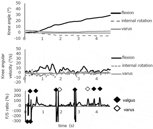
Figure 4b. Knee biomechanics during a slow, single-leg squat from 0° to 30° knee flexion in subject 2. Quantitative analysis of the F/S spikes detected five valgus KWs and six varus KWs at 0-5° knee flexion during the descending phase of the first squat. This case presented many KWs at very low knee flexion
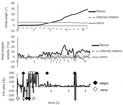
Figure 4c. Knee biomechanics during a slow, single-leg squat from 0° to 30° knee flexion in subject 3. Quantitative analysis of the F/S spikes detected one valgus KW at 15-20° knee flexion and two valgus KWs at 20-25° knee flexion during the descending phase of the first squat. This case presented decreasing knee flexion near 20° knee flexion
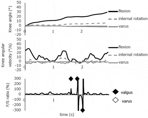
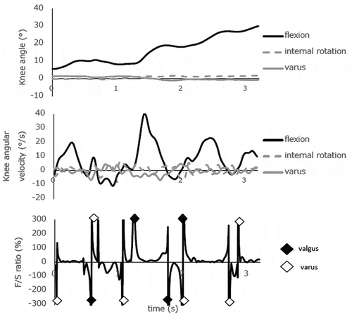
Figure 4d. Knee biomechanics during a slow, single-leg squat from 0° to 30° knee flexion in subject 4. Quantitative analysis of the F/S spikes detected only one valgus KW at 0-5° knee flexion, and one valgus KW at 20-25° knee flexion during the descending phase of the first squat. Notably, the second KW occurred with increasing knee varus angular velocity
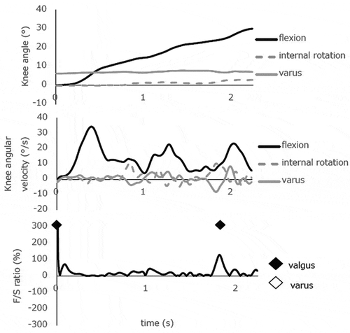
Figure 4e. Knee biomechanics during a slow, single-leg squat from 0° to 30° knee flexion in subject 5. Quantitative analysis of the F/S spikes detected a lot of KW during the entire descent. This case presented an obvious moment with decreasing knee flexion movement
Discussion
The purpose of this study was to develop a method to detect knee wobbling (KW) at low knee flexion during a single-leg squat. After low-pass filtering at 6 Hz, the median number of F/S spikes detected during the descending phase of each squat cycle was 3 times, which correlated well with the number of KWs observed by experienced observer. The median knee flexion angle of F/S spikes at 6-Hz low-pass filtering was 6.8°. Median numbers of valgus and varus F/S spikes during the descending phase were 2.5 and 1.0 times, respectively. Poor inter-observer agreement was found in counting numbers of KW movements visually between experienced and inexperienced observer ().
Table 3. Numbers of F/S spikes at various knee flexion angles during the descending phase of the second squatting cycle
F/S spikes were utilized as an index of sudden knee valgus/varus movement during the descending phase of SSLS. The F/S ratio allowed dynamic characteristics of combined sagittal and frontal knee kinematics to be quantified. The time-F/S spike graph clearly showed rapid increases in F/S ratio (valgus/varus angular velocity relative to knee flexion angular velocity at low knee flexion) during the descending phase. Ten-hertz and 15-Hz filtering frequencies resulted in excessive noise, while 6 Hz appeared optimal for analysis of non-high impact movement. Therefore, with this filtering procedure, KWs and noise could be accurately distinguished. A filtering rate of 6 Hz was also utilized to exclude noise during gait in a previous study (Gao and Zheng Citation2008). SSLS involves slower movement than a normal gait, which justifies the selection of 6-Hz low-pass filtering and elimination of higher frequencies. Our findings showed that the number of F/S spikes after low-pass filtering at 6 Hz was consistent with KWs counted visually. Thus, examining abnormal movement at a low flexion angle could provide an objective parameter to screen for the risk of ACL injury.
Detecting abnormal knee movement at low flexion angle must be included in the screening activity. ACL distance between femoral and tibial attachments was measured, as well as knee kinematics during single-leg squats using MR-based models and biplanar fluoroscopic images (Utturkar et al. Citation2013). ACL distances at full knee extension, 30° flexion, and at the valgus collapse position (30° of flexion with 10° of external rotation of the tibia and maximal internal rotation at the hip) were 30.2, 27.1, and 25.6 mm, respectively. Therefore, the ACL distance was greater at full extension than in the valgus position. Cadaveric studies indicate that the posteromedial bundle of the ACL shows the greatest elongation at full extension (2.92 mm at full extension and 2.16 mm at 90°) (Yoo et al. Citation2010). Since the knee flexion angle during screening measurements must be important in determining stress on the ACL, the knee flexion angle during screening should involve low flexion positions (<25°) (Krosshaug et al. Citation2007; Koga et al. Citation2010). In a DVJ screening task, the knee flexion angle is greater than 30° (Cortes et al. Citation2011; Mok et al. Citation2017). Single-leg landing involves 15°-40° of knee flexion; therefore, the range and standard deviation were large (Schmitz et al. Citation2007; Weinhandl et al. Citation2010). Since there were no landing tasks that could reproduce knee kinematics with a flexion angle less than 25°, slower activities such as SSLS should be utilized to observe abnormal knee kinematics, such as KWs at a flexion angle less than 25° (Kianifar et al. Citation2017; Munro et al. Citation2017). Previous screening studies lacked the ability to detect abnormal knee movement at low flexion angles (Hewett et al. Citation2005; Krosshaug et al. Citation2016). Therefore, SSLS is a better choice to reproduce abnormal knee movement at low knee flexion utilizing F/S spikes.
Even though ‘dynamic knee valgus’ has been proposed as a risk factor for ACL injury (Hewett et al. Citation2004, Citation2005; Sell et al. Citation2006), many studies utilized only snapshots, showing simple knee valgus angles at the initial contact and/or maximal flexion angle during landing (Hewett et al. Citation2005; Krosshaug et al. Citation2016). ‘Valgus collapse’ at low knee flexion was observed during ACL injury in female athletes on video observation (Krosshaug et al. Citation2007). However, valgus collapse was determined by observation without analysing the change in flexion angular velocity. Such analyses could not detect multiplanar dynamic movement of the knee. A previous prospective cohort study from our laboratory showed that five of six athletes who sustained ACL injury presented repeated valgus/varus knee motion or wobbling during landing at baseline (Koresawa et al. Citation2014). This suggested that dynamic knee analyses would be required to capture repeated valgus/varus movement with or without changes in flexion angular velocity. The argument above suggests that F/S spikes should provide an index representing abnormal dynamic knee movement that cannot be detected by snapshot analyses. In this study, although the knee was expected to flex smoothly without frontal movement during SSLS, frontal knee movement occurred as F/S spikes accompanying the decrease in knee flexion angular velocity. Accordingly, F/S spikes during SSLS may detect abnormal movement of the knee.
Although F/S spikes can detect abnormal knee movements during SSLS, it is not yet clear how this movement is related to the ACL injury mechanism, that is, whether the valgus, varus, or some other movement is responsible. Previous video studies showed that valgus load during slight knee flexion led to ACL injury (Krosshaug et al. Citation2007; Koga et al. Citation2010). A video study of non-contact ACL injuries in Australian football found that 10.5% showed giving-way in the varus direction (Cochrane et al. Citation2007). A questionnaire study of ACL injury indicated that non-contact ACL injuries involved both valgus and varus knee movements (Myklebust et al. Citation1998). Ten cases showed tibial external rotation relative to the femur and seven cases showed internal rotation at the time of the ACL injury. In addition, an in vivo study showed that ACL strain increased significantly by 2.4 ± 1.97% as the anterior tibial shear force and knee internal tibial rotation moment increased under load-bearing conditions (Fleming et al. Citation2001). On the other hand, knee valgus-varus and external rotation had little effect on ACL strain. Therefore, these studies showed that valgus and varus loading could cause ACL injury under weight-bearing or with anterior shear force at the proximal end of the tibia at low knee flexion. We observed a greater number of valgus F/S spikes than varus F/S spikes at low flexion angle. We speculate that valgus F/S spikes may be caused by sudden decreases in knee flexion angular velocity, and that the energy in flexion/extension was transferred to another degree-of-freedom, i.e. valgus/varus, inducing a sudden increase in valgus/varus movement. Although valgus loading has been considered the main mechanism of ACL injury, it remains unclear whether varus movement may also be a risk factor. F/S spikes in the valgus or varus direction with reduction of flexion angular velocity is a candidate for an index to detect abnormal motion that may be related to ACL injury and may be useful for future cohort or intervention studies.
KWs identified by F/S spikes were validated using a motion capture system along with MTT. The accuracy of MTT using cadavers has been validated, and root-mean-square errors of MTT were 0.77° for flexion/extension (FE), 2.15° for axial rotation (AR), 0.84° for varus/valgus (VV), 1.82 mm for anterior/posterior translation (AP), 2.02 mm for medial/lateral translation (ML), and 0.99 mm for superior/inferior translation (SI) (Gao and Zheng Citation2008). The authors demonstrated that rotation of three markers fixed on the vertices can be recognized as skin surface rotation. Root-mean-square errors of the point cluster technique (PCT) were 0.79° for FE, 1.79° for AR, 0.4° for VV, 3.98 mm for AP, 2.42 mm for ML, 1.08 mm for SI. In addition, MTT calculates marker motion relative to a triad marker (Gao and Zheng Citation2008). Since a triad has the same skin attachment area as a single marker, the center position of the three vertices can be used for skin surface translation analysis. Local coordinate systems of the reference triads were defined parallel to the anatomical reference frames in the neutral standing posture. Used with dynamic motion capture, MTT allows discrimination between translation due to skin motion and rotation due to joint motion. Although PCT can be utilized to detect small rotational movements of the knee, only single markers are used to determine the segment coordinate system. Since all markers are single markers in PCT, it is impossible to discriminate between translation due to skin motion and rotation due to joint motion. Considering these systems, MTT is advantageous to detect KWs with higher precision. As for validity of this study, subjects included only female college athletes, which was appropriate considering their greater incidence of ACL injury.
This study has three limitations. First, the F/S ratio does not reflect the absolute value of knee flexion and valgus angle, and the amount of stress on the ACL could not be determined. Although KW, which is defined by the F/S ratio as a dynamic frontal knee movement with less knee flexion movement, would increase ACL strain, this study does not prove that KW is a risk factor for ACL injury. In order to establish KW as a risk factor of ACL injury, a prospective cohort study with screening for KW is needed. Second, this study only examined female athletes and whether F/S spikes also quantify KW in male athletes is unknown. Third, this study did not show good inter-observer reliability because it compared between experienced and inexperienced examiners. It is difficult to detect KWs visually because it is complicated to judge simultaneous decreasing knee flexion movement and frontal movement from a single plane. On the other hand, KWs identified visually by a skilled examiner corresponded with those identified by motion capture. These results showed that calculating F/S spikes using motion capture is more accurate than by visual observations. These limitations do not impact the conclusions.
Conclusions
After low-pass filtering at 6 Hz, the number of F/S spikes corresponds with the number of KWs observed during SSLS. F/S spikes are an objective indicator of KWs, including flexion and varus/valgus angular velocity data. On the other hand, visual observations reflect the skill of the observer. Therefore, it is advantageous to use motion capture data with 6-Hz low-pass filtering in quantifying KW. This information will be utilized in a future validation study. Moreover, a future prospective cohort study should demonstrate whether KWs accurately predict the risk of ACL injury.
Acknowledgments
We are extremely grateful to the students from Hiroshima International University who willingly participated in the study and provided valuable data.
Disclosure statement
Only Kazuyoshi Gamada has received only personal fees from GLAB, Higashihiroshima, Japan. The others have no potential conflict of interest.
Correction Statement
This article has been republished with minor changes. These changes do not impact the academic content of the article.
References
- Agel J, Arendt EA, Bershadsky B. 2005. Anterior cruciate ligament injury in national collegiate athletic association basketball and soccer: a 13-year review. The American Journal of Sports Medicine. 33(4):524–530. eng. doi:https://doi.org/10.1177/0363546504269937.
- Agel J, Rockwood T, Klossner D. 2016. Collegiate ACL injury rates across 15 sports: national collegiate athletic association injury surveillance system data update (2004-2005 through 2012-2013). Clinical Journal of Sport Medicine: Official Journal of the Canadian Academy of Sport Medicine. 26(6):518–523. eng. doi:https://doi.org/10.1097/JSM.0000000000000290.
- Bahr R. 2016. Why screening tests to predict injury do not work-and probably never will … : a critical review. British Journal of Sports Medicine. 50(13):776–780. Eng. doi:https://doi.org/10.1136/bjsports-2016-096256.
- Bell AL, Pedersen DR, Brand RA. 1990. A comparison of the accuracy of several hip center location prediction methods. Journal of Biomechanics. 23(6):617–621. eng. doi:https://doi.org/10.1016/0021-9290(90)90054-7.
- Boden BP, Dean GS, Feagin JA Jr., Garrett WE Jr. 2000. Mechanisms of anterior cruciate ligament injury. Orthopedics. 23(6):573–578. Eng. doi:https://doi.org/10.3928/0147-7447-20000601-15.
- Cochrane JL, Lloyd DG, Buttfield A, Seward H, McGivern J. 2007. Characteristics of anterior cruciate ligament injuries in Australian football. Journal of Science and Medicine in Sport. 10(2):96–104. Eng. doi:https://doi.org/10.1016/j.jsams.2006.05.015.
- Cortes N, Onate J, Van Lunen B. 2011. Pivot task increases knee frontal plane loading compared with sidestep and drop-jump. Journal of Sports Sciences. 29(1):83–92. Eng. doi:https://doi.org/10.1080/02640414.2010.523087.
- Fleming BC, Renstrom PA, Beynnon BD, Engstrom B, Peura GD, Badger GJ, Johnson RJ. 2001. The effect of weightbearing and external loading on anterior cruciate ligament strain. Journal of Biomechanics. 34(2):163–170. Eng. doi:https://doi.org/10.1016/S0021-9290(00)00154-8.
- Gao B, Zheng NN. 2008. Investigation of soft tissue movement during level walking: translations and rotations of skin markers. Journal of Biomechanics. 41(15):3189–3195. doi:https://doi.org/10.1016/j.jbiomech.2008.08.028.
- Harris JD, Abrams GD, Bach BR, Williams D, Heidloff D, Bush-Joseph CA, Verma NN, Forsythe B, Cole BJ. 2014. Return to sport after ACL reconstruction. Orthopedics. 37(2):e103–108. Eng. doi:https://doi.org/10.3928/01477447-20140124-10.
- Herzog MM, Marshall SW, Lund JL, Pate V, Mack CD, Spang JT. 2018. Trends in Incidence of ACL reconstruction and concomitant procedures among commercially insured individuals in the United States, 2002-2014. Sports Health: A Multidisciplinary Approach. 10(6):523–531. Eng. doi:https://doi.org/10.1177/1941738118803616.
- Hewett TE, Lindenfeld TN, Riccobene JV, Noyes FR. 1999. The effect of neuromuscular training on the incidence of knee injury in female athletes. A prospective study. The American Journal of Sports Medicine. 27(6):699–706. Eng. doi:https://doi.org/10.1177/03635465990270060301.
- Hewett TE, Myer GD, Ford KR. 2004. Decrease in neuromuscular control about the knee with maturation in female athletes. The Journal of Bone and Joint Surgery American Volume. 86(8):1601–1608. Eng. doi:https://doi.org/10.2106/00004623-200408000-00001.
- Hewett TE, Myer GD, Ford KR, Heidt RS Jr., Colosimo AJ, McLean SG, van den Bogert AJ, Paterno MV, Succop P. 2005. Biomechanical measures of neuromuscular control and valgus loading of the knee predict anterior cruciate ligament injury risk in female athletes: a prospective study. The American Journal of Sports Medicine. 33(4):492–501. eng. doi:https://doi.org/10.1177/0363546504269591.
- Kianifar R, Lee A, Raina S, Kulic D. 2017. Automated assessment of dynamic knee valgus and risk of knee injury during the single leg squat. IEEE Journal of Translational Engineering in Health and Medicine. 5:2100213. Eng. doi:https://doi.org/10.1109/JTEHM.2017.2736559.
- Koga H, Nakamae A, Shima Y, Iwasa J, Myklebust G, Engebretsen L, Bahr R, Krosshaug T. 2010. Mechanisms for noncontact anterior cruciate ligament injuries: knee joint kinematics in 10 injury situations from female team handball and basketball. The American Journal of Sports Medicine. 38(11):2218–2225. eng. doi:https://doi.org/10.1177/0363546510373570.
- Koresawa K. 2013. The relationship between dynamic alignment and anterior cruciate injury risk during single leg landing and squat. Hiroshima (Japan): Hiroshima International University.
- Koresawa K, No Y, Kubota S, Gamada K 2014. Biomechanical risk factor of anterior cruciate ligament injury in adolescent female basketball players: a prospective cohort study [abstract] presented at: the 60th Orthopaeic Research Society Annual Meeting; 2014 Mar 15- 18;New Orleans (U.S).
- Krosshaug T, Nakamae A, Boden BP, Engebretsen L, Smith G, Slauterbeck JR, Hewett TE, Bahr R. 2007. Mechanisms of anterior cruciate ligament injury in basketball: video analysis of 39 cases. The American Journal of Sports Medicine. 35(3):359–367. eng. doi:https://doi.org/10.1177/0363546506293899.
- Krosshaug T, Steffen K, Kristianslund E, Nilstad A, Mok KM, Myklebust G, Andersen TE, Holme I, Engebretsen L, Bahr R. 2016. The vertical drop jump is a poor screening test for ACL injuries in female elite soccer and handball players: a prospective cohort study of 710 athletes. The American Journal of Sports Medicine. 44(4):874–883. eng. doi:https://doi.org/10.1177/0363546515625048.
- Kvist J. 2004. Rehabilitation following anterior cruciate ligament injury: current recommendations for sports participation. Sports Medicine (Auckland, NZ). 34(4):269–280. eng. doi:https://doi.org/10.2165/00007256-200434040-00006.
- Mandelbaum BR, Silvers HJ, Watanabe DS, Knarr JF, Thomas SD, Griffin LY, Kirkendall DT, Garrett W Jr. 2005. Effectiveness of a neuromuscular and proprioceptive training program in preventing anterior cruciate ligament injuries in female athletes: 2-year follow-up. The American Journal of Sports Medicine. 33(7):1003–1010. eng. doi:https://doi.org/10.1177/0363546504272261.
- Mather RC 3rd, Koenig L, Kocher MS, Dall TM, Gallo P, Scott DJ, Bach BR Jr., Spindler KP. 2013. Societal and economic impact of anterior cruciate ligament tears. The Journal of Bone and Joint Surgery American Volume. 95(19):1751–1759. eng. doi:https://doi.org/10.2106/JBJS.L.01705.
- Mok K-M, Bahr R, Krosshaug T. 2017. The effect of overhead target on the lower limb biomechanics during a vertical drop jump test in elite female athletes. Scandinavian Journal of Medicine & Science in Sports. 27(2):161–166. eng. doi:https://doi.org/10.1111/sms.12640.
- Munro A, Herrington L, Comfort P. 2017. The relationship between 2-dimensional knee-valgus angles during single-leg squat, single-leg-land, and drop-jump screening tests. Journal of Sport Rehabilitation. 26(1):72–77. eng. doi:https://doi.org/10.1123/jsr.2015-0102.
- Myklebust G, Maehlum S, Holm I, Bahr R. 1998. A prospective cohort study of anterior cruciate ligament injuries in elite Norwegian team handball. Scandinavian Journal of Medicine & Science in Sports. 8(3):149–153. eng. doi:https://doi.org/10.1111/j.1600-0838.1998.tb00185.x.
- Nagelli CV, Hewett TE. 2017. Should return to sport be delayed until 2 years after anterior cruciate ligament reconstruction? Biological and functional considerations. Sports Medicine (Auckland, NZ). 47(2):221–232. eng. doi:https://doi.org/10.1007/s40279-016-0584-z.
- Padua DA, DiStefano LJ, Beutler AI, de la Motte SJ, DiStefano MJ, Marshall SW. 2015. The landing error scoring system as a screening tool for an anterior cruciate ligament injury-prevention program in elite-youth soccer athletes. Journal of Athletic Training. 50(6):589–595. eng. doi:https://doi.org/10.4085/1062-6050-50.1.10.
- Schmitz RJ, Kulas AS, Perrin DH, Riemann BL, Shultz SJ. 2007. Sex differences in lower extremity biomechanics during single leg landings. Clinical Biomechanics (Bristol, Avon). 22(6):681–688. eng. doi:https://doi.org/10.1016/j.clinbiomech.2007.03.001.
- Sell TC, Ferris CM, Abt JP, Tsai Y-S, Myers JB, Fu FH, Lephart SM. 2006. The effect of direction and reaction on the neuromuscular and biomechanical characteristics of the knee during tasks that simulate the noncontact anterior cruciate ligament injury mechanism. The American Journal of Sports Medicine. 34(1):43–54. eng. doi:https://doi.org/10.1177/0363546505278696.
- Smith HC, Johnson RJ, Shultz SJ, Tourville T, Holterman LA, Slauterbeck J, Vacek PM, Beynnon BD. 2012. A prospective evaluation of the Landing Error Scoring System (LESS) as a screening tool for anterior cruciate ligament injury risk. The American Journal of Sports Medicine. 40(3):521–526. eng. doi:https://doi.org/10.1177/0363546511429776.
- Utturkar GM, Irribarra LA, Taylor KA, Spritzer CE, Taylor DC, Garrett WE, Defrate LE. 2013. The effects of a valgus collapse knee position on in vivo ACL elongation. Annals of Biomedical Engineering. 41(1):123–130. eng. doi:https://doi.org/10.1007/s10439-012-0629-x.
- Weinhandl JT, Joshi M, O’Connor KM. 2010. Gender comparisons between unilateral and bilateral landings. Journal of Applied Biomechanics. 26(4):444–453. eng. doi:https://doi.org/10.1123/jab.26.4.444.
- Wiggins AJ, Grandhi RK, Schneider DK, Stanfield D, Webster KE, Myer GD. 2016. Risk of secondary injury in younger athletes after anterior cruciate ligament reconstruction: a systematic review and meta-analysis. The American Journal of Sports Medicine. 44(7):1861–1876. eng. doi:https://doi.org/10.1177/0363546515621554.
- Yoo YS, Jeong WS, Shetty NS, Ingham SJ, Smolinski P, Fu F. 2010. Changes in ACL length at different knee flexion angles: an in vivo biomechanical study. Knee Surgery, Sports Traumatology, Arthroscopy: Official Journal of the ESSKA. 18(3):292–297. eng. doi:https://doi.org/10.1007/s00167-009-0932-8.