
ABSTRACT
The male and female terminalia of the poorly known sharpshooter Macugonalia semiguttata are described and illustrated for the first time based on specimens from a mountainous region in Rio de Janeiro State, southeastern Brazil (Atlantic Forest). External characters of the head, thorax, and abdomen of this species are also described and illustrated. Macugonalia semiguttata is compared with other seven similar species of the diverse genus Macugonalia Young, 1977. Among the 28 known species of this genus, it is apparently closely related to Macugonalia dallasi (Signoret, 1853), Macugonalia geographica (Signoret, 1855), and Macugonalia elegantula Cavichioli, 2004. Specimens of M. semiguttata were collected from the leaves and branches of shrubs and small trees in an area of low forest (circa 1,700 m a.s.l.), which is intermediate between the typical Atlantic Forest and the vegetation of alpine fields.
Introduction
The diverse and widespread New World Cicadellinae genus Macugonalia Young, 1977 includes 28 known species and occurs from the West Indies (a single rare species from Saint Vincent and the Grenadines), Costa Rica, Panama, and Trinidad Island southward to Bolivia, south and southeastern Brazil, Paraguay, and Argentina [Citation1–3].
According to Young [Citation1, p. 942], Macugonalia is closely related morphologically to Apogonalia Evans, 1947; in addition, species that are superficially similar to those of Macugonalia are found in Willeiana Young, 1977 and Versigonalia Young, 1977. Macugonalia can be distinguished from these three genera, as well as from other Neotropical genera of the Cicadellini, by the following combination of features [Citation1,Citation3]: (1) male pygofer strongly produced posteriorly; (2) subgenital plates usually very short and triangular; (3) aedeagus short, convex dorsally, with a pair of ventroapical lobes and a pair of slender, elongate basiventral processes, the latter extending beyond the aedeagal shaft; (4) paraphyses usually absent or vestigial; (5) female abdominal sternite VII produced posteriorly and with apex rounded or acute (in some species extending almost as far posteriorly as the ovipositor apex); and (6) valvulae II of ovipositor broadened beyond basal curvature.
Some species of Macugonalia are considered of agricultural importance. Macugonalia leucomelas (Walker, 1851) and Macugonalia cavifrons (Stål, 1862) were recorded as vectors of the bacterium Xylella fastidiosa Wells et al., 1987 in plum orchards in Brazil [Citation4,Citation5]. These two species are also potential vectors of X. fastidiosa in citrus cultures in Sergipe and Paraná states, as well as in olive orchards in Minas Gerais and São Paulo states [Citation6–9]. Macugonalia geographica (Signoret, 1855) and M. cavifrons were recorded as possible vectors of this bacterium in olive orchards from Rio Grande do Sul State [Citation10]. Macugonalia umbrosa Young, 1977 and Macugonalia moesta (Fabricius, 1803) were recorded as possible vectors of X. fastidiosa in citrus orchards in Amazonas [Citation11]. Vásquez and Lozada [Citation12] recorded M. moesta associated with six species of medicinal plants from the Allpahuayo Research Center in Peru.
According to Costa [Citation13], the mountainous Pico da Caledônia area (municipality of Nova Friburgo, Rio de Janeiro State, southeastern Brazil) has a characteristic alpine field vegetation from 1,800 m to the highest summit at 2,257 m (Campo de Altitude da Floresta Pluvial Costeira). This area has a typical physiognomic aspect within the Atlantic Forest, including rocky outcrops interspersed with vegetation “islets” or plateaus with continuous vegetation [Citation13]. A diverse and characteristic flora is present, including endemic species isolated on the mountain tops [Citation13]. However, unfortunately, it has suffered considerable anthropogenic changes related to the constructions of communication towers and a heliport at the summit, as well as a service road that goes up to 1,800 m [Citation13]. The specimens of M. semiguttata (Signoret, 1853) studied here have been collected on the vegetation along this road.
Macugonalia semiguttata (Signoret, 1853) is known only from Brazil [Citation1,Citation14–16]. This taxon was included by Young [Citation1] in his key to the species of Macugonalia. However, he provided no description or illustrations of M. semiguttata. Thus, both the male and female terminalia remained unknown. In this paper, we provide information necessary for an accurate identification of this sharpshooter. We describe and illustrate both sexes in detail based on specimens from the Pico da Caledônia area and compare it with other similar species of the genus.
Materials and methods
The specimens herein studied are deposited in the Departamento de Entomologia, Museu Nacional, Universidade Federal do Rio de Janeiro (MNRJ, Rio de Janeiro), Coleção Entomológica Prof. José Alfredo P. Dutra, Departamento de Zoologia, Instituto de Biologia, Universidade Federal do Rio de Janeiro (DZRJ, Rio de Janeiro), and Coleção Entomológica Padre Jesus de Santiago Moure, Departamento de Zoologia, Setor de Ciências Biológicas, Universidade Federal do Paraná (DZUP, Curitiba). In quotations of label data, a reversed virgule (\) separates lines on a label.
Techniques for preparation of terminalia structures followed those of Oman [Citation17] for males and Mejdalani [Citation18] for females. Dissected parts were stored in small vials with glycerin, as suggested by Young and Beirne [Citation19]. Morphological terminology adopted here followed mainly Young [Citation1], except for the head [Citation18,Citation20,Citation21] and female terminalia [Citation22,Citation23]. Use of the term gonoplac followed [Citation18]. Photographs of the body, in dorsal and lateral views, of the face, and of the male terminalia were taken with a Leica M205 C stereomicroscope and processed with LAS 4.6 software. Photographs of the female terminalia were made using a digital camera attached to a Motic SMZ-171 stereomicroscope. Details of the ovipositor valvulae I and II were photographed with a Nikon Eclipse E200 light microscope. Most of the female images presented here were produced by CombineZP, a free software developed by Alan Hadley, from the in-focus areas of the original photographs (http://combinezp.software.informer.com).
Results
Macugonalia semiguttata (Signoret, 1853) (–14)
Figure 1–4. Macugonalia semiguttata (Signoret, 1853). 1–3, body, in dorsal and lateral view, and face, anterior view (male). 4, original illustration provided by Signoret (1853, pl. 12, Figure 4) showing diagnostic color features of the species.
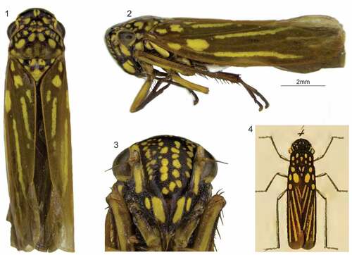
Figure 5. Macugonalia semiguttata (Signoret, 1853), specimen photographed on a shrub beside the road to the Pico da Caledônia, Nova Friburgo, Rio de Janeiro State (collection site is 1,737 m a.s.l.); photograph taken by André Almeida Alves, 6 December 2019.
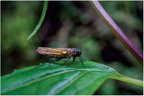
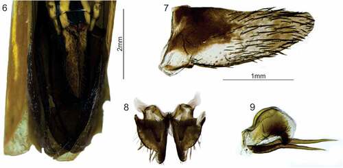
Figure 6–9. Macugonalia semiguttata (Signoret, 1853), male. 6, apical portion of abdomen, ventral view. 7, pygofer, lateral view. 8, subgenital plates, connective, and styles, dorsal view. 9, aedeagus, lateral view.
Tettigonia semiguttata Signoret [Citation14, p. 367] [original description]; Young and Beier [Citation24, p. 574] [lectotype designated].
Apogonalia semiguttata Metcalf [Citation25, p. 292] [catalogued].
Macugonalia semiguttata Young [Citation1, p. 944] [new combination]; McKamey [Citation16, p. 170] [catalogued].
Length
Male 10.8 mm (n = 1), females 11.6–11.8 mm (n = 2).
Head (). Moderately produced anteriorly, in dorsal view; median length of crown approximately 1/2 of interocular width and 1/3 of transocular width; anterior margin broadly rounded; without carina at transition from crown to face. Ocelli located behind imaginary line between anterior eye angles, each closer to adjacent anterior eye angle than to median line of crown. Surface of crown convex, without transverse concavity across ocelli and without sculpturing or setae. Frontogenal suture extending onto crown and attaining ocellus. Antennal ledge, in dorsal view, slightly protuberant; in lateral view, with anterior margin oblique and convex. Face without vertical lenticular sclerite bordering frontogenal suture below antenna; frons convex, muscle impressions distinct; epistomal suture obscure medially; clypeus moderately swollen, its upper portion continuing profile of frons and lower portion more horizontal.
Thorax ()
Pronotum with width less than transocular width of head; lateral margins approximately parallel; posterior margin slightly concave or almost rectilinear; dorsolateral carina complete; disk not sculptured, not pubescent. Mesonotum with scutellum not transversely striate. Forewing with membrane clearly delimited, including all apical cells; veins distinct; with three closed anteapical cells of which bases of inner two are usually transversally aligned, base of outer anteapical cell located slightly more proximally than those of inner two cells; with four apical cells, base of fourth located more proximally than base of third; without anteapical plexus of veins; forewings of female, in rest position, exceeding apex of ovipositor. Hind wing with vein R2 + 3 incomplete. Hind leg with femoral setal formula 2:1:1 or 2:1:1:1; length of first tarsomere greater than combined length of two more distal tarsomeres, with two parallel rows of small setae on plantar surface.
Color (, 5)
Ground color of head, pronotum, and mesonotum dark brown to black, with various irregular yellow spots. Forewing brown; clavus with three to five basal spots and one longitudinal stripe on vein Pcu, yellow; corium with two spots on basal portion and two elongate stripes (one close to costal margin on vein R and another close to claval sulcus on vein CuA), yellow. Face dark brown to black; frons with various irregular yellow spots centrally and laterally; gena, lorum, and clypeus with large yellow spots; labrum brownish-yellow to brown. Lateral and ventral portions of thorax dark brown to black with yellow spots; legs yellowish-brown to dark brown; hind femur sometimes with distinct ventral yellow area. Ground color of abdomen dark brown to black, with large yellow spots on sternites and laterotergites.
Male terminalia
Pygofer (), in lateral view, strongly produced posteriorly, subtriangular; without processes; apex narrowly rounded; surface with many macrosetae distributed mostly on posterior 1/2 of disk. Subgenital plate (), in ventral view, very short, not attaining 1/4 of pygofer length; subtriangular, distinctly narrowed on apical 1/4; apex narrowly rounded; with sparse microsetae and single row of few large macrosetae along external margin; plates separated from each other throughout their length. Style (), in dorsal view, extending posteriorly beyond apex of connective; with lateral lobe on median outer portion; apex obtuse, with tiny dentiform projection directed inwards. Connective (), in dorsal view, Y-shaped; arms broad, strongly divergent; stalk with median keel. Aedeagus (), in lateral view, short; shaft convex dorsally, lobate; with pair of elongate slender processes arising basiventrally, greatly exceeding shaft apex; in ventral view, shaft with pair of apicoventral small projections; gonopore located ventroapically. Paraphyses absent.
Female terminalia
Sternite VII (), in ventral view, strongly produced posteriorly, with elongate, strong distal projection reaching median 1/3 of pygofer; apex of projection subacute. “Internal” sternite VIII without distinct sclerites. Pygofer (), in lateral view, strongly produced posteriorly; posterior margin narrowly rounded; macrosetae distributed mostly on posterior 1/3 of disk and extending anteriorly along ventral margin. Valvifer I (, Vli), in lateral view, expanded dorsally, guttiform. Valvula I, in ventral view, with inner basiventral emargination; blade (), in lateral view, distinctly expanded dorsally along distal 2/3; apex acute; ventral interlocking device (, VID) elongate, extending posteriorly slightly beyond basal 1/2 of blade; dorsal sculptured area (, DSA) extending from basal portion of blade to apex; ventral sculptured area (, VSA) located on apical portion of blade and with elongate section positioned more basally; both sculptured areas formed mostly by linear structures. Valvula II (), in lateral view, moderately expanded beyond basal curvature; dorsal margin convex, with approximately 50 subtriangular teeth (–c, TOO) that become progressively smaller towards apex (teeth at basal ascending portion of blade of irregular form); denticles distributed on teeth and on dorsal and ventral apical portions of blade (ventral dentate apical portion longer than dorsal one); preapical prominence absent; apex obtuse; blade with numerous ducts (, DUC) extending toward teeth and apex. Gonoplac (ovipositor sheath) of the usual Cicadellinae type: in lateral view (), with distal 1/2 distinctly expanded, slightly narrowed at apical portion toward obtuse apex; surface with denticuli and few setae on posterior portion and extending anteriorly along ventral margin.
Figure 10–14. Macugonalia semiguttata (Signoret, 1853), female. 10, apical portion of abdomen, lateral view (the sternite VII is dislocated downward and the ovipositor is exposed). 11, sternite VII, ventral view. 12, valvifer I and valvula I, lateral view; 12a, dorsal sculptured area at apical portion; 12b, apex. 13, valvula II, lateral view; 13a–b, teeth at median and apical portions; 13 c, apex. 14, valvifer II and gonoplac, lateral view. DEN: denticle; DSA: dorsal sculptured area; DUC: duct; Evii: sternite VII; GON: gonoplac; OVI: ovipositor valvulae I and II; RAM: ramus; TOO: tooth; VID: ventral interlocking device; Vli: valvifer I; Vlii: valvifer II; VSA: ventral sculptured area.

Material examined
Atlantic Forest, southeastern Brazil, Serra dos Órgãos mountain range. One male and six females: “RJ [Rio de Janeiro State] Nova Friburgo\Estrada [para o] Pico da\Caledônia [22°20ʹ44.8”S, 42°35ʹ13.2”W, 1,737 m a.s.l.]\6/XII/2019\Mejdalani, Pecly,\Quintas, Ferreira, Alves” (MNRJ). One male and six females: “BR: RJ – Nova Friburgo\Estrada p/ o Pico da\Caledônia\06.XII.2019\Pecly, Quintas, Almeida, Ferreira & Mejdalani cols.” (DZRJ). One male and three females: same data as preceding (DZUP).
Discussion
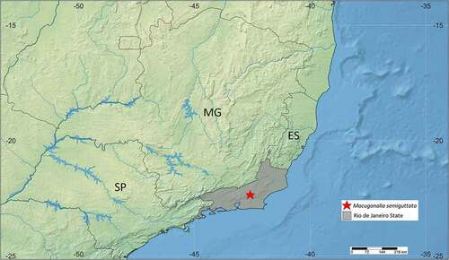
Among the known Macugonalia species, M. semiguttata has the dorsal lobe and pair of processes of the aedeagus more similar to those of Macugonalia contristata Young, 1977, Macugonalia elegantula Cavichioli, 2004, Macugonalia geographica (Signoret, 1855), Macugonalia nefasta Cavichioli, 2004, Macugonalia picta (Distant, 1908), and Macugonalia sobrina (Stål, 1862). A combination of subtle features distinguishes the aedeagus of M. semiguttata from those of the listed species, viz., the dorsal lobe is rounded, elongate, and not truncate posteriorly, whereas the processes are very elongate, extending posteriorly well beyond the aedeagal shaft, slender, and slightly curved ventrally at median portion (). The dorsal dark brown to black ground color with yellow markings of M. semiguttata () is similar to those of M. elegantula and Macugonalia dallasi (Signoret, 1853). In addition to the aedeagal features just given, M. semiguttata can be distinguished from the seven mentioned species, as well as from other species of the genus, by the following combination of characteristics: (1) position of yellow stripes and spots on forewings (; see details in the color description provided above); (2) large yellow spots on abdominal sternites and laterotergites; (3) subgenital plates () distinctly narrowed on apical 1/4; and (4) styles () with apex obtuse, with tiny dentiform projection directed inward. Although the original color illustration provided by Signoret [Citation14, pl. 12, ], here reproduced as , is somewhat schematic, it shows the basic diagnostic features of M. semiguttata, allowing a precise identification of our specimens from the Serra dos Órgãos mountain range. This is the single known precise record of the species (), which previously was known only from “Brazil” [Citation1,Citation16,Citation25]. Specimens of M. semiguttata () were collected from the leaves and branches of shrubs and small trees in an area of low forest (about 1,700 m a.s.l.), which is intermediate between the typical Atlantic Forest and the vegetation of alpine fields.
Figure 15. Collection site of Macugonalia semiguttata (Signoret, 1853) in the Serra dos Órgãos mountain range, Rio de Janeiro State (22°20ʹ44.8”S, 42°35ʹ13.2”W, circa 1,700 m a.s.l.). This is the single known precise record of the species. ES: Espírito Santo State; MG: Minas Gerais State; SP: São Paulo State.
As in most other genera of the Cicadellinae, the morphology of the female terminalia in Macugonalia is still poorly known. Recently, a detailed description of the female terminalia of M. geographica, including the sternite VII, pygofer, and valvulae II, was provided by Carvalho and Mejdalani [Citation3]. Additionally, detailed information on the valvulae I and II of M. leucomelas was given by Mejdalani [Citation18]. The present description of M. semiguttata is apparently the most complete so far provided for the genus. The general form of the valvulae II of M. semiguttata () is quite similar to that of M. geographica. However, the valvulae II of these two species are quite distinct from those of M. leucomelas, because the blade of the latter is strongly expanded at the basal portion, so that the dorsal margin is distinctly declivous toward the apex (see illustrations of M. leucomelas in [Citation18]). Also, the number of valvula II teeth is distinctly smaller in M. leucomelas than in M. semiguttata and M. geographica. Therefore, although the female terminalia of cicadellids are usually considered morphologically more conservative than the male terminalia [Citation26], our preliminary comparisons suggest that the valvulae II can provide useful features for our understanding of the taxonomy and phylogeny of this diverse, geographically widespread, and complex Neotropical genus of sharpshooters.
Disclosure statement
No potential conflict of interest was reported by the author(s).
Additional information
Funding
References
- Young DA. Taxonomic study of the Cicadellinae (Homoptera: Cicadellidae). Part 2. New World Cicadellini and the genus Cicadella. Bull NC Agric Exp Stn. 1977;239:1–1135.
- Cavichioli RR. Macugonalia Young (Hemiptera, Cicadellidae, Cicadellinae): duas novas espécies de Minas Gerais, Brasil. Rev Bras Zool. 2004;21:123–126.
- Carvalho RA, Mejdalani G. Taxonomic notes on Macugonalia geographica (Signoret) with descriptions of the male and female genitalia (Insecta: Hemiptera: Cicadellidae: Cicadellini). Stud Neotrop Fauna Environ. 2005;40:143–148.
- Molina RO, Santos KSD, Nunes WMDC. Comparison of protocols for the extraction of genomic DNA from sharpshooters (Hemiptera: Cicadellidae) for the detection of Xylella fastidiosa. Arq Inst Biol. 2018;85:1–5.
- Müller C, Esteves MB, Kleina HT, et al. First sharpshooter species proven as vectors of Xylella fastidiosa subsp. multiplex in Prunus salicina trees in Brazil. Trop Plan Pathol. 2021;1–6. doi:10.1007/s40858-021-00430-8
- Almeida RPP, Blua MJ, Lopes JRS, et al. Vector transmission of Xylella fastidiosa: applying fundamental knowledge to generate disease management strategies. Ann Entomol Soc Am. 2005;98:775–786.
- Azevedo RL, Lima MF. Cigarrinhas dos citros, vetoras da bactéria Xylella fastidiosa Wells et al.: pragas potenciais para a citricultura sergipana. EntomoBrasilis. 2015;8:1–7.
- Molina RO, Santos KS, Gonçalves ACA, Nunes, WMC. Distribuição espaço-temporal de cigarrinhas (Hemiptera: Cicadellidae) vetores da Xylella fastidiosa em pomares cítricos. Revista Agro@mbiente On-line. 2016;10:145–152.
- Froza JA. Levantamento de espécies de cigarrinhas (Hemiptera: Auchenorrhyncha) com ênfase em possíveis vetores de Xylella fastidiosa em pomares de oliveira na Serra da Mantiqueira [M.Sc. dissertation]. Piracicaba (SP): Escola Superior de Agricultura “Luiz de Queiroz”, Universidade de São Paulo; 2017.
- Paris P, Azevedo Filho WS, Poletto G, et al. Ocorrência de cigarrinhas (Cicadellidae: Cicadellinae) potenciais vetoras de Xylella fastidiosa associadas à cultura da videira na Serra Gaúcha. Poster presented at XXII Congresso Brasileiro de Fruticultura; 2012 Out 22–26; Bento Gonçalves, RS.
- Feitosa MCB. Comunidade e dinâmica populacional de cigarrinhas (Hemiptera: Cicadellidae: Cicadellinae) e seus parasitoides de ovos (Hymenoptera: Chalcidoidea) em pomares cítricos no Amazonas, Brasil [D.Sc. dissertation]. Manaus (AM): Instituto Nacional de Pesquisas da Amazônia; 2017.
- Vásquez J, Lozada P. Las especies de cigarritas (Homoptera, Cicadellidae) asociadas a las plantas medicinales y ornamentales en Allpahuayo, Iquitos-Perú. Folia Amazón. 2014;23:199–204.
- Costa DPD. Hepáticas do Pico da Caledônea [sic], Nova Friburgo, Rio de Janeiro, Brasil. Acta Bot Bras. 1992;6:3–39.
- Signoret V. Revue iconographique des Tettigonides. Ann Soc Entomol Fr. 1853;1: 323–374(pls.8–12).
- Zanol KMR, de Menezes M. Lista preliminar dos cicadelídeos (Homoptera, Cicadellidae) do Brasil. Iheringia (S Zool). 1982;61:9–65.
- McKamey SH. Taxonomic catalogue of the leafhoppers (Membracoidea). Part 1. Cicadellinae. Mem Amer Ent Inst. 2007;78:1–394.
- Oman PW. The Nearctic leafhoppers (Homoptera: Cicadellidae). A generic classification and check list. Mem Ent Soc Wash. 1949;3:1–253.
- Mejdalani G. Morfologia externa dos Cicadellinae (Homoptera, Cicadellidae): comparação entre Versigonalia ruficauda (Walker) (Cicadellini) e Tretogonia cribrata Melichar (Proconiini), com notas sobre outras espécies e análise da terminologia. Rev Bras Zool. 1998;15:451–544.
- Young DA, Beirne BP. A taxonomic revision of the leafhopper genus Flexamia and a new related genus (Homoptera: Cicadellidae). Tech Bull US Dep Agric. 1958;1173:1–53.
- Hamilton KGA. Morphology and evolution of the rhynchotan head (Insecta: Hemiptera, Homoptera). Can Ent. 1981;113:953–974.
- Mejdalani G. Morfologia da cabeça de Versigonalia ruficauda (Walker, 1851), com notas sobre a terminologia (Homoptera, Cicadellidae, Cicadellinae). Rev Bras Ent. 1993;37:279–288.
- Nielson MW. A revision of the genus Cuerna (Homoptera, Cicadellidae). Tech Bull US Dep Agric. 1965;1318:1–48.
- Hill BG. Comparative morphological study of selected higher categories of leafhoppers (Homoptera: Cicadellidae) [Ph.D. dissertation]. Raleigh (NC): North Carolina State University; 1970.
- Young DA, Beier M. Types of Cicadellinae (Homoptera: Cicadellidae) in the Natural History Museum in Vienna. Ann Naturhistor Mus Wien. 1963;67:565–575.
- Metcalf ZP General catalogue of the Homoptera. Fascicle VI. Cicadelloidea. Part 1. Tettigellidae. Washington, Agricultural Research Service, U.S. Department of Agriculture. 1965;1–730 pp.
- Carvalho RA, Mejdalani G. Remarkable morphological features of taxonomic interest in the female genitalia of five Erythrogonia species (Hemiptera: Cicadomorpha: Cicadellidae). Zootaxa. 2014;3872:275–290.