
Abstract
Oncocytic lipoadenoma is an extremely rare benign salivary gland tumor with few reported cases. We provide a comprehensive description of key radiographic features suggesting the diagnosis of oncocytic lipoadenoma and review its clinical and pathologic features. We present a case of a 70-year-old woman with a slowly enlarging submandibular mass. Magnetic resonance imaging showed a heterogeneous fat-containing lesion without aggressive features. The solid components enhanced with gadolinium. Fine needle aspiration biopsy showed oncocytic cells. After surgical resection, histology showed a well-encapsulated mass with a mixture of oncocytes and adipocytes consistent with oncocytic lipoadenoma. While the usual differential diagnosis of oncocytes in a salivary gland aspirate includes oncocytoma, Warthin’s tumor, oncocytic carcinoma, and oncocytic metaplasia/hyperplasia, specific radiographic features on MRI of a well-circumscribed soft tissue and fat-containing mass, can direct preoperative diagnosis and treatment planning. Awareness of this entity is helpful during the evaluation of patients with salivary gland neoplasms.
Introduction
Oncocytic lipoadenoma is an extremely rare benign tumor of the salivary glands. This entity belongs to a family of fat-containing tumors of the salivary glands including lipoma, non-oncocytic sialolipoma, and pleomorphic adenoma/myoepithelioma with extensive lipometaplasia.[Citation1] Oncocytic lipoadenoma of a salivary gland was first described in 1998 by Hirokawa et al., who characterized it as a benign tumor composed of an admixture of mature fat cells and oncocytes in a submandibular gland.[Citation2] Since the first description, there have been 15 cases reported in the English literature, often using different names such as oncocytic sialolipoma or adenolipoma.[Citation1,Citation3–7]
A recent review of fat-containing salivary tumors by Agaimy reports that while salivary lipomas occur primarily among elderly men, oncocytic lipoadenomas do not have a gender discrepancy and have a wider range of age at presentation. The parotid gland is the most frequent location, followed by intraoral salivary glands and, less commonly, the submandibular gland. Surgical resection is the mainstay of treatment.[Citation1]
We present a rare case of oncocytic lipoadenoma of the submandibular gland. We provide a comprehensive description of key radiographic features that suggest the diagnosis and review the clinical and pathologic features of this tumor.
Case report
A 70-year-old Caucasian woman presented with a two-year history of a slowly growing, non-tender left submandibular mass. The patient was otherwise asymptomatic and the mass was identified incidentally during a thyroid ultrasound to investigate a multinodular goiter two years prior to our evaluation. On physical examination she had a soft, mobile, and enlarged left submandibular gland without other palpable masses. There were no overlying skin changes or cranial nerve deficits. The patient had further diagnostic workup with a magnetic resonance imaging (MRI) as well as a fine needle aspiration (FNA) biopsy. Both the imaging and cytologic findings were highly suggestive of oncocytic lipoadenoma.
She subsequently underwent uncomplicated surgical resection of the left submandibular gland. Intraoperative findings included a very soft, 3 cm, fatty mass attached to the anteroinferior surface of a normal-appearing submandibular gland. The gland and the attached tumor were easily dissected away from the surrounding tissues and no significant inflammation was encountered.
Imaging findings
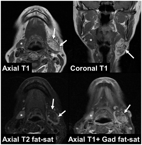
MRI demonstrated a 3.2 × 1.7 cm well-circumscribed, heterogeneous and partially enhancing mass confined to the left submandibular gland, with normal glandular tissue at its medial aspect. The mass was heterogeneous, but had a predominantly bright signal on T1 imaging that was lost on fat-saturated sequences, indicating a lipomatous component. The other solid components of the lesion enhanced with contrast. The contralateral submandibular gland appeared normal ().
Figure 1. Magnetic resonance imaging. White arrows indicate tumor. * indicates normal contralateral gland.
Cytologic findings
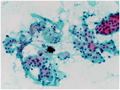
Palpation-guided FNA was performed with suction using a 23-gauge needle and 10 ml syringe. Two passes yielded three smears which were fixed with ethanol and stained with Papanicolaou stain. Aspirate smears were sparsely to moderately cellular and showed predominantly oncocytic cells with round to oval monomorphic nuclei and abundant dense granular cytoplasm. Oncocytes were admixed with adipocytes and fibrous tissue. The diagnosis of ‘oncocytic lesion’ was rendered with a differential diagnosis of oncocytoma, oncocytosis and, less likely, oncocytic carcinoma ().
Figure 2. Photomicrograph showing clusters of oncocytic epithelium with associated adipocytes (Pap stain 400×).
Gross findings
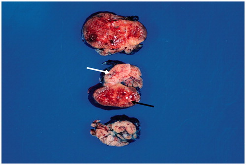
Examination of the surgical specimen revealed an encapsulated, well circumscribed, red-tan lobular mass measuring 4.3 × 3.0 × 2.1 cm attached to an otherwise unremarkable submandibular gland. On cut sections, the parenchyma of the mass was mottled yellow-tan to brown-red with a lobulated appearance ().
Figure 3. Photograph of the surgical specimen demonstrating tumor and adjacent normal salivary gland (white arrow). The well-demarcated, encapsulated tumor contains both yellow-tan adipose tissue and dark red oncocytic components (dark arrow).
Histologic findings
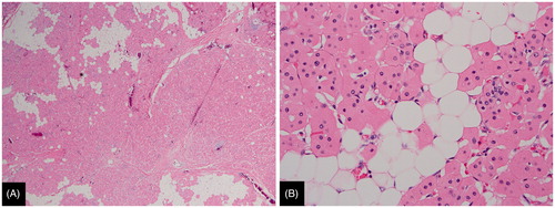
Microscopically, the tumor was composed of a mixture of oncocytes and mature adipose tissue. The oncocytic epithelium formed small glands and tubules which were not associated with ducts. No typical acinar elements or myoepithelial proliferation was identified. The oncocytes had abundant fine granular eosinophilic cytoplasm and a single small round nucleus. No atypia, mitotic activity, necrosis, or hemorrhage was identified. There was no sebaceous metaplasia. The adjacent submandibular gland showed no abnormalities. The diagnosis of oncocytic lipoadenoma was made based on these findings ().
Figure 4. (A). Histologic section of oncocytic lipoadenoma showing solid islands of pink oncocytic cells admixed with mature adipose tissue (H&E, 40×). (B). High power view showing the oncocytic cells which exhibit abundant fine granular eosinophilic cytoplasm and a single small round nucleus. Oncocytes are admixed with mature fat (H&E, 400×).
Discussion
Oncocytic lipoadenoma is a rare benign tumor of the salivary glands. We present a case of a 70-year-old woman with a submandibular gland tumor in whom the diagnosis of oncocytic lipoadenoma was highly favored based on unique imaging characteristics. MRI is superior to CT scan in its ability to define tissue contours and also to better characterize the nature of soft tissue masses in the neck. MRI is also better able to demonstrate salivary masses that are occult on CT scan and define features that distinguish benign and malignant salivary tumors.[Citation8]
While prior reports have described the histologic findings of oncocytic lipoadenoma and similar fat-containing salivary tumors,[Citation1–7,Citation9] relatively little has been written regarding the imaging characteristics on computed tomography, ultrasound, and MRI.[Citation4,Citation7,Citation9] With regard to MRI findings, Mitsimponas et al. present a limited description of a heterogeneous lesion with intermediate signal intensity on T1 and T2. Based on these imaging features, this tumor was initially assigned the non-specific diagnosis of ‘parotid adenoma’.[Citation9] In this current case, MRI clearly identified both soft tissue and lipomatous elements of a lesion that was intimately associated with the submandibular gland, but without features of invasion on imaging. While the previously published MRI features of oncocytic lipoadenoma have been limited, we report a more comprehensive description which matches what would be expected based on the composition of this tumor.
These imaging findings were further supported by the FNA biopsy findings of oncocytes admixed with adipocytes. The usual differential diagnosis of oncocytes in a salivary gland aspirate includes oncocytoma, Warthin’s tumor, oncocytic carcinoma, and oncocytic metaplasia/hyperplasia.[Citation7] Histologic evaluation in this case showed the characteristic mixture of fat cells and oncocytes without atypia typically seen in oncocytic lipoadenoma.
Conclusion
Oncocytic lipoadenoma is a rare benign tumor of the salivary glands with only a small number of cases reported in the literature. Specific radiographic features on MRI of a well-circumscribed soft tissue and fat-containing mass, coupled with cytopathologic findings of oncocytes can direct preoperative diagnosis and treatment planning. Awareness of this entity is helpful during the evaluation of patients with salivary gland neoplasms.
Disclosure statement
The authors report no conflicts of interest. The authors alone are responsible for the content and writing of this article.
References
- Agaimy A. Fat-containing salivary gland tumors: a review. Head Neck Pathol. 2013;7:90–96.
- Hirokawa M, Shimizu M, Manabe T, et al. Oncocytic lipoadenoma of the submandibular gland. Hum Pathol. 1998;29:410–412.
- Agaimy A, Ihrler S, Markl B, et al. Lipomatous salivary gland tumors: a series of 31 cases spanning their morphologic spectrum with emphasis on sialolipoma and oncocytic lipoadenoma. Am J Surg Pathol. 2013;37:128–137.
- Pusiol T, Franceschetti I, Scialpi M, et al. Oncocytic sialolipoma of the submandibular gland with sebaceous differentiation: a new pathological entity. Indian J Pathol Microbiol. 2009;52:379–382.
- Kato M, Horie Y. Oncocytic lipoadenoma of the parotid gland. Histopathology. 2000;36:285–286.
- Klieb HB, Perez-Ordonez B. Oncocytic lipoadenoma of the parotid gland with sebaceous differentiation. Study of its keratin profile. Virchows Arch. 2006;449:722–725.
- Aouad R, Matar N, Sader-Ghorra C, et al. Pathology quiz case 1. Oncocytic lipoadenoma of the parotid gland. Arch Otolaryngol Head Neck Surg. 2008;134:446–448.
- Christe A, Waldherr C, Hallett R, et al. MR imaging of parotid tumors: typical lesion characteristics in MR imaging improve discrimination between benign and malignant disease. Am J Neuroradiol. 2011;32:1202–1207.
- Mitsimponas KT, Agaimy A, Schlittenbauer T, et al. Oncocytic lipoadenoma of the parotid gland: a report of a new case and review of the literature. Int J Clin Exp Pathol. 2012;5: 1000–1006.