
Abstract
Inspired by Jackass (a tv-show about self-injuring stunts), some friends topped off a drinking party with live fishes from their aquarium. After the goldfishes had gone down smoothly, a bronze catfish was ingested. Unaware of the morphology and anti-predator behaviour of this species, a healthy but intoxicated 28-year-old man got a surprise. The catfish erected and locked the spines of its pectoral fins and got lodged in the hypopharynx. After several hours, he presented himself at the emergency department with dysphonia and dysphagia. The fish had to be removed endoscopically. Intubation and admittance to the intensive care unit was necessary due to laryngeal oedema. Two weeks postoperatively, the patient made a full recovery and donated the fish to the Natural History Museum Rotterdam. The publicity generated by public exhibition of the ‘do-not-swallow-fish’ emphasised the official Jackass warning: ‘.. do not attempt any of the stunts you’re about to see’.
Introduction
Fish bones are the most common foreign bodies causing airway obstruction [Citation1]. Obstruction after whole fish ingestion is rare. Relatively few cases have been reported, mostly describing cases with a life-threatening course and often a fatal outcome. Pinheiro et al. presented a fatal case of a 50-year-old fisherman who accidentally choked on a live fish [Citation2]. Tam et al. mentioned that a fisherman who kissed his first catch that subsequently slipped in his mouth, obstructed the hypopharynx, caused acute respiratory distress but was saved by emergency evacuation of the fish [Citation3]. Kumar and Surianarayanan’s classic ‘Accidental Entry of Fish into Throat While Bathing in a Pond’ [Citation4] is also exemplary for the involuntary nature of many cases of fish obstructing the upper airway.
Intentional foreign body ingestion is commonly seen in adult (institutionalised) patients with psychiatric disorders, intellectual or mental disabilities or in case of significant alcohol and/or drug abuse [Citation5]. A schizophrenic patient who choked to death after ingesting an entire dead sole (Solea solea) is the single known case in this category involving a fish [Citation6].
Ali and Metha [Citation7] reviewed 75 cases of live fish aspiration, reported in the scientific literature between 1567 and 2015. Of these fishes, 56 ended-up in the upper airway and hypopharynx. Only four cases involved voluntary ingestion of live fishes, resulting in three fatalities. Deliberate ingestion of live fish from home aquaria is unknown from the scientific literature, but well-known in popular culture, such as the British-American heist comedy film ‘A Fish Called Wanda’ [Citation8].
Here we present the first case of intentional ingestion of a live pet catfish causing obstruction of the upper airway that needed acute medical intervention.
Case report
Based on verbal testimonies and a two-minute home video of the incident supplied by the patient, the following chain of events was reconstructed.
Inspired by Jackass (an American tv-show, with ten stuntmen performing extreme stunts, including various dangerous, crude, self-injuring pranks), a 28-year old man and his friends had developed a tradition to swallow live pet fish from their home aquarium. Such case was in the afternoon of 3 April 2016, when the patient and his friends drank excessive amounts of beer and used 3,4-methylenedioxymethamphetamine (ecstasy).
The first batch of live fishes (goldfish Carassius gibelio auratus) passed smoothly, but the final fish did not. A two-minute home video shows the following sequence of events: (00:00) drinking and shouting [“grote vis, grote vis!” (“big fish, big fish!”)]; (00:13) person a drinks from a glass containing clear water and a live fish; (00:17) person a spits out water and fish, catches fish in his hands and throws fish on table; (00:27) fish flounders in distress on table; (00:33–37) agonised fish handed over by person b to patient; (00:45–48) patient gulps beer from bottle and subsequently engulfs the fish; (00:49) patient unable to drink more beer as fish apparently got stuck in his throat; (00:55) patient gags vigorously; (00:57) patient clearly in distress, vomits liquids; (01:01–29) patient in extreme distress, uses two fingers to induce gag reflex, but apparently fish remains stuck; (01:30–45) person c administers wrongly applied Heimlich manoeuvre; (01:46–58) patient still gagging; (01:59) patient spews blood in bucket.
After several hours of unsuccessful self-applied treatment with more beer, honey and ice cream, the patient finally presented himself to the emergency department.
Physical examination
At the presentation, we saw a patient with acute dysphonia and dysphagia. He was not dyspnoeic, although he mentioned a swollen throat and stated difficulty swallowing his own saliva. His vital functions were normal and there were no abnormalities during intra-oral examination and no subcutaneous emphysema could be palpated in the head and neck area. Nasal fibre-endoscopy indeed revealed a fish-like structure just lateral of the left arytenoid with supraglottic hematoma and oedema but no glottic stenosis.
Additional investigations
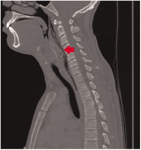
CT-scan of the head and neck area (Figure ) showed a foreign body of about 5 cm in length and 1.5 cm across at the level of the hypopharynx and the proximal oesophagus. The foreign body consisted of a hyper-dense border with internal air-filled levels. Three sharp prominences were visible on the anterolateral left, lateral and dorsal side. The last of these reached within the retropharyngeal space that was air filled extending until the carotid space on both sides.
Figure 1. (Mid) sagittal section of CT-scan of the head and neck area revealing a foreign body (arrow) of ±5 cm in length and 1.5 cm across at the level of the hypopharynx and the proximal oesophagus.
Treatment
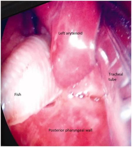
After orotracheal intubation (single-lumen tube 6.5 mm) endoscopic examination under general anaesthesia was performed. This revealed a dead fish in the hypopharynx (Figure and additional Supplementary video). The foreign body was removed by using a grasping forceps. Additional gastro-oesophagoscopy showed oedema and a haematoma of the left piriform sinus. The oesophagus showed no signs of perforation. Due to the presence of laryngeal oedema and haematoma the patient was left intubated and admitted to the intensive care unit.
Figure 2. Direct laryngoscopy shows a fin-like structure lateral of the left arytenoid with supraglottic oedema. Intubation with a single-lumen tube.
Outcome and follow up
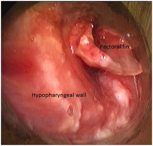
The microbiologist advised intravenous piperacillin/tazobactam to treat a possible infection with Pseudomonas aeruginosa, which often inhabit freshwater aquaria. Two days later, a follow-up CT scan still showed a small foreign body at the posterior wall of hypopharynx (Figure ). A second endoscopic procedure was necessary to remove it (Figure ). After removal, the oedema reduced and the patient could be extubated safely and was transferred to the ENT ward. Full recovery was reached after two weeks of intravenous antibiotics.
Figure 3. Sagittal CT slice shows a residual foreign body (arrow) at the posterior wall of hypopharynx.

Figure 4. Remaining pectoral fin in the left hypopharynx.
Examination and identification of the foreign bodies
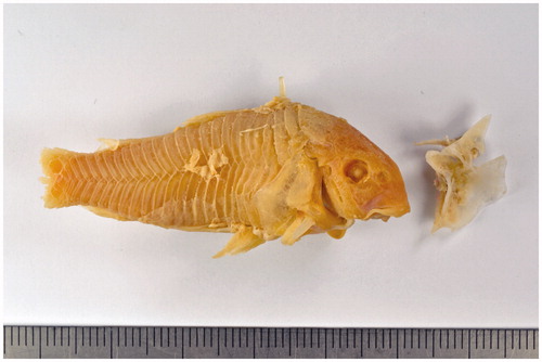
Both foreign bodies were preserved in 70% ethanol and presented to the Natural History Museum Rotterdam for identification. The first, 53 mm long, object could, without doubt, be identified as a full-grown Corydoras aeneus (T.N. Gill, 1858), lacking the tail fin [Citation9]. This is a freshwater catfish belonging to the family Callichthyidae (Actinopterygii, Siluriformes). This species is a cheap, widely available and popular pet fish, colloquially known as Bronze Catfish, Cory Catfish or simply ‘Cory’ [Citation10]. The second object was the left pectoral fin and spine, belonging to the same specimen. The patient donated the complete set to the Natural History Museum, where it is catalogued as NMR 9979-01801 (Figure ). The whereabouts of the missing tail remained unknown.
Figure 5. Bronze catfish (Corydoras aeneus), lacking the tail (left) and pectoral fin (right) as recovered from the patient; preserved in the collection of the Natural History Museum Rotterdam.
Conclusion and discussion
Ingestion of a live fish resulting in airway obstruction is rare and in most cases, fatal occasion [Citation2]. The present case is the first reported intentional ingestion of a bronze catfish. The osteology and anti-predator behaviour of C. aeneus rate this species as a bad choice for a drinking game. The dorsal, pectoral and adipose fins contain sharp barbs and spines; beneath each eye there is a spike adding to the fish’ defence mechanism [Citation11]. The spines of the pectoral fins release a generally harmless poison when attacked [Citation12]. Besides, when bronze catfishes are under attack or in distress, the fish erects and locks the pectoral fin spines and releases the venom. In the present case the catfish, clearly in distress after being taken from its safe surroundings, being tossed around and showered by beer in a man’s throat, had erected the spines and ended-up being stuck in the patient’s hypopharynx. The defence mechanism proved very effective, as the left pectoral fin was lodged into such a depth, that it could not be removed (and was overlooked) during the first endoscopic procedure.
Furthermore, this case illustrates how a reckless drinking game, imitating Jackass [Citation13] and/or ‘A Fish Called Wanda’ [Citation8], can turn into a dangerous and critical medical situation with serious consequences. When confronted with an ingested fish stuck in the upper airways, identification of the specimen is vital for further treatment. As shown in this case, the expertise of the local Natural History Museum was of great help. The fish became part of the museum’s ‘Dead Animal Tales’ exhibition, showing how, where and why animals and humans collide, often with dramatic consequences for both parties [Citation14]. The publicity generated by donation and public exhibition of the fish [Citation15] emphasised the official Jackass warning: ‘.. do not attempt any of the stunts you’re about to see’.
Supplemental Material
Download MP4 Video (5.3 MB)Disclosure statement
No potential conflict of interest was reported by the authors.
References
- Nandi P, Ong GB. Foreign body in the oesophagus: review of 2394 cases. Br J Surg. 1978;65:5–9.
- Pinheiro J, Cordeiro C, Vieira DN. Choking death on a live fish (Dicologoglossa cuneata). Am J Forensic Med Pathol. 2003;24:177–178.
- Tam T, Weinberg L, Edington J. Airway obstruction from accidental ingestion of a live fish. BMJ Case Rep. 2013;2013:010486.
- Parida PK, Surianarayanan G. Accidental entry of fish into throat while bathing in a pond. Case Rep Med. 2013;2013:604687.
- O'Sullivan ST, Reardon CM, McGreal GT. Deliberate ingestion of foreign bodies by institutionalised psychiatric hospital patients and prison inmates. IJMS. 1996;165:294–296.
- Busardò FP, Mannocchi G, Pugnetti P, et al. A very unusual accidental mechanical asphyxia of choking with a whole Solea solea. J Forensic Sci. 2017;62:511–514.
- Ali SR, Mehta AC. Alive in the airways: live endobronchial foreign bodies. Chest. 2017;151:481–491.
- Cleese J, Crichton C. A fish called Wanda. Beverly Hills (CA): Metro-Goldwyn-Mayer Production; 1988.
- Fuller IAM, Evers HG. Identifying corydoradinae catfish – Corydoras. Rodgau (Germany): Aqualog Verlag; 2005.
- Fuller IAM. Breeding corydoradinae catfish, 2nd ed. Ian Fuller Enterprises; 2012.
- Huysentruyt F, Adriaens D. Descriptive osteology of Corydoras aeneus (Siluriformes: Callichthyidae). Cybium. 2005;29:261–273.
- Greven H, Flasbeck T, Passia D. Axillary glands in the armoured catfish Corydoras aeneus (Callichthyidae, Siluriformes). Verhandlungen Der Gesellschaft Für Ichthyologie. 2006;5:65–69.
- Clives S, editor. Jackass: 10 years of stupid. New York (NY): Powerhouse Cultural Entertainment; 2010.
- Moeliker K. Verslikvis bijgezet in expo ‘Dode dieren met een verhaal’ [Do-not-swallow-fish added to ‘Dead Animal Tales’ exhibition]. Straatgras. 2016;28:41.
- Whipple T. The drinker and the fish is a hard story to swallow. Times (London). 2017;72114:7. [January 7th]