
Abstract
Environment radiations in vault rooms resulting from Axesse linac use was assessed at Medical University Hospital using Thermoluminescent dosimeters (TLDs) during the use of the new radiation treatment known as volumetric modulated arc therapy (VMAT) in the treatment of nasopharyngeal cancer (NPC). The TLD-100H was calibrated using linac 6 MV photons. A total of 75 measurement points of the TLD-100H were utilized for environmental monitoring. The measured environment radiations were then visualized as three-dimensional graphical representations. Environment radiations were visualized using colored three-dimentional graphical representations. The radiations in NPC treatment of Rando phantom was found to reach levels up to 370 mSv/treatment. Many researchers consider TLD, which is the most cost-effective method to environment radiations. The minimum detectable dose (MDD) was also identified to demonstrate the reliability of the TLD approach. Quantitative results that provide practical guidance with regarded radiation protections. Potentially hazardous of secondary radiations from the operating linac is evaluated with regard to its potential health effects on both patients and the public.
This work tried to evaluate the environment radiation and minimal detectable limit of TLD inside the LINAC maze.
KEY POINTS
1. Introduction
A novel method, volumetric modulated arc therapy (VMAT) with an Axesse (Elekta 2538) linac, has proved to be a very effective treatment in many cancer cases. However, Axesse generates high-energy radiations that exposed patients to undesirable radiations during oncology therapy treatments.[Citation1–4] Patients are exposed to significant radiations due primarily to out-of-field 6 MV X-rays. These radiations are not discussed in ordinary treatment plan, and few researches have focused on the spread of secondary radiations into the environment around the Department of Radiation Oncology (DRO).[Citation3] In addition, a detailed survey is needed to measure the secondary radiations around the vault room of medical university. The problem of 6 MV photons spread into the vault room may require additional attention. Thermoluminescent dosimetry (TLD-100H), 3.0 × 3.0 × 1.0 mm3 (Harshaw Chemical Company, Solon, OH) has been employed to measure three-dimensional distributions of environment radiations.[Citation5,Citation6] Intensive effort can allow quantitative measurement of environment radiations, as well as the effective provision of data to elaborate radiation safety.
2. Materials and methods
2.1. Axesse linac
Many linacs are operating around the world for oncology therapy now. The linac, Axesse (Elekta 2538) of Chung Shan Medical University Hospital (CSMUH) is designed for VMAT. Dose conformity can increase to the tumor during faster delivery time, fewer monitor units and superior dose distribution.[Citation3] Between 9500 and 10,500 patients who with various pathologies, NPC, have been treated at CSMUH in 2014. Unfortunately, no previous studies have addressed the secondary environment radiations in the vault rooms that can occur as a result of Axesse linac use. A detailed survey of environment radiations in vault rooms is important to both patients and the public.[Citation3,Citation4]
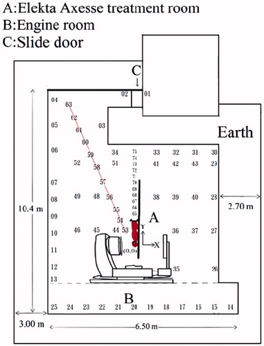
demonstrates vault room’s plane view of Axesse linac. The tumor center is A (0, 0) represented origin and located exactly on Rando phantom’s tumor center. Vault room is 5.9 m × 10.4 m × ×.8 m. B area is machine room of linac. A sliding door used to shield secondary radiations made of 10 PE cm thick and 1 Pb cm thick. Sliding door is located at C (maze end). The maze, which is at the upper side of the treatment room located between the restricted areas. A metal slab and concrete (3.0 m thick) located on the left side of the primary barriers. The right primary wall barriers are fabricated entirely of 2.70-m-thick concrete. A 0.250 cm thick Pb slab is located on the left side of the primary barriers. Restricted areas are approximately rectangular, 3.36 m × 1.85 m × 3.8 m. However, these secondary X-ray radiations minor are a health consideration for both patients and the publics.[Citation5]
Figure 1. Measurement locations and plane view in the vault room of Axesse at the CSMUH. The sliding door (C) was installed at the maze end which can shield from secondary radiations and Rando phantoms as patient surrogate.
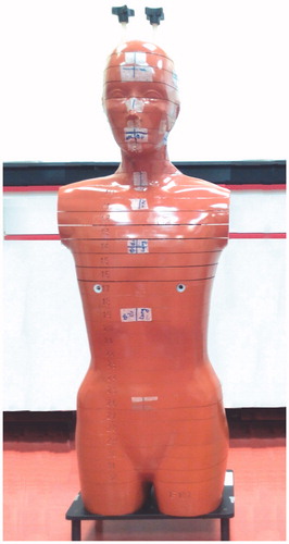
The Rando phantom (Alderson Radiation Therapy Phantom) (Radiology Support Devices Inc., Long Beach, CA) with height 175 cm and weight 73.5 kg. Rando phantom was made of polyurethane, human skeleton and lung soft tissue, suitable for dose measurements ().[Citation3,Citation7]
Figure 2. Outer appearance of Rando phantom.
shows the planned target volume (PTV) (red) and the gross tumor volume (GTV) (orange). The organs at risk of Rando phantom include the right eye (blue), right optic nerve (brown) and left optic nerve (green).
Figure 3. The NPC treatment plan for Pinnacle planning system of Philips Radiation Oncology System (Fitchburg, WI).
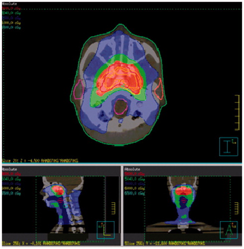
The organ contours and images were scanned from CT and transferred to Axesse’s VMAT. The prescribed doses were specified at the PTV. A total of 6 MV X-ray dose of PTV were displayed in red, then delivered to the phantom during a single NPC treatment.
2.2. Thermoluminescent dosimeters (TLD)
During past 30 years, the TLD can use widely to measure environment radiations. A calibrated TLD readout system evaluates specific doses with better reliability and can be useful to measure environment radiations. These exposure TLDs were evaluated via an automated Harshaw 3500 reader and coupled with an oven during 10 min at 240 °C followed by a rapid cool down. The readout was first, the TLD was heated up to 50 °C and held there during 1 s. Second, it was heated up to 240 °C under a rate of 10 °C/s.[Citation3,Citation4,Citation7,Citation8]
These TLDs have been shown to be accurate dosimeters for linacs operated at 6 MV. Throughout the experiment, the TLDs were always handed with great care, avoiding direct contact with dirt or hand material.[Citation3,Citation4,Citation7,Citation8]
2.3. TLD-100H calibration
The TLD-100H which was selected for its dimensions measured X-ray intensities and dose rates. In order to evaluate batch homogeneity, the TLDs were irradiated according to published procedure TRS-398. Solid water phantom (SWP) slabs, 10 mm in thickness and 30 × 30 cm2 in area, via electronic equilibrium, can be placing above them using a skin source. Distance is 100 cm under a 10 × 10 cm2 field size.[Citation3,Citation4,Citation7] In order to evaluate responses to doses of environment radiations, a batch of 400 TLDs was irradiated at different doses ranging from 0.10 to 11 Gy, which encompasses the range of the prescribed daily fraction dose. A Farmer type ionization chamber with a volume of 0.6 cc, type NE 2571 (PTW, Freiburg, UNIDOS), was positioned in the SWP.[Citation3,Citation4,Citation7]
shows 225 TLDs were randomly suspended 1.22 m above the floor at the DRO for mapping the distributions of environment radiations. Five measurements were averaged. To minimize errors, one bag contains 3 TLDs, so that 225 reusable TLDs represented 75 measurement locations during one NPC treatment. states measurements were acquired five times during oncology therapy of patients with NPC. In addition, environment radiations were measured by 3 TLDs among the control room, 14 m away from the Axesse in the public waiting room. Environment radiations of students’ dormitory were measured by 6 TLDs. Background radiations were measured by 30 TLDs those were not irradiated as in the our previous study.[Citation3,Citation4] Each bag was averaged in every NPC treatment.
2.4. Minimum detectable dose (MDD)
The MDD was evaluated to determine whether the values of environment radiations have the necessary confidence level of 95%. These correlations between confidence level and net counts is discussed this MDD to enhance the use of the TLD-100H method herein.[Citation3,Citation9]
3. Results and discussions
3.1. Calibration
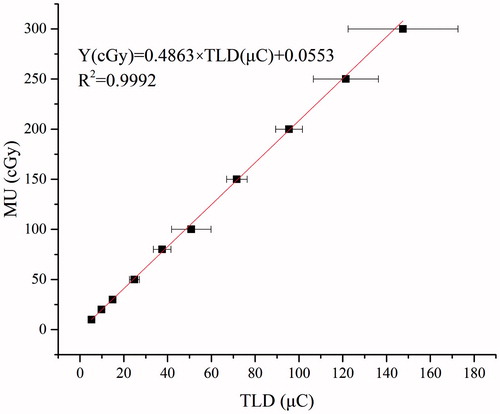
For calibration of X-ray dose and linearity, those TLDs were exposed with using 6 MV of Axesse linac at CSMUH. TLD measurements were performed five times at random locations. shows that the response for TLD-100H to a 6 MV photon showed a linear response in the range studies. Conversion factor was Y(cGy) = 0.4863 × TLD (μ C) + 0.0553. The R2, square of the correlation coefficient, is 0.9992.
Figure 4. TLD-100H response at different calibration doses of 6 MV Axesse linac.
3.2. Environment radiations among DRO
The range of color represented is from 350 mSv/treatment (red) to 7.3 mSv/treatment (grass green) at (0, 1.9) and (−2.5, 2.5), representing the intensity of environment radiations from severely strong to light. Environment radiations at (0, 0.7) is 75 mSv/treatment, Rando gonad, and was approximately 21.4% of that at the hottest A (tumor center). Environment radiations at (0, 1.9) was approximately 2.08% of that at the hottest tumor center. Average environment radiations of relatively and major significant variations varied from 369 (tumor center) to 7.3 mSv/treatment. lists the environment radiations corresponding to the data set of 75 measured points at DRO.
Table 1. Environment radiations among the vault room for DRO of CSMUH.
states the red zones those are the hottest radiations. The other colors are smoothed radiations of different radiations. This is nearly twice the MDD, so a measuring period during one NPC treatment was considered sufficient to achieve reasonably precise measurements. High-energy X-rays are emitted strongly forward coming from the Axesse target herein. Maintenance workers and medical staff could not enter the maze and vault room when the linac is in operation.
Figure 5. Averaged environment radiations of three-dimensional distributions of vault rooms, mapped via colored profiles which can reflect various environment radiation levels during the 6 MV NPC therapy of Axesse linac at CSMUH.
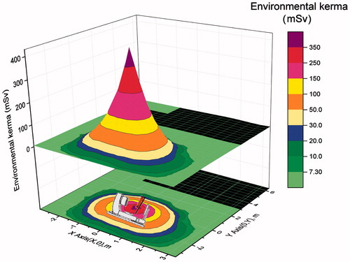
The environment radiation rates detected were very similar to background radiations in the control room, 0.14 ± 0.03 mSv/mo (14 m away from tumor center) and waiting room, 0.16 ± 0.03 mSv/mo (16 m away from tumor center) (cf. ). These values are in good agreement and in the range of 0.07–0.15 μSv/h using TLD-100 techniques, because of the low-background environment radiations in Western Sweden, in Taichung ROC, on Spratly Island in the South China Sea, and in Argentina, Brazil, Spain. The background radiations are higher than that measured in South India.[Citation4,Citation6] Therefore, successfully, the environment radiations generated from the Axesse that operated at 6 MV could be evaluated.
3.3. Minimum detectable dose (MDD) of the TLD technique
Many measurements of environment radiations are uncertain because the resulting data are under the MDD. EquationEquation (1)(1) lists net counts (Δnt) of MDD in the assessment of environment radiations that have significant meaning.
(1)
Where BG is background counts; kβ, standard deviations with an area β, to the tail left; kα, standard deviations of the net count rate that gains a one-tailed area of α.
Accordingly, Δnt that meets the MDD increases with environment radiations. shows the probability density Pn(rn) for BG and gross count rates of environment radiations correlated with BG and Δnt in identifying the glow curve from the Harshaw 3500 reader. Notably, as presented in , α and β in the one-tailed area are different regions.[Citation9] The kβ value represents standard deviations that leave an area β to its left. Either α or β is 0.050 to gain a confidence level of 95% during the statistical result. Furthermore, kα and kβ are equal to 1.645 herein.[Citation9] The MDD of TLD-100H was 4.4 mSv/treatment.
Figure 6. The probability density Pn(rn) for background (BG) and gross count of environment radiations.
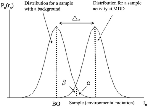
Most BG is associated with fluctuations of reader noise in the detected environmental γ-ray background. In total, 167 of the 225 data determined at the Axesse vault room of DRO failed the 95% confidence criterion to identify environment radiations using the TLD-100H approach. The majority of environment radiations values measured at the vault room reveal that negligible photons actually survive from the vault room and expand to its outer edge.
4. Conclusion
This is the first attempt to evaluate secondary radiations spread into the vault room using TLD-100H. Three-dimensional distributions of measured environment radiations were mapped and indicated that this secondary radiations reveals dramatic variations among tumor center’s position, and that the environment radiations is highest up to 370 mSv/treatment. Organs closer to the tumor center will receive highly gentle environment radiations. The calculated secondary radiations clearly show that environment radiations decreased rapidly with increasing distance. Good linearity was exhibited between each TLD. TLD-100H method states high sensitivity and stability. According to 167 measured data, the confidence level and the reported values of environment radiations reached 95%. These results could provide practical guidance with environmental protections.
Acknowledgments
The authors would like to thank the radiotherapists of CSMUH for their efficient supports.
Disclosure statement
The authors report no conflicts of interest. The authors alone are responsible for the content and writing of this article.
Funding
Financial assistances were partly supported by the CSMU under Contract no. CSH-2016-A-014.
References
- Zhu JJ, Shan JJ, Sun LB, et al. Study of the radiotherapy sensitization effects and mechanism of capecitabine (Xeloda) against non-small-cell lung cancer cell line A549. Genet Mol Res. 2015;14:16386–16391.
- Zhai XJ, Cheng HR, Long HL, et al. Effects of docetaxel plus three-dimensional conformal radiation therapy on microvessel density and apoptosis expression in local advanced squamous non-small-cell lung cancer. Genet Mol Res. 2015;14:5399–5406.
- Tseng HC, Liu WS, Tsai HH, et al. Radiation dose for normal organs by helical tomotherapy for lung cancer. Appl Radiat Isot. 2015;102:35–41.
- Changlai SP, Tsai HH, Tsai SC, et al. Environmental radiation detected at Lin Shin hospital in Taichung during the Fukushima nuclear power plant accident. J Radioanal Nucl Chem. 2012;291:859–863.
- International Commission on Radiological Protection, Recommendation of the ICRP, ICRP Publication 60, Annals of the ICRP, Vol. 21, No. 1–3, Oxford: Pergamon Press; 1991.
- Chung C, Chen CY, Wei YY, et al. Monitoring of environmental radiation on the spratly Island in the South China Sea. J Radioanal Nucl Chem. 1995;194:291–296.
- Tsai SY, Chen CY, Chen JC. Evaluating effective dose for indigenous phantoms with different weights undergoing computed tomography examination for coronary artery calcium. IEEE Trans Nucl Sci. 2013; 60:2147–2154.
- Chu KH, Lin YT, Hsu CC, et al. Evaluation of effective dose for a patient under Ga-67 nuclear examination using TLD, water phantom and a simplified model. J Radiat Res. 2012;53:989–998.
- Turner JE. Atoms, radiation, and radiation protection. 3rd ed. New York (NY): John Wiley & Sons, Inc.; 2007. p. 315–318.