
Abstract
OCCUPATIONAL APPLICATIONS When arm supports are provided during endodontic work, the use of the arm supports reduces static and median neck/shoulder muscle activity but does not seem to affect peak muscle activity. In addition, providing arm supports reduces the time working with unsupported upper arm angles <60°, but does not affect the time working unsupported with upper arm angles >60°. These results illustrate that in dentistry work in general, and in endodontic work specifically, dentists should be encouraged not only to optimize but also use their work environment to effectively reduce their workload.
TECHNICAL ABSTRACT Background: Endodontic dentists treat dental nerves and tissues by performing precision tasks using a microscope. Their work is associated with a high prevalence of musculoskeletal symptoms. Purpose: This study was conducted to investigate the effect of the availability and use of arm supports on neck and shoulder load during endodontic work. Methods: A group of 11 endodontists performed an endodontic task under three arm support conditions: (1) using flexible arm supports; (2) using fixed arm supports; and (3) without arm supports. Surface electromyography, posture, and self-reported discomfort in the neck and shoulders were assessed as outcome measures. Results: Use of arm supports reduced static and median neck/shoulder muscle activity, but did not affect peak muscle activity. The presence of arm supports reduced the relative time working unsupported with upper arm angles <60°, but did not affect the time working unsupported with upper arm angles >60° or self-reported discomfort. Conclusions: When arm supports are provided during endodontic work, their use decreases neck/shoulder muscle activity and reduces the time working with non-neutral postures of the upper extremities. However, the clinical relevance of these findings needs to be evaluated in longitudinal field studies.
KEYWORDS:
INTRODUCTION
Dental work is associated with a high prevalence of upper extremity musculoskeletal disorders (UEMD) such as regional neck and shoulder pain, shoulder tendonitis, neuropathy, tension neck syndrome, and trapezius myalgia (Morse, Bruneau, & Dussetschleger, Citation2010). These disorders are usually associated with high levels of discomfort and disability that limit functional daily activities, which indicates a high level of severity. The prevalence of UEMDs among dentists ranges between 17%–73% for neck symptoms and 20%–65% for shoulder symptoms (Morse et al., Citation2010). In endodontic work, a specialized dentist diagnoses, treats, and examines the dental nerve and dental tissue. To optimally visualize the oral cavity, 90% of the endodontists in the United States (Creasy, Mines, & Sweet, Citation2009) use a microscope during their work, and endodontists in the Netherlands indicate working with a microscope in 95% of their cases (Shemesh, Nuni, Duijst, Schermerhorn, & Wesselink, Citation2017). The microscope (see ) can usually be adjusted such that the endodontist can sit upright. It has a focusing range of 200–415 mm, which can be continuously adapted to the requirements of the task. Note that a root canal treatment, which is a common endodontic procedure using a microscope, usually takes over 1 hour. In addition, and probably as a consequence, back, neck, and shoulder symptoms were reported by 68% of a population of Dutch endodontists and 22% of the endodontists had to miss work days due to their musculoskeletal symptoms (Shemesh et al., Citation2017).
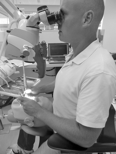
FIGURE 1 An endodontist working in a dental unit on a phantom head using a microscope.
Endodontic work involves exposure to several risk factors for UEMDs. Using the microscope, precision tasks are performed, involving a high degree of visual and manipulative elements, sometimes in combination with the exertion of high forces and psychosocial stress (Droeze & Jonsson, Citation2005; Finsen, Christensen, & Bakke, Citation1998; Morse et al., Citation2010). High precision demands in work tasks have been suggested to be a risk factor for UEMDs (Milerad & Ericson, Citation1994; B. Visser et al., Citation2003), although this has not been sufficiently confirmed in high-quality longitudinal studies (Huysmans, Citation2008). However, experimental studies show that an increase in precision demands often is associated with an increase in muscle activity (Van Galen, Muller, Meulenbroek, & Van Gemmert, Citation2002; van Galen & van Huygevoort, Citation2000; Van Gemmert & Van Galen, Citation1997). Besides precision demands, endodontic work involves repetitive shoulder and hand movements (Alexopoulos, Stathi, & Charizani, Citation2004), the exertion of relatively high forces with the hands and fingers, and non-neutral working postures (Hayes, Cockrell, & Smith, Citation2009), all of which are risk factors for UEMDs (Mayer, Kraus, & Ochsmann, Citation2012; van der Windt et al., Citation2000; van Rijn, Huisstede, Koes, & Burdorf, Citation2009, Citation2010).
The poor working posture of endodontists, and dentists more generally, is often the result of the workplace organization and an improper positioning of both the patient and dental worker (Hayes et al., Citation2009; Morse et al., Citation2010). For dental work, the working posture is commonly a trade-off between the neutral posture, in which the physical activities can be optimally performed, and the posture needed to have optimal vision into the oral cavity. For endodontic work, finding the optimal working posture can be even more difficult because of the use and the design of the microscope. To reduce the physical workload, adjustments should be made in the workplace and work environment to be able to optimize the working posture. Multiple ergonomic aids are designed to improve the working posture, though the effectiveness is often not scientifically evaluated.
Based on studies in related professions, such as general dentistry and microscope work (Kofler, Kreczy, & Gschwendtner, Citation2002), it can be expected that in endodontic work the activity of neck and shoulder muscles may decrease with the use of arm supports. Visser et al. (Citation2000) studied the effect of using arm supports in visual display unit work on physical workload. They reported significantly lower levels of upper trapezius muscle activation when typing with the use of arm supports was compared to typing with no arm supports. When optimally adjusted and used, arm supports were able to bear a part of the weight of the arm. In particular, arm supports with larger shells, larger arms, and better hinges appeared to be superior in reducing the activity of the trapezius muscle. Arm supports might, therefore, be an effective preventive measure in endodontic work to reduce symptoms in the neck and shoulders. The objective of the present study was thus, to investigate the effects of performing a simulated endodontic task using a microscope, both with and without the availability of arm supports, on muscle activity, posture, and discomfort.
METHODS
Participants
A group of 11 endodontists and postgraduate endodontology students participated in the study (8 and 3, respectively). Seven of the participants were female and four were male. Participants had mean (standard deviation [SD]) age of 40.2 (6.4) years, stature of 1.75 (0.07) m, and nine were right-handed. At the time of the experiment, the endodontists had worked 23.6 (4.9) hours per week in clinical practice. Three endodontists were used to working with arm supports in their practice. Participants had at least 2 years of experience in working with a microscope (mean = 9.6, SD = 5.8 years). A history of upper extremity symptoms was reported by four of the endodontists, though at the time of the experiment none of them worked fewer clinical hours as a consequence of the symptoms. All participants gave informed consent prior to the experiment and the study was approved by the local ethical committee.
Tasks and Procedures
The Academic Centre for Dentistry Amsterdam (ACTA) provided a dental unit for measurements in the endodontic department. The dental unit consisted of a standard set of dental furniture with a patient chair, a dental lamp, and one or more displays for instruments. Before commencement of the experimental conditions, the dentist's stool and arm supports, patient chair, instrument tray holder, and microscope were adjusted to the dentists' neutral work posture according to ISO 11226. The dentist stool (Ghopec II, JPG Ergonomics, Utrecht, The Netherlands; [De Bruyne et al., Citation2016]) was chosen because of its (vertical and horizontal) adjustable lumbar support and the adjustable inclination angle of the seat, which provides a minimal horizontal distance between the trunk and the work area.
FIGURE 2 (A) The dentist stool that was used in the present study. (B) The fixed arm supports on another dentist stool (only the arm supports were used on the dentist stool shown in part A). (C) The flexible arm supports that were also fixed to the dentist stool shown in part A.
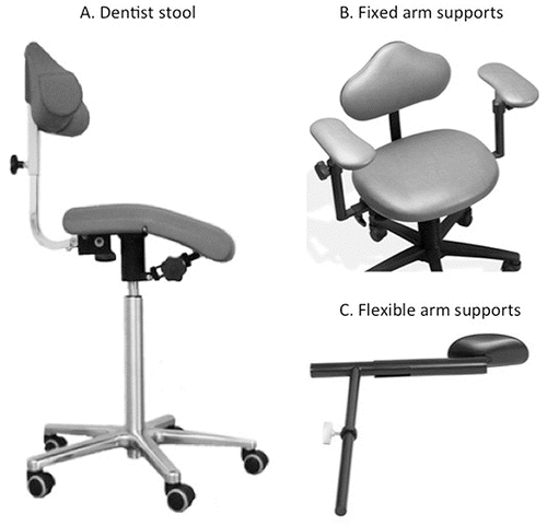
Two types of arm supports that are commonly used in practice were tested in the present study, and these were fixed to the Ghopec stool: (1) fixed arm supports (which are usually fixed to the dental microsurgeon stool of Global Surgical Corporation, St. Louis, MO, USA.; ); and (2) flexible arm supports (arm rest BQE 02, Back Quality Ergonomics, Den Haag, The Netherlands; ). The fixed arm supports had a soft padding and could be adjusted in height, which was done once for each participant before the start of the measurements. The flexible arm supports, which consisted of a hard plastic shell, could move in the horizontal (forward and backward with respect to the endodontist) and vertical (up and down) directions during the procedure and thus, could follow the movements of the arm and provide support for the arm of the endodontist. The microscope used (OPMI ProErgo, Zeiss, Jena, Germany) had multiple functions to adjust the microscope to a neutral work posture.
To standardize the experimental task, a phantom head was affixed to the patient chair, with an imitation tooth (Frasaco, Germany) placed in the jaw. During the task, participants made use of a common set of endodontic instruments. The specific task required participants to make an endodontic access in element 1.1 (dental notation for the large upper-right front tooth), and file with hand files (starting with file 25) toward a master apical file 45, with a step back toward file 60. This task is very common in the work of endodontists, and simulated the treatment necessary when a large cavity damages the root of the tooth. Endodontists treat this by electrically drilling into the tooth and root followed by electrically filing the root. Subsequently the root is filed by hand. The task was performed in a front upper tooth, which usually takes in practice about 5 to 10 minutes depending on the complexity of the tooth and the severity of the inflammation. Performing this task required the participant to exert force with the dominant hand while using an electrical dental drill or file. The part of the task where the participant files by (dominant) hand is a fine motor task, which requires force exertion with the thumb and index finger. The other arm/hand is used for stabilization. All participants were familiar with the endodontic task.
The three arm support conditions (flexible, fixed, or none) were presented to the participants in a random order, and each condition was performed for 5 minutes. The researcher explained how to use the arm supports during the simulated work task, and data collection only started after the participants indicated that they felt comfortable working with the specific arm support after some practice. Before and after each task, the endodontists rated their local perceived discomfort of their upper body. Between each arm support condition the participants had to rest for at least 2 minutes.
Data Acquisition
Muscle Activity
In preparation for an electromyography (EMG) assessment, the skin of the participants at the electrode locations was shaved, cleaned with alcohol, and surface electrodes were attached. The activity of four muscles was monitored bilaterally: upper trapezius, middle trapezius, anterior deltoid, and lateral deltoid. Bipolar disposable electrodes (Blue Sensor Electrodes, USA) were used, and electrode locations were according to Franssen (Franssen, Citation1995). The reference electrode was placed on the C7 spinous process. EMG signals were band-pass pre-filtered (10–400 Hz, second order) and AD-converted at a sample rate of 1000 Hz (22 bits) using a portable electrophysiological amplifier system (Porti-17™, Twente Medical Systems International BV, The Netherlands, input resistance >1012 Ω, CMRR >90 dB) and stored on a computer.
Posture Observation
A portable observation system, task recording and analysis on computer (TRAC; Frings-Dresen & Kuijer, Citation1995) was used to assess upper arm angle, the use of arm support, and neck angle; assessment were done every 15 seconds over the 5-min duration of each arm support condition. This and comparable observation systems have been shown to be a reliable and valid method for the direct and indirect assessment of tasks, activities, and postures at the workplace (Kilbom, Citation1994; Takala et al., Citation2010; van der Beek & Frings-Dresen, Citation1998; van der Beek, van Gaalen, & Frings-Dresen, Citation1992). Direct observations were performed in real-time by one observer at the dental unit (i.e., not using video recordings) while the participant was working. Preceding these observations, the observer was trained to improve intra-observer reliability. During a week of training, the percentage of agreement and the Cohen's kappa (within the observer) were assessed for all the variables. At the end of the training period, it was ensured that all variables had a percentage of agreement of at least 80% and a Cohen's kappa of at least 0.50, which is acknowledged to be an acceptable standard of observer reliability (van der Beek et al., Citation1992).
Upper arm angle was defined as the angle between the vertical (in line with gravity) and the upper arm. Three categories, based on ergonomic guidelines (Hokwerda, Wouters, de Ruijter, & Zijlstra-Shaw, Citation2006; Peereboom & de Langen, Citation2008), were determined for the upper arm angle: “<20°,” “20–60°,” and “>60°.” The relative frequency of the use of arm supports was assessed using the following four categories: “use arm rest left,” “use arm rest right,” “both arm rests used,” “no arm rests used.” The neck angle was defined as flexion high in the neck, in the sagittal plane with respect to the vertical axis. Three categories were defined: “<0°,” “0–25°,” and “>25°.”
Local Perceived Discomfort
After completing each arm support condition, participants rated their perceived pain, symptoms, or discomfort for eight different areas (head, neck, left and right shoulder, left and right upper arm, left and right lower arm) using a standardized picture of an upper body. For each area, participants graded their perceived discomfort using a slightly modified Borg CR-10 scale (Borg, Citation1990). With respect to the original CR-10 scale, the anchored verbal expressions were not changed—but translated to Dutch—and ranged between 0 (nothing at all) and 10 (extremely strong). However, the CR-10 was not used to assess perceived exertion but perceived discomfort.
Data Analysis
Electromyographic signals of the eight muscles were full-wave rectified and low-pass filtered at 2 Hz (second order Butterworth) using MATLAB (The MathWorks, Inc.) to smooth the signal. For each arm support condition, and for each of the bilateral muscles, the 10th, 50th, and 90th percentiles (i.e., P10, P50, and P90) of the amplitude probability distribution function (APDF) were determined as indicators of static, median, and peak levels of muscle activity, respectively (Hagg, Luttmann, & Jager, Citation2000; Jonsson, Citation1978, Citation1982).
Percentages of the total task time in each category were calculated for upper arm angle, separated for the 15-second observations when arm support was or was not used (the three categories of arm support were combined to two categories). For neck angle, similar percentages were determined, but independent of whether the available arm supports were used at the 15-second observations.
For each upper body region and for each arm support condition the prevalence of discomfort (value > 0) was determined. Except for one participant, discomfort was reported only in the neck and the left shoulder. Therefore, only these two regions were used for further analysis, for which the median and range of reported discomfort (values 0–10) were determined for each arm support condition.
Statistical Analysis
To identify the effect of arm support on neck and shoulder muscle activity, the three percentiles (P10, P50, and P90) were compared between the three arm support conditions. The data were first log-transformed to meet parametric assumptions, then one-way, repeated measures analyses of variance (ANOVAs) were performed separately for each of the three outcome measures (percentiles) and for each muscle. Bonferroni corrections were used for post-hoc testing. In all statistical analyses, a p-value <0.05 was considered significant, and analyses were performed using SPSS (v 20.0, IBM).
Three analyses were performed to address whether the three arm support conditions affected upper arm and neck angles. In the first analysis, the effect of the flexible versus fixed arm supports on upper arm angle was tested, while using the arm supports. As the datasets were not normally distributed, non-parametric Wilcoxon signed-rank tests were used. In the second analysis, the effect of the three arm support conditions on upper arm angle was testing, while not using the arm supports. For this second analysis, non-parametric Friedman ANOVA were used and post-hoc pairwise comparisons were performed using Wilcoxon signed-rank tests. To avoid family-wise error in post-hoc testing, a p-value of <0.017 (=0.05/3) was considered significant in pairwise comparisons. In the third analysis, using non-parametric Friedman ANOVAs, the percentage of time working at the three neck angle categories were compared between the arm support conditions, independent of whether the available arm support was actually used or not.
Data concerning local perceived discomfort appeared not to be normally distributed. Therefore, non-parametric Friedman ANOVAs were used to test whether local perceived discomfort at the neck and left shoulder was affected by arm support condition.
RESULTS
Muscle Activity
As an example of the results concerning muscle activity, shows the distributions of activity levels in the middle trapezius muscle. A significant effect of arm support condition was found on the activity of the right upper trapezius muscle, right middle trapezius, left and right anterior deltoid, and right lateral deltoid (). Pairwise comparisons showed that the right middle trapezius P10 activity was reduced by 33% when flexible arm supports were provided compared to no arm supports. Right anterior deltoid activity was significantly lower with the fixed arm supports compared to working without arm supports, with a 68% reduction of P10.
FIGURE 3 Distributions of the absolute amplitudes of EMG of the middle trapezius muscle (left and right side) for the P10, P50, and P90 of the APDF.
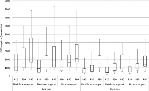
TABLE 1 Results of the effect of arm support conditions on the P10, P50, and P90 of the log-transformed muscle activity of eight muscles.
Arm support significantly influenced median (P50) activity in the right middle trapezius, right anterior deltoid, and left anterior deltoid. Pairwise comparisons showed that the P50 was lower (23%) only for the right middle trapezius muscle when working with the flexible arm supports compared to no arm supports. Arm support condition did not significantly affect peak levels of activity (P90) for any of the eight muscles monitored.
Posture Observations
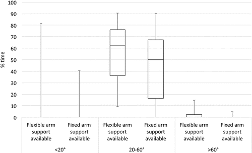
In the first analysis, the effect of the two arm support conditions on the upper arm angle when using arm supports () was tested. When using arm supports, participants worked with an upper arm angle of 20°–60° about 50% of the total task time. Participants had an upper arm angle of <20° or >60°during a small portion (<10%) of the total task time. There were no significant differences between the use of the flexible arm support and the use of the fixed arm supports in any of the upper arm angle categories when using the arm supports (with p = 0.285, p = 0.182, and p = 0.109 for the <20°, 20°–60°, and >60° categories, respectively).
FIGURE 4 Distributions of the percentages of time for upper arm angle when using the two arm support conditions (flexible arm supports, fixed arm supports), when participants were using arm support.
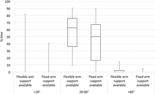
The second analysis was performed to test the effect of arm support conditions on the upper arm angle when the arm supports were not used (). When arm supports were provided, but not used, about 1% of the total task time was spent in upper arm angle category <20°, while this was about 10% of the total task time when there were no arm supports provided (p = 0.012). There was a significant difference in upper arm angle category <20° between flexible arm supports and no arm supports (p = 0.014). In the upper arm angle category 20°–60°, there was a significant effect of arm support condition on percentage of total time spent within this category (p = 0.014) when arm supports are not used. When arm supports were provided but not used, 14%–30% of the total task time the arm angle was between 20 and 60 degrees, compared to almost 60% in the no arm support condition. Using a conservative approach, though, pairwise comparisons did not show differences to be significant. In the upper arm angle category >60°, no significant effect of arm support condition was found (p = 0.424). Participants in all arm support conditions had an upper arm angle of >60° for approximately 20% of the total task time when not using arm supports.
FIGURE 5 Distributions of the percentages of time for upper arm angle in each of the three arm support conditions (flexible arm supports, fixed arm supports, no arm supports), when participants were not using arm support.
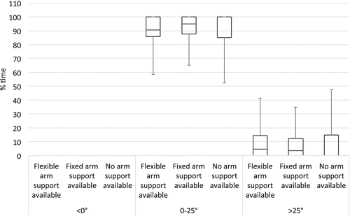
No significant effects of arm support condition were found on neck flexion, with p = 0.867 for both the neck flexion categories of 0°–25° and >25°. Neck angles were between 0°–25° for approximately 90% of the time in all arm support conditions (). Neck extension (neck flexion <0°) was not observed.
FIGURE 6 Distributions of the percentages of time for neck angle in the three arm support conditions (flexible arm supports, fixed arm supports, no arm supports).
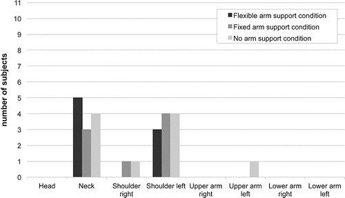
Regarding localized discomfort, only one participant reported discomfort in areas other than the neck or the left shoulder (). No significant effects of arm support condition were found on discomfort in either the neck (p = 0.368) or left shoulder (p = 0.223). The intensity of the reported discomfort ranged from 0.5 to 3.0.
FIGURE 7 The number of participants reporting discomfort for each of the eight body regions and three arm support conditions (flexible arm supports, fixed arm supports, no arm supports).
DISCUSSION
The present study was designed to investigate the effect of arm supports on the activity of muscles of the neck and shoulder region in endodontic dentistry using a microscope. Use of arm supports decreased the static and median activity of several muscles, mostly on the right side of the upper body. Posture observations showed that use of arm support was effective in decreasing the time without support in the upper arm categories <20° and 20°–60°, although there was not a clear indication that one type of arm support was preferable. In the upper arm angle category >60°, there was no effect of the presence of arm support on the relative time that the arm was not supported. Self-reported discomfort was reported at the neck and left shoulder, and this remained unchanged when working with arm supports.
Muscle Activity
When working with arm supports, there was a decrease in static (P10) and median (P50) activity of the right trapezius muscle. The trapezius muscle has frequently been the object of investigation when evaluating musculoskeletal workload in many occupations and has pathophysiological importance for many professions (Huysmans, Citation2008). Therefore, decreasing the trapezius load during work such as performed by endodontists may help prevent the development of musculoskeletal disorders. More distinct results regarding the influence of arm supports on workload were found in other intervention studies. In general dentistry, Visser (Citation1999) studied the influence of arm supports on muscle activity around the neck and wrists, found a reduction in trapezius muscle activity while working with arm supports, and concluded that moveable arm rests were most effective in decreasing the trapezius muscle load. Among visual display workers, Visser et al. (Citation2000) also found lower trapezius muscle activity when arm supports were used during computer work. Results of these studies in related professions are consistent with the outcome of the present study, in that a reduction in trapezius muscle activity could be achieved by the use of arm supports.
While the presence of arm support was found to affect static (P10) and median (P50) muscle activity here, peak activity (P90) did not decrease when arm supports were provided. An explanation for this may be that during activities in which near maximum muscle activity is required, specifically when substantial force needs to be exerted with the arm or hand, the arms are most of the time not in a position where they can be supported by arm supports. It is likely that during endodontic work that requires a high level of force to be exerted by the hands and fingers, the arm is lifted off the arm support to be able to exert force with a relatively neutral wrist posture and to use the upper body. If the exertion of high forces coincides with larger upper arm angles and not using arm supports, this might also explain our finding that the presence of arm support did not affect the time spent working in the arm angle category >60°.
Posture Observations
Posture observation results showed that the presence of arm support in the upper arm categories <20° and 20°–60° was an effective measure in decreasing the time spent without support. In other words, the arm supports were used when provided, and this likely explains the reduction in static and median muscle activity found in the present study. Ergonomic guidelines indicate that unsupported upper arm angles of 20°–60° and >60° should be avoided (Peereboom & de Langen, Citation2008). The present study shows that if arm supports are provided, the relative time that endodontic work required upper arm between 20° and 60° can be reduced from 60% to less than 30%. This reduction is likely to be the cause of the observed reduction in muscle activity, which indicates that the use of arm supports can reduce the load on the neck and shoulder region because of their weight bearing function.
As stated previously, the presence of arm supports did not affect the time spent working in the arm angle category >60°. Besides the earlier explanation (that for the exertion of high forces with hands and fingers endodontists may not want their arm to be supported), it is also difficult to maintain a neutral working posture while working with the microscope, because with upper arm angles >60° the arm supports would be located too low. Although this might be technically solved by introducing arm supports that are also able to move up and down, the effectiveness of such an approach can be questioned when endodontists do not use arm support when exerting high forces.
Self-Reported Discomfort
We observed no significant effect of arm support conditions on neck or left shoulder discomfort. This is consistent with the previously mentioned study of Visser (Citation1999), though dentists preferred working with arm supports. An explanation could be the short adaptation period provided for the use of arm supports, since in both studies the arm supports were only used during the experiment. A longer adaptation period could result in a reduced discomfort of the neck region.
Limitations and Strengths
The endodontic task (i.e., preparation of element 1.1) was chosen based on the advice of dental professionals. This task is a common procedure in the endodontic practice, as described by Creasy et al. (Citation2009). Among American endodontists, 96.7% performed endodontic surgery in the anterior region and, more specifically, 91.8% on the anterior teeth. Although the task was thus, considered representative for endodontic work, a more backward positioned element in the mouth could have taken more effort to treat (Creasy et al., Citation2009). This more complicated and time-consuming procedure could have resulted in a more pronounced effect in terms of the effectiveness of arm supports on neck and shoulder load.
A comparison of the three experimental conditions using a standardized endodontic task increased the internal validity of the study; however, the endodontic task had a duration of only 5 minutes. It can thus be questioned whether the results of this study can be extrapolated to endodontic work (i.e., performing endodontic work for a whole working day). This would especially be true with respect to the assessment of discomfort. For the 5-minute duration used here, discomfort levels up to 3 (on a 0–10 point scale) were reported by nearly half (5 out of 11) of the participants. However, the experimental design makes it impossible to make inferences about the possible differences between experimental conditions in discomfort over the course of a day. In addition, it might be that differences in perceived discomfort between working using arm support and working without arm support are attenuated by work rate or performance quality. Participants might have reduced their working rate to reduce the development of discomfort. Neither work rate nor quality of the procedure were, however, quantified during the experiment.
A recommendation of Visser (Citation1999) was to include dentists with experience with the use of arm supports. The limited number of endodontists and low prevalence of using arm supports in the endodontic practice constrained us from including a sufficient number of participants. To compensate partially for this, we adjusted the equipment and gave instructions to the participants to adopt a neutral work posture. Participants were instructed to use the arm supports as they preferred. Three of the 11 participants were experienced in working with arm supports. The positive effect of arm support use could thus, again be underestimated. More specific instructions about optimal use of equipment should be given in future research. Four of the 11 participants reported having upper extremity symptoms in the past but were free of symptoms at the time of the experiment. It is, though, difficult to predict how this would have affected the results of the present study. Given their experience, these four participants might be more aware of their working posture and might have trained an optimal working posture. This might have attenuated the potential effect of working with arm supports.
The strength of this study was the combination of outcome measures used. A combination of subjective ratings, posture observation, and assessment of muscle activity presents an overall assessment of the influence of arm supports on the workload. In addition, the measurements were assessed in a controlled situation, though still in a recognizable dental setting for the endodontists, in which they were able and encouraged to work as usual.
Practical Implications
Thornton et al. (Citation2008) found a significant relationship between the occurrence of musculoskeletal symptoms and three work-related variables in dentistry: equipment utilization, work efficiency, and general health. This illustrates that the problem of musculoskeletal complaints in dentistry is multi-factorial. Improving one factor, equipment utilization, could contribute to relieving these problems. Still, there are other factors that should also be improved. For instance, besides optimizing the work environment, it is equally important to train the endodontists to work in a neutral work posture and to use their work environment to facilitate this posture during work.
With the development of materials and techniques, for example working with a microscope and the four-handed dentistry technique, endodontic work becomes more efficient and static. Arm posture improved with arm supports here, but it is not clear to what level. Additional assessments of muscle activity, in particular using normalized EMG levels, would have resulted in information concerning the relative static, median, and peak muscle activity levels during the task. Although not normalizing EMG does not affect the conclusions of the present study concerning the comparison of working with and without the availability of arm supports, normalized EMG could have been used to ergonomically evaluate the endodontic task. In addition, it could be explored whether using arm support affects the amount of variation in arm posture in endodontic work over a working day. Hence, in future research, it would be interesting to investigate the dynamic characteristics of upper arm movements in endodontic work during a total workday. Assuming that variation in work postures and decreased static muscle activity reduces the risk of symptoms (Huysmans, Citation2008), full-shift observations of endodontic work could provide new insights regarding how to reduce the risk of musculoskeletal symptoms in endodontics and also in preserving efficiency.
CONCLUSIONS
When arm supports are provided during endodontic work, the present study indicates that the use of the arm supports decreases the static and median activity of neck/shoulder muscles, but does not affect peak muscle activity. In addition, the presence of arm support reduces the relative time working with upper arm angles <60° without support, but does not affect the time working unsupported with upper arm angles >60° or self-reported discomfort. However, the clinical relevance of these findings has to be evaluated using longitudinal field studies. Furthermore, while the presence of arm supports during endodontic work appears to positively affects the level of workload, it is not clear whether it affects the amount of variation in posture in endodontic work. Investigating the frequency and duration characteristics of the workload of endodontists, in addition to the level of workload, might give additional information on whether arm supports can be used as effective preventive ergonomic measures in endodontic work.
REFERENCES
- Alexopoulos, E. C., Stathi, I. C., & Charizani, F. (2004). Prevalence of musculoskeletal disorders in dentists. BMC Musculoskeletal Disorders, 5, 16. https://doi.org/10.1186/1471-2474-5-16
- Borg, G. (1990). Psychophysical scaling with applications in physical work and the perception of exertion. Scandinavian Journal of Work & Environmental Health, 16(Suppl 1), 55–58.
- Creasy, J. E., Mines, P., & Sweet, M. (2009). Surgical trends among endodontists: The results of a web-based survey. Journal of Endodontics, 35(1), 30–34. https://doi.org/10.1016/j.joen.2008.10.008
- De Bruyne, M. A., Van Renterghem, B., Baird, A., Palmans, T., Danneels, L., & Dolphens, M. (2016). Influence of different stool types on muscle activity and lumbar posture among dentists during a simulated dental screening task. Applied Ergonomics, 56, 220–226. https://doi.org/10.1016/j.apergo.2016.02.014
- Droeze, E. H., & Jonsson, H. (2005). Evaluation of ergonomic interventions to reduce musculoskeletal disorders of dentists in the Netherlands. Work, 25(3), 211–220.
- Finsen, L., Christensen, H., & Bakke, M. (1998). Musculoskeletal disorders among dentists and variation in dental work. Applied Ergonomics, 29(2), 119–125.
- Franssen, J. L. M. (1995). Handboek oppervlakte-elektromyografie [Manual for surface electromyography]. Utrecht: De Tijdstroom.
- Frings-Dresen, M. H. W., & Kuijer, P. P. F. M. (1995). The TRAC-system: An observation method for analysing work demands at the workplace. Safety Science, 21, 163–165.
- Hagg, G. M., Luttmann, A., & Jager, M. (2000). Methodologies for evaluating electromyographic field data in ergonomics. Journal of Electromyography and Kinesiology, 10(5), 301–312.
- Hayes, M., Cockrell, D., & Smith, D. R. (2009). A systematic review of musculoskeletal disorders among dental professionals. International Journal of Dental Hygiene, 7(3), 159–165. https://doi.org/10.1111/j.1601-5037.2009.00395.x
- Hokwerda, O., Wouters, J. A. J., de Ruijter, R. A. G., & Zijlstra-Shaw, S. (2006). Ergonomic requirements for dental equipment. Guidelines and recommendations for designing, constructing and selecting dental equipment. Groningen, The Netherlands: Academisch Centrum Mondzorg Groningen.
- Huysmans, M. A. (2008). From precision demands to neck and upper extremity pain (Doctoral disseration). Amsterdam: VU University Amsterdam.
- Jonsson, B. (1978). Quantitative electromyographic evaluation of muscular load during work. Scandinavian Journal of Rehabilitation Medicine Supplement, 6, 69–74.
- Jonsson, B. (1982). Measurement and evaluation of local muscular strain in the shoulder during constrained work. Journal of Human Ergology (Tokyo), 11(1), 73–88.
- Kilbom, A. (1994). Assessment of physical exposure in relation to work-related musculoskeletal disorders-what information can be obtained from systematic observations? [Review]. Scandinavian Journal of Work & Environmental Health, 20 Spec No, 30–45.
- Kofler, M., Kreczy, A., & Gschwendtner, A. (2002). “Occupational backache”—surface electromyography demonstrates the advantage of an ergonomic versus a standard microscope workstation. European Journal of Applied Physiology, 86(6), 492–497. https://doi.org/10.1007/s00421-002-0576-6
- Mayer, J., Kraus, T., & Ochsmann, E. (2012). Longitudinal evidence for the association between work-related physical exposures and neck and/or shoulder complaints: A systematic review. International Archives of Occupational & Environmental Health, 85(6), 587–603. https://doi.org/10.1007/s00420-011-0701-0
- Milerad, E., & Ericson, M. O. (1994). Effects of precision and force demands, grip diameter, and arm support during manual work: An electromyography study. Ergonomics, 37(2), 255–264.
- Morse, T., Bruneau, H., & Dussetschleger, J. (2010). Musculoskeletal disorders of the neck and shoulder in the dental professions. Work, 35(4), 419–429. https://doi.org/10.3233/WOR-2010-0979
- Peereboom, K. J., & de Langen, N. C. H. (2008). Handboek fysieke belasting. Een complete methode voor het inventariseren en oplossen van knelpunten [Manual for physical workload. A complete method for assessing and solving bottlenecks]. Den Haag, The Netherlands: Sdu Uitgevers.
- Shemesh, H., Nuni, E., Duijst, E. T. M., Schermerhorn, W., & Wesselink, P. (2017). Prevalence of back and neck complaints among Dutch and Israeli endodontists. Manuscript submitted for publication.
- Takala, E. P., Pehkonen, I., Forsman, M., Hansson, G. A., Mathiassen, S. E., Neumann, W. P., & Winkel, J. (2010). Systematic evaluation of observational methods assessing biomechanical exposures at work. Scandinavian Journal of Work & Environmental Health, 36(1), 3–24.
- Thornton, L. J., Barr, A. E., Stuart-Buttle, C., Gaughan, J. P., Wilson, E. R., Jackson, A. D., & Smarkola, C. (2008). Perceived musculoskeletal symptoms among dental students in the clinic work environment. Ergonomics, 51(4), 573–586. https://doi.org/10.1080/00140130701728277
- van der Beek, A. J., & Frings-Dresen, M. H. (1998). Assessment of mechanical exposure in ergonomic epidemiology. Occupational and Environmental Medicine, 55(5), 291–299.
- van der Beek, A. J., van Gaalen, L. C., & Frings-Dresen, M. H. (1992). Working postures and activities of lorry drivers: A reliability study of on-site observation and recording on a pocket computer. Applied Ergonomics, 23(5), 331–336.
- van der Windt, D. A., Thomas, E., Pope, D. P., de Winter, A. F., Macfarlane, G. J., Bouter, L. M., & Silman, A. J. (2000). Occupational risk factors for shoulder pain: A systematic review. Occupational and Environmental Medicine, 57(7), 433–442.
- Van Galen, G. P., Muller, M. L., Meulenbroek, R. G., & Van Gemmert, A. W. (2002). Forearm EMG response activity during motor performance in individuals prone to increased stress reactivity. American Journal of Industrial Medicine, 41(5), 406–419. https://doi.org/10.1002/ajim.10051
- van Galen, G. P., & van Huygevoort, M. (2000). Error, stress and the role of neuromotor noise in space oriented behaviour. Biological Psychology, 51(2–3), 151–171.
- Van Gemmert, A. W., & Van Galen, G. P. (1997). Stress, neuromotor noise, and human performance: A theoretical perspective. Journal of Experimental Psychology: Human Perception and Performance, 23(5), 1299–1313.
- van Rijn, R. M., Huisstede, B. M., Koes, B. W., & Burdorf, A. (2009). Associations between work-related factors and specific disorders at the elbow: A systematic literature review. Rheumatology (Oxford), 48(5), 528–536. https://doi.org/10.1093/rheumatology/kep013
- van Rijn, R. M., Huisstede, B. M., Koes, B. W., & Burdorf, A. (2010). Associations between work-related factors and specific disorders of the shoulder-a systematic review of the literature. Scandinavian Journal of Work & Environmental Health, 36(3), 189–201.
- Visser, B., de Korte, E., van der Kraan, I., & Kuijer, P. (2000). The effect of arm and wrist supports on the load of the upper extremity during VDU work. Clinical Biomechanics (Bristol, Avon), 15(Suppl 1), S34–38.
- Visser, B., de Looze, M. P., Veeger, D. H., Douwes, M., Groenesteijn, L., de Korte, E., & van Dieen, J. H. (2003). The effects of precision demands during a low intensity pinching task on muscle activation and load sharing of the fingers. Journal of Electromyography and Kinesiology, 13(2), 149–157.
- Visser, M. H. (1999). Interventieonderzoek naar de effecten van armsteunen bij tandartsen. Eindproject postdoctorale beroepsopleiding ergonomie bij arbeid [Intervention study of the effect of using arm supports in dentistry. Thesis ergonomics postgraduate program]. Amsterdam, The Netherlands: VU University Amsterdam.