
Abstract
Reports of physical abnormalities in wild bird populations are rare and often anecdotal, whereas reliable estimates of their frequency are important to understand the strength of selection against them. In 2005–2022, we studied various aspects of breeding biology of Eurasian reed warblers and monitored a total of 1194 nests. In July 2022, we observed a deformed nestling of this species in a brood of three young. The nestling displayed a soft distended lump around the cervical area. After further inspection, we concluded that the nestling suffered from subcutaneous emphysema, which resulted in its death on the following day. The nestling body was collected and examined for the presence of injuries, parasitic nematodes and bacteria. We discuss possible factors that might have caused the nestling’s emphysema and conclude that Clostridium bacteria found in the nestling tissues were the most likely cause of its symptoms and death. This was the only case of subcutaneous emphysema recorded in our population among 1995 nestlings of the same age (8th day after hatching), making up 0.05% of all.
Introduction
Avian respiratory system is one of the most complex and unique organs among land vertebrates. It comprises a pair of lungs which perform a respiratory function, and a system of air sacs which support the mechanical aspect of ventilation and take no direct part in gas exchange (Maina Citation2008, Citation2022; Powell Citation2022). Branching into several interconnected sections, air sacs are avascular thinly walled structures prone to ruptures resulting in hyperinflation of an area under the skin known as subcutaneous emphysema (Lee et al. Citation2011; Maina Citation2022).
Subcutaneous emphysema results the most often from injury by a blunt or sharp object and is caused by air leaking from damaged air sacs (Gibbons & Horton Citation2000; Lee et al. Citation2011). However, it may also be induced by a number of etiologic causes including infections, malnutrition or parasitic infestation (Radan & Rautenstein-Arasi Citation1950; Literak et al. Citation2003; Lee et al. Citation2011; Gornatti-Churria et al. Citation2018). The condition is most commonly described in companion birds and poultry (Lee et al. Citation2011; Sudhakara et al. Citation2013; Gornatti-Churria et al. Citation2018), whereas reports from the wild are very scarse (Gochfeld Citation1974; Stewart et al. Citation2011). Here, we present a rare case of subcutaneous emphysema observed in a reed warbler nestling.
Materials and methods
In 2005–2022, we studied reed warblers in the Stawy Milickie (Milicz fish-ponds) nature reserve (SW Poland). Each year we monitored the 3 ha study plot (centre at 51°32’17.5“N 17°20’23.4“E) and tried to find all nests of the species there. The majority of nests (ca. 85%) were discovered at the building stage by observing nest-building and mate-guarding behaviour. During all study years, we found and monitored a total of 1194 reed warbler nests, of which 823 survived until hatching. Nests were visited usually every second day. Starting from 2006 we recorded parental effort during the incubation stage, and on the 8th day after hatching using a digital camera JVC Everio GZ330. Other details of the study area and research methods are presented elsewhere (Orłowski et al. Citation2016; Wierucka et al. Citation2016; Halupka et al. Citation2021).
In February 2023, the nestling body was dissected and examined for the presence of parasitic nematodes (before, we took tissue samples for microbiological analysis). The standard parasitological dissection was provided under stereoscopic microscope. Additionally, the respiratory system, as well as the body cavity, was flushed with physiological fluid and checked for the presence of helminths.
Subsequently, the nestling’s tissues were screened for the presence of Clostridium bacteria. Isolation and identification of C. difficile was carried out according to standard procedures (Pituch et al. Citation2002; Silva et al. Citation2013). The material was incubated in Shaedler Bulion liquid medium with Vitamin K3 and incubated for 24 h at 37°C. After 24 h of incubation, the material was taken from the liquid medium and inoculated reducibly into petri dishes with Shaedler Agar medium supplemented with 5% sheep blood, incubated under anaerobic conditions at 37°C (and at the same time at 28°C) for 48 h. Bacterial identifications were carried out using ANAII biochemical tests (Argenta) and chromID Clostridium chromogenic medium (Biomerieux). Reference C. difficile strains were used as positive controls.
Results
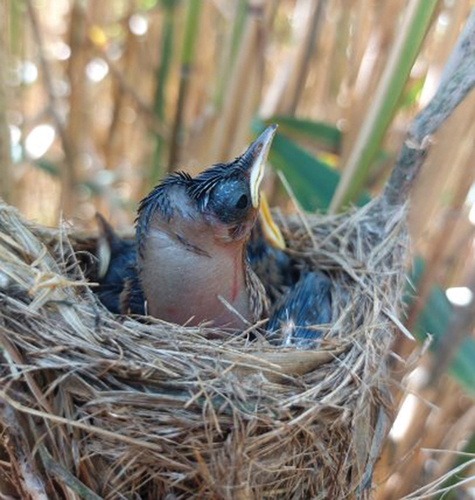
On 3 July 2022, at 11.40 one of us (LH) planned to set a camera near a nest (ID 48) to record feeding intensity of parental birds. The nest contained three nestlings at the age of 8 days. While checking the nest content, the observer noticed that one nestling looked unusually (1 day earlier, during a standard nest visit at ca. 11.30 all young in the focal nest looked normally). Its neck was wide and swollen, and a balloon-shaped lump was visible under its transparent skin. At 11.52, before video-recording, the observer took a few pictures of the nestling (). The recording lasted for 2 h, starting from 11.57. Despite a strange appearance, the focal nestling seemed to be in a good condition. It begged for food very intensively, and frequently craned its neck more than its siblings. The nestling received food as often as one of its siblings (32 feedings during 2 h), and slightly more often than the third young (27 feedings). On the following day (4 July) we checked the nest at ca. 11.30 a.m. The nestling was alive and looked the same as before. However, an hour later we found it dead in the nest. The nestling was collected and kept frozen for future examination. The two remaining nestlings successfully fledged 3 days later.
Figure 1. Eurasian reed warbler nestling with subcutaneous emphysema around the cervical area (photo by Lucyna Hałupka).
In February 2023, we examined the nestling body to find out a possible reason for its emphysema and subsequent death. The parasitological dissection revealed no nematode larvae in the nestling body. We also did not find any internal wounds. However, microbiological analysis revealed the presence of bacteria of the genus Clostridium in the nestling’s tissues.
This was the only case of subcutaneous emphysema in a nestling found in our population among 1995 nestlings of the same age, i.e. that survived until the 8th day after hatching, making up 0.05% of all. The case was observed in one nest among 646 that survived until this stage, 0.15%.
Discussion
Reports of subcutaneous emphysema from wild bird populations are extremely rare (Gochfeld Citation1974; Nievas et al. Citation2007; Stewart et al. Citation2011). One of the earliest documented instances from the wild include the American Tree Sparrow Spizella arborea inflated nearly to twice its normal size (Middleton Citation1951). Literak et al. (Citation2003) examined 158 adult Great Tits and one (0.63%) showed the signs of subcutaneous emphysema. To our knowledge, only two studies reporting subcutaneous emphysema from nestlings of wild bird species exist. Stewart et al. (Citation2011) reported that subcutaneous emphysema occurred in 0.03% of all nestlings of the studied House Sparrows Passer domesticus. Gochfeld (Citation1974) investigated 11,442 young of five seabird species, and found some cases of subcutaneous emphysema in four of them. The proportion of nestlings with emphysema ranged from 0% to 0.96% in different species, on average 0.16%.
Subcutaneous emphysema may be a result of ruptured air sacs, which usually occurs after accidents, e.g. collisions with glass window pane (Maina Citation2022). In the House Sparrow, population described by Stewart et al. (Citation2011) subcutaneous emphysema in nestlings was attributed to ruptured air sacs caused by infanticide attempts by non-parental males. Similar reason was found in the study of Gochfeld (Citation1974), where nestlings in dense seabird colonies were attacked (pecked) by territorial adults. Yet, to our knowledge, there have been no reports of interspecific attacks on nestlings in reed warbler. Furthermore, we did not handle the nestlings, and none of the young had any signs of injuries resulting from an unsuccessful predation attempt.
Literak et al. (Citation2003) links parasitic infestation with nematodes to air sac lesions and subcutaneous emphysema in great tit Parus major. The described great tit individual suffered a respiratory insufficiency stemming from Diplotriaena henryi found in air sacs, which contributed to its subsequent death (but see Stanicka et al. Citation2021). However, Diplotriaena spp. migrate to the air sacs approximately after 40 days after hatching, making it impossible to penetrate the air sacs of an eight-day-old nestling (Sterner et al. Citation2008). Therefore, we perceived a parasitic infection of the focal nestling as a very unlikely reason for its subcutaneous emphysema, and our examination confirmed this.
Some authors imply that another cause may include nutritional deficiencies (Lee et al. Citation2011). Apparently the young was in a good condition, and the feeding frequency by the parents was high compared to other broods of the same age (Duziak Citation2010), therefore eliminating the malnutrition as a possible cause.
Infectious agents, implicated as a possible cause of subcutaneous emphysema, are anaerobic bacteria, primarily Clostridium spp., colonizing tissues under the skin which, in poultry, may result in a significant mortality (Gornatti-Churria et al. Citation2018). The condition occurs when wounds are infected with aerobic bacteria consuming oxygen and thus creating suitable environment for colonization by anaerobic bacteria. Subcutaneous emphysema occurs when gas produced by bacteria accumulates under the skin. Most infections are usually linked to skin lesions induced by conspecifics or predators (Nievas et al. Citation2007, Gornatti-Churria et al. Citation2018). However, Nievas et al. (Citation2007) speculates that even small wounds produced during feathering might create a suitable environment for colonization by anaerobic bacteria. Some reports also indicate that clostridial infections might occur spontaneously without losing the integrity of the skin (Shirasaka & Benno Citation1982). Our examination revealed the presence of Clostridium bacteria in the nestling body. As we did not find any wounds or visible skin lesions, it is likely that Clostridium colonised the nestling’s body during the growth of feathers (Nievas et al. Citation2007) that punctured the skin a few days before (Hałupka et al. Citation2018).
Subcutaneous emphysema is an uncommon and sporadic finding in wild birds. It might be induced by various causes which make it difficult to determine the etiology of the condition. Hence, further research into the causes and frequency of subcutaneous emphysema in wild birds is required.
Disclosure statement
No potential conflict of interest was reported by the author(s).
Additional information
Funding
References
- Duziak K. 2010. Feeding frequency in the Reed Warbler (Acrocephalus scirpaceus). MSc Thesis, Wrocław University.
- Gibbons PM, Horton S. 2000. What is your diagnosis? Journal of Avian Medicine and Surgery 14(1):60–64. DOI: 10.1647/1082-6742(2000)014[0060:WIYD]2.0.CO;2.
- Gochfeld M. 1974. Prevalence of subcutaneous emphysema in young terns, skimmers and gulls. Journal of Wildlife Diseases 10(2):115–120. DOI: 10.7589/0090-3558-10.2.115.
- Gornatti-Churria CD, Crispo M, Shivaprasad HL, Uzal FA. 2018. Gangrenous dermatitis in chickens and turkeys. Journal of Veterinary Diagnostic Investigation 30(2):188–196. DOI: 10.1177/1040638717742435.
- Halupka L, Borowiec M, Neubauer G, Halupka K. 2021. Fitness consequences of longer breeding seasons of a migratory passerine under changing climatic conditions. Journal of Animal Ecology 90(7):1655–1665. DOI: 10.1111/1365-2656.13481.
- Hałupka L, Sztwiertnia H, Marczuk M, Dziachan A, Kosmowska A, Klimczuk E, Halupka K. 2018. Ageing nestlings of the reed warbler acrocephalus scirpaceus. Ringing & Migration 33(1):1–9. DOI: 10.1080/03078698.2018.1546485.
- Lee S-Y, Kim H-J, Kim J-W. 2011. Deflation treatment for subcutaneous emphysema in a Goffin cockatoo (Cacatua goffini). Journal of Veterinary Clinics 28:519–521.
- Literak I, Barus V, Hauptomanova K, Halouzka R. 2003. The nematode Diplotriaena henryi (Nematoda: Diplotriaenoidea) as the possible cause of subcutaneous emphysema and respiratory insufficiency in a great tit (parus major). Helminthologia 40:23–25.
- Maina JN. 2008. Functional morphology of the avian respiratory system, the lung-air sac system: Efficiency built on complexity. Ostrich 79:117–132. DOI: 10.2989/OSTRICH.2008.79.2.1.575.
- Maina JN. 2022. Perspectives on the structure and function of the Avian respiratory system: Functional efficiency built on structural complexity. Frontiers in Animal Science 3. DOI: 10.3389/fanim.2022.851574.
- Middleton RJ. 1951. A sick tree Sparrow, Spizella a. arborea. The Auk: Ornithological Advances 68:111–112.
- Nievas VF, Leotta GA, Vigo GB. 2007. Subcutaneous clostridial infection in Adelie Penguins in Hope Bay, Antarctica. Polar Biology 30:249–252. DOI: 10.1007/s00300-006-0179-5.
- Orłowski G, Hałupka L, Klimczuk E, Sztwiertnia H. 2016. Shell thinning due to embryo development in eggs of a small passerine bird. Journal of Ornithology 157(2):565–572. DOI: 10.1007/s10336-015-1295-1.
- Pituch H, Obuch-Woszczatyński P, Van Den Braak N, Van Belkum A, Kujawa M, Luczak M, Meisel-Mikolajczyk F. 2002. Variable flagella expression among clonal toxin A−/B+Clostridium difficile strains with highly homogeneous flagellin genes. Clinical Microbiology and Infection 8(3):187–188. DOI: 10.1046/j.1469-0691.2002.00394.x.
- Powell FL. 2022. Respiration. In: Scanes CG, Dridi S, editors. Sturkie’s Avian Physiology. 7th ed. San Diego, CA: Academic Press. pp. 445–484. DOI: 10.1016/B978-0-12-819770-7.00033-5.
- Radan M, Rautenstein-Arasi N. 1950. Anaerobic subcutaneous emphysema of poultry. Nature 166(4219):442. DOI: 10.1038/166442a0.
- Shirasaka S, Benno Y. 1982. Isolation of Clostridium septicum from diseased chickens in broiler farms. Japanese Journal of Veterinary Science 44:807–809.
- Silva ROS, Santos RLR, Pires PS, Pereira LC, Duarte MC, de Assis RA, Lobato FC. 2013. Detection of toxins A/B and isolation of Clostridium difficile and Clostridium perfringens from dogs in Minas Gerais, Brazil. Brazilian Journal of Microbiology 44:133–137. DOI: 10.1590/S1517-83822013005000008.
- Stanicka A, Zając KS, Jefimow M, Wojciechowski MS. 2021. Diplotriaena obtusa (Nematoda: Filariidae) infection in first-year Sylvia atricapilla from Poland – molecular evidence. The European Zoological Journal 88(1):1144–1151. DOI: 10.1080/24750263.2021.1998679.
- Sterner III MC, Cole RA, Atkinson CT, Thomas NJ Hunter DB. 2008. Diplotriaena, Serratospiculum and Serratospiculoides. In: Atkinson CT, Thomas NJ, Hunter DB, editors. Parasitic diseases of wild birds. Ames, Iowa: John Wiley & Sons, Inc. pp. 434–438. DOI: 10.1002/9780813804620.ch25.
- Stewart IRK, Wetzel DP, Hatch MI. 2011. Physical deformities and subcutaneous emphysema in a population of house sparrows. North American Bird Bander 36:14–18.
- Sudhakara RB, Sivajothi S, Ananda RP. 2013. Subcutaneous emphysema in a pullet. International Journal of Livestock Research 3:77–80.
- Wierucka K, Hałupka L, Klimczuk E, Sztwiertnia H, Avilés JM. 2016. Survival during the breeding season: Nest stage, parental sex, and season advancement affect reed warbler survival. PLoS ONE 11(3):e0148063. DOI: 10.1371/journal.pone.0148063.