
 ?Mathematical formulae have been encoded as MathML and are displayed in this HTML version using MathJax in order to improve their display. Uncheck the box to turn MathJax off. This feature requires Javascript. Click on a formula to zoom.
Abstract
This study aimed to investigate the effects of alteplase on patient neurological function and serum levels of stromal cell-derived factor-1 (SDF-1), claudin-5, neuron-specific enolase (NSE), coagulation factor VIII (FVIII), von Willebrand factor (vWF), tumor necrosis factor-α (TNF-α), and interleukin-1β (IL-1β). We randomly categorized 85 patients into a control group (n = 42) and an observation group (n = 43). Patients in the control group received routine and batroxobin treatments, whereas those in the observation group received routine and alteplase thrombolytic treatments. Two groups were compared in terms of therapeutic effect; serum levels of SDF-1, claudin-5, and NSE; coagulation indicators of FVIII and vWF; thrombin time (TT); serum inflammatory factors of TNF-α, IL-1β, and interleukin-6 (IL-6); and adverse reactions. The total effective rate of the observation group was higher than that of the control group. With the extension of treatment time (6 h-2 weeks), NIHSS score decreased in both groups, and the NIHSS score of the observation group at 6, 12, 24 h, 7 d and 2 weeks after treatment was lower than that of the control group. In conclusion, Alteplase shows enhanced clinical efficacy in patients with acute cerebral infarction.
Introduction
Acute cerebral infarction is a common disease, and accounts for 70%–80% of all cerebrovascular diseases (Lin et al. Citation2018). It is typically, caused by a sharp reduction or interruption of blood supply to the brain, acute cerebral infarction leads to damage and necrosis of brain tissues. The pathogenesis of acute cerebral infarction is relatively complicated. The occurrence and development of acute cerebral infarction are influenced by coagulation and inflammatory factors and several other cell factors, including coagulation factor VIII (FVIII), von Willebrand factor (vWF), tumor necrosis factor-α (TNF-α), and interleukin-1β (IL-1β), among others. (Bong et al. Citation2017; Misumi et al. Citation2017) Reportedly, stromal cell-derived factor-1 (SDF-1) and claudin-5 closely correlated to acute cerebral infarction (He et al. Citation2017). For instance, SDF-1, a protein within the family of chemotactic factors, can induce the migration of CD34 hematopoietic stem/progenitor cells. Considering that the migration activity of CD34 hematopoietic stem/progenitor cells is proportional to the concentration of SDF-1, SDF-1 may be the key target to prevent atherosclerosis and restenosis. Furthermore, SDF-1 also plays a crucial role in the regulation of the inflammatory reaction (Wan and Ma Citation2017; Xu et al. Citation2018). Claudin-5 belongs to a group of tight junction proteins called Claudins, and it has an important role in cell proliferation, cell differentiation, and maintenance of cell polarity. These tight junction proteins play a key role in maintaining the blood–brain barrier; additionally, the changes in its structure are closely related to the prognosis of cerebral infarction (Zhang et al. Citation2017). Few reports in the literature have examined how intravenous thrombolytic treatment after acute cerebral infarction changes SDF-1 and claudin-5 levels. In this study, we investigated the effect of alteplase on the treatment of acute cerebral infarction and its influence on coagulation factors, inflammatory factors, and serum SDF-1 and claudin-5.
Material and methods
General patient data
We included 85 patients with acute cerebral infarction, who were admitted to our hospital between January, 2017 and June, 2019. Inclusion criteria were as follows: (1) patients suffering from acute cerebral infarction for the first time; (2) those with normal limb function before illness; (3) those whose National Institutes of Health Neurological Deficit Score (NIHSS) scores were >4; (4) those without cerebral hemorrhage based on CT examination; (5) those aged 30–75 years; (6) those who were admitted to hospital within 4.5 h after onset; (7) those whose infarction volume was <15 cm3; and (8) those who signed the letter of consent. Exclusion criteria were as follows: (1) patients presenting with malignant tumors; (2) those presenting with complications such as diabetes, malignant hypertension, peptic ulcer, and other severe diseases; (3) those with cognitive impairment; (4) those who had undergone any surgical intervention within the past 3 months; (5) those with coagulation disorders; (6) those who suffered a myocardial infarction within the past 3 months; (7) those with an allergic constitution; (8) those with autoimmune diseases; and (9) those with an active infection. The enrolled patients were randomly assigned into either a control group (n = 42) or an observation group (n = 43). This study was approved by the Ethics Committee of the Third Affiliated Hospital of Qiqihar Medical University, and all participants signed the informed consent.
Methods
All patients received symptomatic and supportive treatments such as oxygen inhalation, dehydration, nutritional support, and aspirin (Bayer HealthCare Co., Ltd., Approval No.: J20130078) 100 mg, qd. Patients in the control group were administered 10 BU of batroxobin (Penglai Nuokang Pharmaceutical Co., Ltd., SFDA approval number: H20051840) and 250 ml of normal saline through intravenous drip. When fibrinogen exceeded 400 mg/dl, patients were injected with 5 BU of batroxobin every other day. Patients in the observation group were injected with 10% of 0.9 mg/kg alteplase through intravenous injection and 90% of 0.9 mg/kg alteplase through intravenous drip once a day. Both groups were treated for 2 weeks.
Evaluation criteria
| 1. | Evaluation of therapeutic effect: The therapeutic effect was judged according to the score-reducing rate of NIHSS. The therapeutic effects were categorized as curative (score-reducing rate of NIHSS, ≥90%; disability level, 0); excellent (score-reducing rate of NIHSS, 46%–89%; disability level, 1–3); valid (score-reducing rate of NIHSS, 18%–45%); invalid (score-reducing rate of NIHSS or increasing rate of NIHSS scores, <18%); and worsened (increasing rate of NIHSS scores, >18%). Furthermore, the total effective rate was measured using the following equation
| ||||
| 2. | Neurological function: The neurological function, with scores ranging 0–42, was evaluated through NIHSS, including consciousness, contemplation, vision, facial paralysis, upper and lower limb movement, limb ataxia, sensation, language, dysarthria, and neglect. Between 0 and 42, the scores were categorized as mild represented scores (1–6), moderate represented scores (7–14), and severe represented scores (15–42). (3) We collected 5 ml of venous blood before and after treatment and centrifuged at 3,000 rpm for 30 min. The double antibody ABC-ELISA was used to measure the SDF-1 level and the ELISA method was used to measure claudin-5 and NSE levels. Kits were purchased from America’s R&D Company. | ||||
| 3. | Subsequently, 3 ml of venous blood was collected and placed in an anticoagulation tube. The auto-coagulation analyzer (Sysmex Corporation, Japan) was used to detect FVIII, vWF, and TT. | ||||
| 4. | Then, 5 ml of venous blood was collected before and after treatment and centrifuged at 3,000r/min for 30 min. The ELISA method was used to measure the serum inflammatory factors TNF-α, IL-1β, and interleukin-6 (IL-6). Kits were purchased from Switzerland’s Roche Group. | ||||
| 5. | The Logic7 Doppler ultrasound diagnostic apparatus (GE, Waukesha, WI, USA) was used to examine the maximum peak velocity (Vs), resistance index (RI), and pulsatility index (PI) on both sides of the brain. | ||||
| 6. | The Barthel index, which includes nine activities such as washing, dressing, and eating, was used to evaluate daily activity. With scores ranging from 0 to 100, the higher scores indicated a better daily activity. | ||||
| 7. | Consecutively, the two groups were compared in adverse reactions. | ||||
Statistical analysis
SPSS23.0 statistical software was used for analysis. The measurement data were represented by ± s in t test and the enumeration data were represented by % in χ2 test. Statistical significance was defined by P < 0.05.
Results
Clinical data
There were no statistically significant intergroup differences in sex, age, infarction location, infarction volume, time interval from onset to admission, NIHSS scores, and complications (P = 0.757, 0.678, 0.954, 0.706, 0.702, 0.693, and 0.943), which were comparable, as shown in Table .
Table 1. Comparison of clinical/pathological characteristics between two groups ().
Comparison of therapeutic effect
After treatment, the total effective rate of the observation group was 90.70%, greater than 73.81% of the control group, which indicated a statistically significant difference (P = 0.041). This implied that alteplase effectively enhances the clinical efficacy of acute cerebral infarction patients, as shown in Table .
Table 2. Comparison of clinical efficacy between two groups [n (%)].
Comparison of neurological function
With the extension of treatment time (6 h-2 weeks), the NIHSS scores were decreased in both groups, and the NIHSS scores of the observation group at 6 h (P = 0.016), 12 h, 24 h, 7 d and 2 weeks (P < 0.001 in all cases) after treatment were lower than those of the control group. This implied that alteplase effectively improves the neurological function of acute cerebral infarction patients, as shown in Table .
Table 3. Comparison of NIHSS scores between two groups (, score).
Comparison on serum levels of SDF-1, claudin-5, and NSE
There was no statistical difference in the serum levels of SDF-1, claudin-5, and NSE between the two groups before treatment (P = 0.276, 0.723, and 0.751). The serum levels of SDF-1, claudin-5, and NSE reduced after treatment in the two groups; and the observation group exhibited lower levels of SDF-1, claudin-5, and NSE than the control group, which indicated a statistically significant difference (P < 0.001 in all cases). This implied that alteplase effectively reduces the serum levels of SDF-1, claudin-5, and NSE leading to improving the condition of acute cerebral infarction patients, as shown in Figure and Table .
Figure 1. Comparison on serum levels of SDF-1, claudin-5, and NSE between two groups ( ± s). Notes: A: SDF-1; B: Claudin-5; C: NSE. ***P< 0.001 means intergroup comparison with that before treatment; ###<P< 0.001 means intragroup comparison with that of control group after treatment.
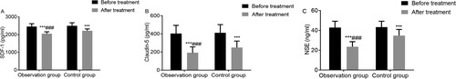
Table 4. Comparison of P values of each indicator between two groups.
Comparison of coagulation function
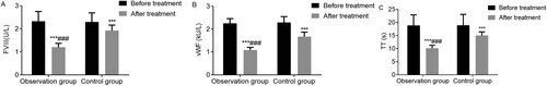
There was no statistical difference in coagulation function between the two groups before treatment (P = 0.734, 0.437, and 0.929). After treatment, FVIII and vWF were decreased and TT was shortened in both groups; and the observation group showed lower FVIII, vWF and TT than the control group (P < 0.001 in all cases). This implied that alteplase effectively improves the coagulation function of acute cerebral infarction patients, as shown in Figure and Table .
Figure 2. Comparison of coagulation function between two groups ( ± s). Notes: A: FVIII; B: Vwf; C: TT. ***P< 0.001 means intergroup comparison with that before treatment; ###P< 0.001 means intragroup comparison with that of control group after treatment.
Comparison of serum inflammatory factors
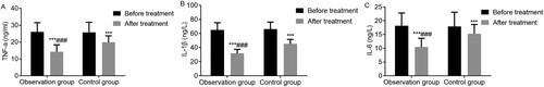
There was no statistical difference in the serum levels of TNF-α, IL-1β, and IL-6 between the two groups before treatment (P = 0.786, 0.573, and 0.793). After treatment, the serum levels of TNF-α, IL-1β, and IL-6 were decreased in both groups; and the observation group showed lower levels of TNF-α, IL-1β, and IL-6 than the control group (P < 0.001 in all cases). This implied that alteplase effectively reduces the level of serum inflammatory factors in acute cerebral infarction patients, as shown in Figure and Table .
Figure 3. Comparison of serum inflammatory factors between two groups ( ± s). Notes: A: TNF-α; B: IL-1β; C: IL-6. ***P< 0.001 means intergroup comparison with that before treatment; ###P< 0.001 means intragroup comparison with that of control group after treatment.
Comparison of cerebral hemodynamics
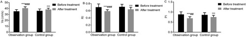
There was no statistical difference in Vs, RI and PI between the two groups before treatment (P = 0.226, 0.570, and 0.447). After treatment, Vs increased and RI and PI decreased in both groups; and the observation group showed higher Vs and lower RI and PI than the control group (P < 0.001 in Vs and RI, P = 0.002 in PI). This implied that alteplase effectively improves the cerebral blood circulation of acute cerebral infarction patients, as shown in Figure and Table .
Figure 4. Comparison of cerebral hemodynamics on both sides of the brain between two groups ( ± s). Notes: A: Vs; B: RI; C: PI. ***P< 0.001 means intergroup comparison with that before treatment; and ###P< 0.001 and ##P< 0.01 mean intragroup comparison with that of control group after treatment.
Comparison of Barthel index scores
There was no statistical difference in the Barthel index scores between the two groups before treatment (P = 0.832). The Barthel index scores of observation group were higher than those of control group 2 weeks, 1 month, and 3 months after treatment (P < 0.001 in all cases). This implied that alteplase effectively improves the daily activity of acute cerebral infarction patients, as shown in Table .
Table 5. Comparison of Barthel index scores between two groups (±s, scores).
Comparison of adverse reactions
The total incidence of adverse reactions was 13.95% in observation group and 11.90% in control group, which indicated that there was no obvious difference in the incidence of adverse reactions between the two groups (P = 0.778), as shown in Table .
Table 6. Comparison of adverse reactions between two groups [n (%)].
Discussion
Acute cerebral infarction is a common disease in middle-aged people that is characterized by necrosis, which is caused by ischemia and hypoxia. Currently, acute cerebral infarction patients account for 68%–80% of more than 7 million cerebrovascular disease patients (Choi et al. Citation2018). The pathogenesis of acute cerebral infarction mainly includes atheromatous changes in blood vessels and thrombogenesis. Thrombogenesis accounts for 75% of the patients with cerebrovascular diseases (Song et al. Citation2018). In cases of large metabolic exhaustion of brain tissues and poor tolerance to ischemia and hypoxia, irreversible neuron damage will occur after acute cerebral infarction. Thrombolytic treatment is an important method to rapidly restore cerebral blood supply in acute cerebral infarction patients. In current clinical practice, the optimum time window to rescue penumbra tissues after cerebral infarctions is 4.5–6 h from onset (Chen and Zhao Citation2018). Alteplase, a thrombolytic drug of genetic recombination, and batroxobin, a defibrase preparation, are recommended drugs for acute cerebral infarction.
Batroxobin has the effects of reducing blood viscosity, decomposing fibrinogen, promoting the release of plasminogen activator, and realizing thrombolysis, and it can inhibit platelet aggregation and thrombogenesis, which has been widely used in the treatment of acute cerebral infarction, vascular cognitive impairment, deep venous thrombosis of lower extremities and other diseases, but it works slowly (Xu et al. Citation2017). Alteplase is mainly composed of glycoprotein. Its lysine residues, in combination with fibrous protein, can activate plasminogen, decompose fibrinogen, dissolve blood clots, and thus restore the blood supply of infarction location (Mould et al. Citation2013; Jia et al. Citation2018).
This study showed that the total effective rate of the observation group (90.70%) was higher than that of the control group (73.81%), and the improvement effect of neurological function in observation group was better than that in control group, which was consistent with similar reports (Tang et al. Citation2017). The results of this study showed that with the extension of treatment time (6 h-2 weeks), the NIHSS scores of the two groups were decreased, and those of the observation group at 6, 12, 24 h, 7 d and 2 weeks after treatment were lower than those of the control group (P < 0.05), suggesting that alteplase takes effect faster and has better effect than batroxobin on improving nerve function within the same time after treatment. However, the effect of batroxobin is relatively slow, and the state of neurological defect is slowly improved after the application of batroxobin.
The development of acute cerebral infarction is a complicated process that is accompanied by changes of multiple cell factors (Sun et al. Citation2017). SDF-1, which has similar biological effects as vascular endothelial growth factors, can rapidly motivate bone marrow stem cells and circulating endothelial progenitor cells to move to the lesion location and thus play a promoting role in the repair of tissue/organic damage. SDF-1 is not only an important cell factor for damage repair, but also an inflammatory chemotactic factor (Shinohara et al. Citation2017). After ischemic damage of local tissues, the body will activate the repair mechanism to up-regulate the expression of SDF-1 (Gon et al. Citation2017). The tight junction protein claudin-5 is a key factor in regulating blood–brain barrier permeability and plays a crucial role in maintaining the structure and foundation of the blood–brain barrier. The expression level of claudin-5 is related to the damage of blood–brain barrier structure and function (Ansar et al. Citation2014). Studies have shown that claudin-5 expression was up-regulated and the blood–brain barrier permeability increased after cerebral ischemia/reperfusion injury (Sun et al. Citation2015a). NSE, which is mainly distributed in neurons and neuroendocrine cytoplasm, is a vital marker to evaluate neuron damage in clinical practice. Cerebral ischemia causes damage to the nerve tissue and cell membrane. Consequently, the NSE in brain tissues is released from cells into the circulating blood through blood–brain barrier to increase the level of serum NSE. In this study, SDF-1, claudin-5, and NSE of observation group decreased after treatment and were lower than those of control group. A possible explanation may be that the blood supply was restored in blood-starved tissues after the thrombolysis with alteplase, and then the structure and function of the blood–brain barrier and damaged brain tissues and nerve tissues were repaired, which reduced the levels of SDF-1, claudin-5, and NSE. Furthermore, alteplase has a strong thrombolytic effect that could reduce damage, promote the repair of damaged tissues and thus reduce the levels of SDF-1, claudin-5, and NSE.
Coagulation and fibrinolysis are in an equilibrium state in normal physiology. The disruption of this state is a risk factor for acute cerebral infarction. FVIII is a regulatory factor that activates the coagulation mechanism of the body and vWF is a polymer of macromolecular protein. FVIII can combine with vWF to form compounds that mediate the aggregation and adhesion of blood platelets. Reportedly, increase of FVIII and vWF, as a risk factor for cerebral infarction, was closely correlated with the severity and poor prognosis of cerebral infarction (Tampieri et al. Citation2015). TT is indicative of anticoagulant status in the body. Extension of TT indicates hyperfibrinolysis, which is a significant indicator of coagulation function (Brüning and Al-Khaled Citation2015). Alteplase may play a role in improving patients’ coagulation function by inhibiting the FVIII and vWF coagulation factors and inhibiting the adhesion and aggregation of blood platelets.
The inflammatory reaction participates in the occurrence and development of acute cerebral infarction. Studies have shown that ischemic brain damage, which is complicated by inflammatory reaction in early stages, is mediated by multiple inflammatory cytokines (Fugate and Rabinstein Citation2015). TNF-α, IL-1β, and IL-6 are inflammatory factors that play an important role in the process of inflammatory cascade reaction in cerebral infarction patients. TNF-α and IL-1β are proinflammatory factors that participate in the inflammatory reaction of ischemia/reperfusion; and IL-6, secreted by mononuclear giant cells and endothelial cells, has a neurotropic effect on mononuclear macrophages. Under normal conditions when the expression level of serum IL-6 is relatively low, necrotic histiocytes will generate a large number of antigens after cerebral infarction, which causes strong immunoreaction and stimulates mononuclear giant cells and lymphocytes to release IL-6 in quantity. Studies have shown that serum levels of TNF-α, IL-1β, and IL-6 in acute cerebral infarction patients were higher than those in patients of the normal control group (Sun et al. Citation2015). As shown in this study, TNF-α, IL-1β, and IL-6 of the observation group were lower than those of the control group after treatment, which was consistent with previous studies (Tabata et al. Citation2014). A possible explanation is that thrombolytic treatment with alteplase could relieve hypoxia and ischemia at the infarction location, prevent necrotic tissues from generating antigens, relieve immunoreaction, reduce inflammatory cascade reaction and thus reduce the levels of TNF-α, IL-1β and IL-6. Meanwhile, this study showed that Vs, RI, and PI of the observation group were improved and became progressively better than those of control group after treatment and the Barthel index scores of the observation group were higher than those of the control group, which was consistent with other reports (Sobolewski et al. Citation2014; Ji et al. Citation2017). However, this may be attributable to the fact that the thrombolytic treatment with alteplase improved the cerebral blood circulation, rescued the dying brain tissues, reduced the neuron necrosis, protected the nervous system, and therefore enhanced the daily activities of patients. When considering safety of the two treatment groups, the main adverse reaction was bleeding in both groups and the incidence had no statistical difference, indicating that the safety factors were equivalent in the two groups. Bleeding after taking the medication should be observed closely in clinical practice for symptomatic treatment.
In conclusion, alteplase can effectively enhance the clinical efficacy of acute cerebral infarction patients by improving neurological function and cerebral hemodynamics. Its mechanism of action may be associated with reducing the serum levels of SDF-1, claudin-5, NSE, and inflammatory factors and regulating coagulation function.
Disclosure statement
No potential conflict of interest was reported by the author(s).
Data availability statement
Due to the nature of this research, participants of this study did not agree for their data to be shared publicly, so supporting data is not available.
Additional information
Funding
References
- Ansar S, Chatzikonstantinou E, Thiagarajah R, Tritschler L, Fatar M, Hennerici MG, Meairs S. 2014. Pro-inflammatory mediators and apoptosis correlate to rt-PA response in a novel mouse model of thromboembolic stroke. PloS one. 9(1):e85849.
- Bong JB, Kang HG, Choo IS. 2017. Acute cerebral infarction after pyrethroid ingestion. Geriatr Gerontol Int. 3(17):510–511.
- Brüning T, Al-Khaled M. 2015. Risk of symptomatic intracerebral hemorrhage after thrombolysis with rt-pa: the sedan score. CNS Neurosci Ther. 21(3):296.
- Chen Y, Zhao Y. 2018. Curative efficacy of penehyclidine combined with edaravone on acute cerebral infarction and their effects on serum TNF-α and NDS score in rats. Eur Rev Med Pharmacol Sci. 22:223–228.
- Choi J-I, Ha S-K, Lim D-J, Kim S-D, Kim S-H. 2018. S100ss, matrix metalloproteinase-9, D-dimer, and heat shock protein 70 are serologic biomarkers of acute cerebral infarction in a mouse model of transient MCA occlusion. J Korean Neurosurg Soc. 61(5):548.
- Fugate JE, Rabinstein AA. 2015. Absolute and relative contraindications to IV rt-PA for acute ischemic stroke. Neurohospitalist. 5(3):110–121.
- Gon Y, Sakaguchi M, Oyama N, Mochizuki H. 2017. Diagnostic utility of contrast-enhanced 3D T1-weighted imaging in acute cerebral infarction associated with graves disease. J Stroke Cerebrovasc Dis. 26(2):e38–e40.
- He X, Li D, Cui C, Wen L. 2017. Clinical significance of serum MCP-1 and VE-cadherin levels in patients with acute cerebral infarction. Eur Rev Med Pharmacol Sci. 21(4):804–808.
- Jia C, Wei C, Hu M, Xu J, Niu K, Zhang C, Lv P, Li L, Dong Y. 2018. Correlation between antiplatelet therapy in secondary prevention of acute cerebral infarction and cerebral microbleeds: a susceptibility-weighted imaging (SWI) study. J Xray Sci Technol. 26(4):623–633.
- Ji B, Zhou F, Han L, Yang J, Fan H, Li S, Li J, Zhang X, Wang X, Chen X. 2017. Sodium tanshinone IIA sulfonate enhances effectiveness Rt-PA treatment in acute ischemic stroke patients associated with ameliorating blood-brain barrier damage. Transl Stroke Res. 8(4):334–340.
- Lin Z-J, Qiu H-Y, Tong X-X, Guo Y, Han M-F, Yang C-S, Lin K-H, Wu J, Li X, Yang Y. 2018. Evaluation of efficacy and safety of reteplase and alteplase in the treatment of hyper-acute cerebral infarction. Biosci Rep. 38(1):BSR20170730.
- Misumi I, Nagao A, Iwamoto K, Honda T, Ishii M, Ueyama H, Maeda Y, Ishizaki M, Kurisaki R, Okazaki T. 2017. Acute multiple cerebral infarction in a patient with an accessory mitral valve. Intern Med. 56(2):153–155.
- Mould WA, Carhuapoma JR, Muschelli J, Lane K, Morgan TC, McBee NA, Bistran-Hall AJ, Ullman NL, Vespa P, Martin NA. 2013. Minimally invasive surgery plus rt-PA for intracerebral hemorrhage evacuation (MISTIE) decreases perihematomal edema. Stroke. 44(3):627.
- Shinohara Y, Kato A, Kuya K, Okuda K, Sakamoto M, Kowa H, Ogawa T. 2017. Perfusion MR imaging using a 3D pulsed continuous arterial spin-labeling method for acute cerebral infarction classified as branch atheromatous disease involving the lenticulostriate artery territory. AJNR Am J Neuroradiol. 38(8):1550–1554.
- Sobolewski P, Brola W, Wiszniewska M, Szczuchniak W, Fudala M, Domagalski M, Śledzińska-Dźwigał M. 2014. Intravenous thrombolysis with rt-PA for acute ischemic stroke within 24 h of a transient ischemic attack. J Neurol Sci. 340(1-2):44–49.
- Song T-J, Chang Y, Chun M-Y, Lee C-Y, Kim A-R, Kim Y, Kim Y-J. 2018. High dietary glycemic load is associated with poor functional outcome in patients with acute cerebral infarction. J Clin Neurol. 14(2):165–173.
- Sun X, Berthiller J, Trouillas P, Derex L, Diallo L, Hanss M. 2015a. Early fibrinogen degradation coagulopathy: a predictive factor of parenchymal hematomas in cerebral rt-PA thrombolysis. J Neurol Sci. 351(1-2):109–114.
- Sun X, Berthiller J, Derex L, Trouillas P, Diallo L, Hanss M. 2015b. Post-thrombolysis haemostasis changes after rt-PA treatment in acute cerebral infarct. Correlations with cardioembolic aetiology and outcome. J Neurol Sci. 349(1-2):77–83.
- Sun B, Li X, Liu X, Ge X, Lu Q, Zhao X, Pu J, Xu J, Zhao H. 2017. Association between carotid plaque characteristics and acute cerebral infarction determined by MRI in patients with type 2 diabetes mellitus. Cardiovasc Diabetol. 16(1):111.
- Tabata E, Yasaka M, Wakugawa Y, Okada Y. 2014. Recombinant tissue-type plasminogen activator (rt-PA) therapy in an acute stroke patient taking dabigatran etexilate: a case report and literature review. Intern Med. 53(14):1515–1517.
- Tampieri A, Giovannini E, Rusconi AM, Cristoni L, Bendanti D, Cenni P, Lenzi T. 2015. Safety and feasibility of intravenous rt-PA in the emergency department without a neurologist-based stroke unit: an observational study. Intern Emerg Med. 10(2):181–192.
- Tang S, Luo C, Zhang K, Li K, Fan X, Ning L, Xue P. 2017. Effects of dl-3-n-butylphthalide on serum VEGF and bFGF levels in acute cerebral infarction. Eur Rev Med Pharmacol Sci. 21(19):4431–4436.
- Wan J-L, Ma Z-W. 2017. The value of mean platelet volume for prognosis of patients with acute cerebral infarction. Clin Lab. 63(11):1801–1807.
- Xu X, Li C, Wan T, Gu X, Zhu W, Hao J, Bao H, Zuo L, Hu H, Li G. 2017. Risk factors for hemorrhagic transformation after intravenous thrombolysis in acute cerebral infarction: a retrospective single-center study. World Neurosurg. 101:155–160.
- Xu W, Xie N, Zhang C, Huang Q. 2018. Imaging characteristics and pathogenesis of intracranial artery stenosis in patients with acute cerebral infarction. Exp Ther Med. 15(5):4564.
- Zhang Y-Z, Wang J, Xu F. 2017. Circulating miR-29b and miR-424 as prognostic markers in patients with acute cerebral infarction. Clin Lab. 63(10):1667–1674.