Abstract
We report a 67-year-old man with acute uric acid nephropathy, secondary to spontaneous tumor lysis syndrome, that presented itself as a huge intra-abdominal tumor that led to acute renal failure, hyperuricemia, and azotemia. Initial finding of hydronephrosis detected by ultrasonography led us to believe that the azotemia and decreasing amount of urine resulted from obstructive uropathy, a common complication of malignancy, caused by either a direct renal invasion or a urinary outflow tract compression because of a tumor mass effect. However, clinical observations and the response to therapeutic intervention confirmed the diagnosis of spontaneous tumor lysis syndrome, which is a rare cause of acute uric acid nephropathy.
INTRODUCTION
Renal failure is a common complication of malignant disease that occurs in a variety of clinical situations for numerous reasons. The acute tumor lysis syndrome (TLS) is perhaps the most dramatic cause of acute renal failure in cancer patients because it is fulminant in onset, associated with severe metabolic derangement, and is potentially reversible Citation[[1]]. This syndrome commonly occurs after chemotherapy for rapidly developing high-grade lymphomas and has also been reported as secondary to steroid Citation[[2]], interferon usage Citation[[3]], and surgical manipulation Citation[[4]].
Spontaneous tumor lysis syndrome rarely occurs, and most of the associated malignancies belong to the histopathologic type of Burkitt's lymphoma/leukemia, which has extraordinary rapid cell turnover rates. The enormous abdominal mass of our patient was a non-Hodgkin's lymphoma of a small non-cleaved cell. Our patient had the full-blown spontaneous tumor lysis and enjoyed an excellent response to dialysis therapy.
CASE REPORT
A 67-year-old man came to the outpatient department on 25 March 2000 for an evaluation of the general malaise, back pain, and unexplained body weight loss he had endured for three months. His medical history was otherwise unremarkable. Upon examination, he was in moderate distress but neither anemic nor dehydrated. A large mass was palpated in the left middle and lower quadrant of his abdomen. A soft, non-tender lymph node measuring 3 × 4 cm was also discovered in the left inguinal region. His hemoglobin was 127 g/L, white blood cells 8.2 × 10⁹ /L, platelet count 146 × 109 /L, blood urea nitrogen 10.3 mmol/L, creatinine 132.6 μmol/L, uric acid 761.6 μmol/L, and lactate dehydrogenase 257 U/L. A routine urine examination revealed microscopic hematuria without proteinuria. A non-contrast computerized tomography revealed an enormous mass was occupying almost the entire left half of the abdomen. An ultrasonogram displayed that both kidneys were moderately enlarged with normal cortical thickness; moderate left hydronephrosis was observed as well. Although the tumor pushed the left kidney upward and forward, no obvious renal parenchymal infiltration was discovered.
The patient was admitted to our hospital. A presumptive diagnosis of obstructive uropathy was forwarded because of persisting oliguria despite therapy with allopurinol, urinary alkalization, vigorous hydration and diuretics. After a retrograde pyelography revealed there was no obvious evidence of obstruction in both urinary outflows, a left side pelvic-ureter catheter was inserted for the hydronephrosis. Progressive deterioration of his renal function, hyperuricemia, and oliguria ensued: blood urea nitrogen 48.6 mmol/L, creatinine 645.3 μmol/L, calcium 10.9 mg/dL, phosphate 6.0 mg/dL, sodium 124 mmol/L, potassium 5.8 mmol/L, uric acid 1457.8 μmol/L. The serum creatinine kinase level was normal and the plasma protein electrophoresis did not discover any paraprotein. The urinary uric acid-creatinine ratio was 1.13 (1743.4 μmol/L:1547.0 μmol/L).
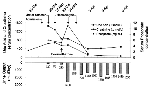
An acute uric acid nephropathy secondary to spontaneous tumor lysis syndrome was diagnosed based on the clinical findings and because the ratio of urinary uric acid to creatinine was greater than one. Dialysis therapy was initiated to remove the uric acid load as well as to correct the rising azotemia. Diuresis developed after three sections of hemodialysis performed in the following four days. The patient's kidney function returned to normal and both the electrolytes and uric acid concentration were within normal ranges one week after the dialysis therapy was begun ().
Figure 1. The patient's clinical course.
A biopsy of the left inguinal lymph node was performed after the first dialysis section. The pathologic diagnosis was a non-Hodgkin's lymphoma of small non-cleaved cell with prominent mitotic features, which our pathologist concluded a Burkitt-like lymphoma.
DISCUSSION
The most common cause of the patient's clinical presentation was an invasive malignancy with acute renal failure and azotemia produced by either direct renal invasion or urinary tract obstruction resulting from the effect of the mass. Although the radiological evidence of a bulky tumor and hydronephrosis pointed towards obstructive uropathy, the actual cause was a uric acid nephropathy associated with spontaneous tumor lysis.
A uric acid nephropathy secondary to spontaneous tumor lysis syndrome was the major cause of renal failure in this patient according to the persisting oliguria after a pelvic-ureter catheter placement, the ratio of urinary uric acid to creatinine was greater than one Citation[[5]], the rapid diuretic response after uric acid removal by hemodialysis, and the completely reversed renal function. The acute uric acid nephropathy indicated that intrarenal hydronephrosis was associated with uric acid precipitates in both the distal nephron and medullary collecting ducts. This phenomenon contrasted the extranephronal urinary tract obstruction caused by either the extrinsic compression from the tumor itself or massive lymphadenopathy, which is a common cause of renal failure in cancer patients.
The acute TLS is characterized by hyperuricemia, hyperkalemia, hyperphosphatemia, hypocalcemia, and acute renal failure. Acute TLS is an avoidable complication that conventionally occurs after treatment for hema-tological malignancies with rapid cell proliferation such as Burkitt's lymphoma/leukemia. Acute spontaneous TLS is uncommon, especially in the pretreatment malignancy, and very few cases of pretreatment metabolic derangements have been spontaneous TLS Citation[6-10]. Our patient had a textbook spontaneous tumor lysis and later enjoyed an excellent response to therapy. This case highlights the importance of remaining apprehensive of spontaneous TLS in patients with acute renal failure associated with hyperuricemia, especially when presented with a bulky tumor.
ACKNOWLEDGMENT
We would like to thank Dr. Lee-Yung Shih (Chief, Division of Hematology and Oncology, Chang Gung Memorial Hospital, Kweishan, Taiwan) for helpful suggestions and review of this manuscript.
REFERENCES
- Arrambide K, Toto R D. Tumor lysis syndrome. Semin Nephrol 1993; 13: 273–280
- Loosveld O J, Schouten HC, Gaillard C A, Blijham G H. Acute tumor lysis syndrome in a patient with acute lymphoblastic leukemia after a single dose of prednisolone. Br J Haematol 1991; 77: 122–123
- Fer M F, Bottino G C, Sherwin S A, Hainsworth J D, Abrams P G, Foon K A, Oldham R K. A typical tumor lysis syndrome in a patient with T-cell lymphoma treated with recombinant leukocyte interferon. Am J Med 1984; 77: 953–956
- Jona J Z. Progressive tumor necrosis and lethal hyperkalemia in a neonate with sacrococcygeal teratoma (SCT). J Perinatol 1999; 19: 538–540
- Kelton J, Kelley W N, Holmes E W. A rapid method for the diagnosis of acute uric acid nephropathy. Arch Intern Med 1978; 138: 612–615
- Cohen L F, Balow J E, Magrath I T, Poplack D G, Zeigler J L. Acute tumor lysis syndrome a review of 37 patients with Burkitt's lymphoma. Am J Med 1980; 68: 486–491
- Azouri J, Afif N, Ghosn M, Nader N, Bou Saba C. Primary breast lymphoma: a case report. J Med Liban 1992; 40: 202–206
- Jasek A M, Day H J. Acute spontaneous tumor lysis syndrome. Am J Hematol 1994; 47: 129–131
- Lotfi M, Brandwein J M. Spontaneous acute tumor lysis syndrome in acute myeloid leukemia? a single case report with discussion of the literature. Leuk Lymphoma 1998; 29: 625–628
- Agha-Razii M, Amyot S L, Pichette V, Cardinal J, Quimet D, Leblanc M. Continuous veno-venous hemodiafiltration for the treatment of spontaneous tumor lysis syndrome complicated by acute renal failure and severe hyperuricemia. Clin Nephrol 2000; 54: 59–63