Abstract
Atrial septal aneurysm (ASA) is a well known morphologic abnormality and has been largely investigated with both transthoracic (TTE) and/or transesophageal echocardiography (TEE) CitationCitationCitation. Its association with other congenital and acquired heart diseases and midsystolic clicks has been reported CitationCitationCitation. ASA also may be associated with an increased risk of embolic events CitationCitation. In many cases, it is an incidental finding. We describe a patient with acute renal failure associated with nephrotoxic drugs and ASA suggesting endocarditis.
CASE REPORT
A 53-year-old diabetic woman with infectious and necrotizan lesions on her feet was hospitalized. The patient had no previous history of rheumatic heart disease, dyspnea, syncope or angina. Physical examination revealed a regular pulse of 88/min, a blood pressure of 130/80 mmHg and a temperature of 37°C. The only cardiovascular finding was the grade 2/4 midsystolic murmur at the left sternal edge. Lungs were clear. Hepatosplenomegaly was not found. On her feet there were purulent, edematous and necrotizan lesions. Peripheral arteries were patent. Diabetic retinopathy and neuropathy were diagnosed by ophtalmologist and neurologist. Baseline laboratory findings were as follows: Hemoglobin 11 g/dl, normochromatic and normocytic erythrocytes, platelets 300000/mm3, white blood cells 10000/mm3, blood urea nitrogen 19 mg/dl, creatinine 1 mg/dl. Chest X-ray and electrocardiogram were unremarkable.
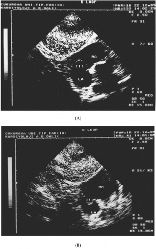
Fever developed on the 3rd hospital day. Cultures from blood, throat, urine and ulcers on her feet were taken. On TTE, a large vegetation was found on the tricuspid valve. Blood cultures were positive for Staphylo-cocus aureus. Vancomycin and amikacin were began. On the 15th day of therapy, anuria developed and the patient was transferred to the nephrology department, and hemodialysis was began. Echocardiographic examination was repeated. On TTE examination using a Toshiba SSH-160A equipment (Toshiba Corporation, Tokyo, Japan), tricuspid valve prolapse was diagnosed as systolic displacement of the septal tricuspid leaflet into the right atrium (). Grade 2 tricuspid regurgitation was detected by color Doppler. Atrial septum was poorly visualized and a dropout on the fossa ovalis region was noticed. Subcostal view showed an atrial septal aneurysm which demonstrated excursions into the left and right atrium during the cardiorespiratory cycle (A and B).
Figure 1. Apical four-chamber two-dimensional echocardiogram of the patient with tricuspid valve prolapse (arrows). Tricuspid valve septal leaflet is protruding into the right atrium (RA) in systole. A dropout on the fossa ovalis region of atrial septum is seen.
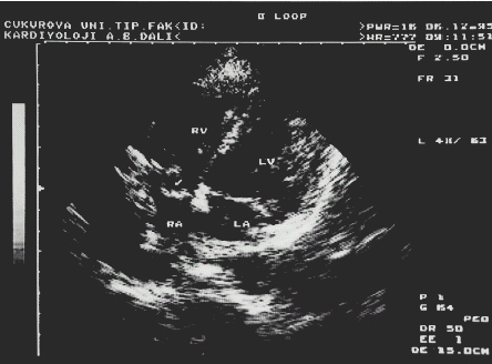
Figure 2. Subcostal view shows atrial septal aneurysm. In (A) the aneurysm (arrows) bulges into the right atrium and in (B) into the left atrium.
ASA was diagnosed as a thin bullousing tissue in the area of fossa ovalis, with the cardiorespiratory cycle and protruding into both atrial cavities. Since valve vegetations were not observed on TTE, the patient underwent TEE examination as a part of diagnostic work up. On TEE, the atrial septum appeared clearly and ASA with excursion of 15 mm into either atrium was seen. The base of the aneurysm was 17 mm in diameter. Interatrial shunting was not found. There were also no vegetations on TEE.
Nephrotoxic drug-induced acute renal failure was diagnosed and antibiotic treatment was stopped. Hemodialysis was started. Anuria persisted and renal function did not recover. Two months later from beginning of anuria renal biopsy was performed. Renal biopsy showed global glomerular sclerosis, glomerular capillary hypertrophy, increment of mesangial matrix and nodular sclerosis, tubular atrophy, interstitial fibrosis and vascular hyalinization. After continous peritoneal dialysis (CAPD), diuresis began. Six months later, her renal function improved and she did not need dialysis. CAPD was stopped. Her creatinine clearance was 20 ml/min and she was symptom free for cardiac status.
DISCUSSION
In 1978, Silver Citation[[6]] provided the pathologic description of ASA as “the septum is securely redundant and it bulges far into the right or left atrial cavity” and found the anomaly in 1% of the autopsies. Initially, in vivo diagnosis of ASA has been made by angiography Citation[[7]], Citation[[8]]. However noninvasive diagnosis of ASA can easily be made in recent years with the advent of both two-dimentional and transesophageal echocardiography Citation[[2]], Citation[[3]], Citation[[4]], even though the combined mitral valve prolapse alone or in combination with tricuspid valve prolapse and ASA have been commonly reported Citation[[4]], Citation[[5]]. The association between ASA and the isolated tricuspid prolapse is seldomly reported. Atrioventricular valve prolapse and ASA association have been attributed to a congenital connective tissue defect that involves endocardial tissue Citation[[5]]. ASA is considered a cardiac abnormality with thromboembolic potential. The mechanism of the embolisation may be either thrombus formation within the aneurysm or a paradoxic embolisation through an interatrial shunting Citation[[2]]. ASA may also mimick or suggest endocarditis. TEE is a reliable and semi-invasive method for diagnosing endocarditis. In our case, although TTE and TEE did not show vegetations, for we could not exclude endocarditis.
Renal function improved after six months of acute insult even though renal biopsy did not show any acute renal injury. Hemodialysis may cause a delayed improvement in renal function. Peritoneal dialysis is more biocompatible and is a better tool than hemodialysis in improving fluid balance Citation[[9]]. In some cases, as in our patient, improvement of acute renal failure may be delayed or, in chronic dialysis, renal function may recover.
REFERENCES
- Pearson A.C., Nagelhout D., Castello R., Gomez C.R., Labovitz A.J. Atrial Septal Aneurysm and Stroke: A Transesophageal Echocardiograhic Study. J. Am. Coll. Cardiol. 1991; 18: 1223–1229
- Schneider B., Hanrath P., Vogel P., Meinertz T. Improved Morphologic Characterization of Atrial Septal Aneurysm by Transesophageal Echocardiography: Relation to Cerebrovascular Events. J. Am. Coll. Cardiol. 1990; 16: 1000–1009
- Belkin R.N., Kisslo J. Atrial Septal Aneurysm: Recognition and Clinical Relevance. Am. Heart J. 1990; 120: 948–957
- Iliceto S., Papa A., Sorino M., Paolo R. Combined Atrial Septal Aneurysm and Nitral Valve Prolapse: Detection by Two-Dimensional Echocardiography. Am. J. Cardiol. 1984; 54: 1151–1153
- Roberts W.C. Aneurysm (Redundancy) of the Atrial Septum (Fossa Ovale Membrane) and Prolapse (Redundancy) of Mitral Valve. Am. J. Cardiol. 1984; 54: 1153–1155
- Silver M.D., Dorsey J.S. Aneurysms of the Septum Primum in Adults. Arch Pathol. Lab. Med. 1978; 102: 62–65
- Thompson J.I., Phillips L.A., Melman K.L. Pseudotumor of the Right Atrium: Report of a Case and Review of Its Etiology. Ann. Intern. Med. 1966; 64: 665–667
- Freedom R.M., Rove R.D. Aneurysm of the Atrial Septum in Tricuspid Atresia: Diagnosis During Life and Therapy. Am. J. Cardiol. 1976; 38: 265–267
- Tielemans C., Dratwa M., Bergmann P., Goldman M., Flamion B., Collard F., Wens R. Continuous Ambulatory Peritoneal Dialysis Vs Haemodialysis: A Lesser Risk of Amyloidosis?. Nephrol. Dial. Transplant. 1998; 3: 291–294