Our patient was a 40-year-old Indian male, non-smoker non-alcoholic, working as a waiter in a furniture shop in Saudi Arabia. This patient was admitted with a history of nausea, vomiting and generalized weakness of 15 days duration. He had no history of diabetes mellitus, hypertension, renal stone disease or drug abuse. He was afebrile, and his blood pressure was 160/100 mmHg standing and 165/102 mmHg lying, with pallor and bilateral pedal edema. Respiratory and cardiovascular examinations were normal. The abdominal examination did not reveal any organomegaly or bruit. The rest of the examination including nervous system and funduscopy was normal. Blood tests showed Hemoglobin—8.2 gm/dl, Hematocrit 24.6% with normocytic normochromic picture, Urea 200 mg/dl, creatinine 14 mg/dl, calcium (Ca) 7.5 mg/dl, Phosphates (Ph) 7 mg/dl, alkaline phosphatase (Alk.Ph) 100 u/l, sodium 140 meq/l, potassium 4.0 meq/l and albumin 4 gm/dl. Arterial blood gases showed compensated metabolic acidosis. Abdominal ultrasound revealed bilateral small kidneys. The serum intact PTH was within normal limits (80 pmol/l), and no abnormalities were detected on skeletal survey. A diagnosis of end stage renal disease with mild hypertension was made. Hemodialysis was initiated along with calcium carbonate, 1–25 dihydroxy-cholecalciferol and dietary advice. However, he had poor drug and dietary compliance, despite being regular on hemodialysis. A dietitian was consulted at different stages, but the patient turned down all advice. Three years later, he developed pain and swelling around left shoulder and left hip. Both swellings were tender and firm to hard in consistency.
The movements of these joints were painful and restricted. The radiography of left shoulder and left hip showed extensive periarticular calcifications (. The other joints did not show any abnormality. Other laboratory investigations revealed elevated product of serum calcium × phosphate (>80 mg/dl), elevated serum Intact PTH (800 pmol/l) but normal serum alkaline phosphatase (table). There was no evidence of aluminium intoxication. A diagnosis of secondary hyperparathyroidism was confirmed, and a subtotal parathyroidectomy was planned and advised, but again refused by the patient. Luckily, during the same year, he had a cadaveric renal transplant and was placed on triple immunosuppressive therapy. He had initially good recovery with immediate diuresis, but on the 3rd post-operative day, the renal function started to deteriorate.
Table. Serum Bone Panel
Figure 1. X-ray showing extensive periarticular calcification involving left shoulder and soft tissues around it.
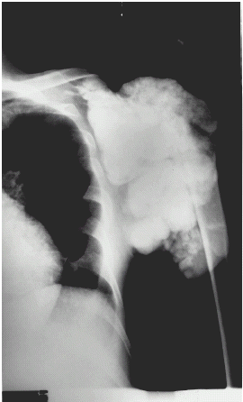
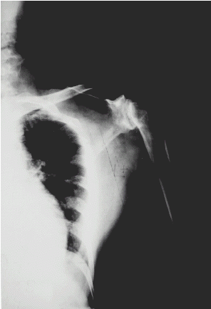
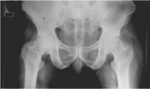
Figure 3. X-ray showing complete disappearance of calcification after successful renal transplantation.
The renal scan (DTPA) showed reduced uptake, reduced perfusion and delayed transit time suggestive of acute rejection. The transplanted kidney biopsy had demonstrated mesangial proliferation and lymphocyte cell infiltration in the interstitium. However, he responded to anti-rejection therapy. The arthropathy of left shoulder and left hip with periarticular calcification gradually subsided ( without any specific treatment. The signs and symptoms as well as serum bone panels also returned to normal (table). At present, he is stable with normal functioning graft (S.Cr. 1.6 mg/dl).
Figure 2. X-ray showing extensive periarticular calcification involving soft tissue around left hip.
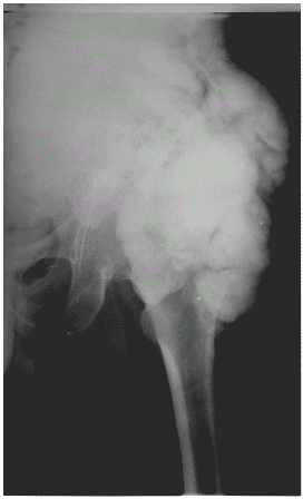
Figure 4. X-ray showing complete disappearance of calcification after successful renal transplantation.
Thus, the final diagnosis was “End stage renal disease with metastatic calcification due to persistent hyperphosphatemia and spontaneous resolution after successful renal transplant”.
The retention of phosphate begins very early in advanced renal failure due to reduction in filtered phosphate load from normal value of 80–95% to as low as 15% in advanced renal failure Citation[[1]], Citation[[2]]. This was aggravated in this patient due to his poor compliance to dietary advice and phosphate binders. Metastatic calcification is most likely to occur when the serum calcium and phosphate product exceeds 70–80 mg/dl Citation[[3]], as has been shown in this case. This may also occur due to a persistent increase in PTH, which, continues to cause calcium phosphate release from bone Citation[[4]]. After reaching this stage, the lowering of the plasma phosphate concentration and thereby the resolution of metastatic calcification can only be achieved by parathyroidectomy, which was refused by the patient. Resorption of soft tissue calcium phosphate deposits, due to persistent hyperphosphatemia, occurs after successful renal transplantation and is often associated with hypercalcemia Citation[[5]]. This was well noted in our case.
This case study demonstrates that extensive metastatic joint calcification is reversible following kidney transplantation. It is rare nowadays to see such complications due to prevention and/or early diagnosis and management of secondary hyperparathyroidism.
REFERENCES
- Delmez J.A., Slatopolsky E. Hyperphosphatemia: Its Consequences and Treatment in Chronic Renal Failure. Am. J. Kidney Dis. 1992; 19: 303–317
- Slatopalsky E., Robson A.M., Elkan I., Bricker M.S. Control of Phosphate Excretion in Uremic Man. J. Clin. Invest. 1968; 47: 1865
- Milliner D.S., Zinmeister A.R., Lieberman E., Landing B. Soft Tissue Calcification in Pediatric Patients with End-Stage Renal Disease. Kidney Int. 1990; 38: 931–936
- Liach F. Parathyroidectomy in Chronic Renal Failure: Indications, Surgical Approach, and the Use of Calcitriol. Kidney Int. Suppl. 1990; 29: 62–68S
- Julian B.A., Quarles L.D., Niemann K.M. Musculoskeletal Complications After Renal Transplantation. Pathogenesis and Treatment. Am. J. Kidney Dis. 1992; 19: 99–120