Abstract
Renal infection, including acute and chronic pyelonephritis, focal bacterial nephritis, renal and perinephric abscess, pyonephrosis and others, represents a spectrum of interrelated conditions. In recent years, computed tomography, ultrasound, nuclear scintigraphy, excretory urography (IVP) and magnetic resonance imaging have offered varying degrees of utility in evaluating renal infection. Although imaging in acute pyelonephritis has been extensively studied, this condition is a rare initial presentation as a renal cystic feature. This investigation presents a case of acute pyelonephritis, with an atypical initial imaging manifestation in renal cystic feature, which became a heterogeneous mass during follow up, and disappeared after treatment. Two conclusions can be drawn: first, a renal cystic lesion may be an initial presentation of acute pyelonephritis; secondly, the abnormalities of the ultrasonography imaging study of renal pyelonephritis persisted and progressed despite the improvement in clinical symptoms and laboratory findings, and displayed complete resolution of imaging abnormalities several months later.
INTRODUCTION
Renal infection, including acute and chronic pyelonephritis, focal bacterial nephritis, renal and perinephric abscess, pyonephrosis, and others, represents a spectrum of interrelated conditions.Citation[[1]] With the significant advances in imaging techniques that have developed in recent years, selecting an appropriate study can shorten the time for appropriate evaluation and facilitate therapy. In recent years, computed tomography, ultrasound, nuclear scintigraphy, excretory urography (IVP) and magnetic resonance imaging have varying degrees of utility in evaluating renal infection. Ultrasound imaging, which is noninvasive, rapid, and inexpensive, and involves no radiation exposure, has been observed to be especially useful in identifying renal abscesses.Citation[[2]] Many papers have presented imaging studies in acute pyelonephritis, but it is rare as an initial presentation of renal cystic feature. This study presents a case of acute pyelonephritis, with atypical initial imaging manifestation in renal cystic feature, which became a heterogeneous mass during follow up, and disappeared after treatment.
CASE REPORT
A 69 year old female housekeeper, with an asthma history of more than 10 years took no regular medication control due to infrequent attack. She was quite well until 1 week before admission. She complained of a sudden onset of high fever and chill associated with right flank pain. She visited a local hospital for help, and was admitted under the impression of urinary tract infection. Although the fever and chill subsided, three days later, renal ultrasonography and abdominal computed tomography revealed an abnormal right renal lesion. She was then referred to our hospital for further evaluation. Right side flank pain was noted on admission. She suffered no hematuria, no dysuria, no body weight loss, no bone pain, and no hemoptysis. Blood pressure was 91/50 mmHg and body temperature was 37.2°C. She was alert, with a normal breathing pattern, but physical examination revealed right costovertebral angle tenderness. The laboratory investigation revealed blood urea nitrogen 14 mg/dL, creatinine 1.1 mg/dL, sodium 145 mmol/L, potassium 3.5 mmol/L, glucose 91 mg/dL, WBC 4600/mm3, hemoglobin 11.0 g/dL, and urinalysis yielded protein 75 mg/dL, occult blood 4 +, RBC 3–5/HPF, WBC >100/HPF.
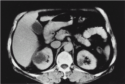
Ultrasonography imaging study during admission revealed a bilateral normal size kidney with an irregular cortical contour. Two hypoechoic lesions (1.5 and 1.2 cm) with posterior wall enhancement were noted over the upper pole of the left kidney, and another similar lesion (2.1 cm) was also discovered over the upper pole of the right kidney ((a)). Examination found no hydronephrosis, no renal stone, and no renal mass. The initial sonographic impression was bilateral renal cysts. Angiography study was performed later, the right renal angiography revealed a hypervascular or avascular lesion in the upper pole of right kidney, measuring about 1 cm in size (). When viewed with CT, the impression favored a simple cyst rather than a cystic tumor or abscess. Left renal angiography revealed no obvious organic renal lesion. A follow up investigation of abdominal computed tomography was performed 3 days later, displaying regressive change of the cystic mass in the right renal cortex compared with the previous film, but no significant change in the left renal cyst (2.5 cm) (). The patient received antibiotic treatment with cefazolin and gentamicin for two weeks. After an improvement in her right lower back pain, she was discharged with an oral antibiotic and returned for a follow up renal ultrasonography study about 10 days later. This time both kidneys exhibited normal size and smooth contours. The ultrasonography detected a heterogeneous mass in the upper portion of the right kidney, but no renal stone ((b)). Because her symptoms improved, the patient was kept on oral antibiotic treatment without surgical drainage. Another ultrasonography renal imaging study was performed 3 months later, with both kidneys normal in size and contour. The previous heterogeneous mass in the right kidney had disappeared ((c)).
Figure 1. Ultrasonography imaging study (a) it revealed a hypoechoic lesions (2.1 cm) with posterior wall enhancement over the upper pole of the right kidney during admission. (b) a heterogeneous mass in the upper portion of the right kidney after antibiotic treatment. (c) 3 months later, the previous heterogeneous mass in the right kidney had disappeared.
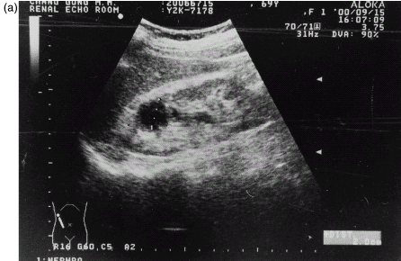

Figure 2. The right renal angiography revealed a hypovascular or avascular lesion in the upper pole of right kidney, measuring about 1 cm in size.
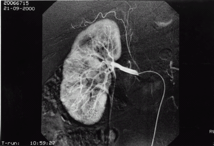
Figure 3. Abdominal computed tomography reveal the cystic mass in the right renal cortex.
DISCUSSION
This case presents a series of changes of imaging study in acute pyelonephritis. Many choices of imaging study are available, and many variables influence test choice, including clinical symptoms and signs, laboratory findings, and most important, suspected diagnosis. Ultrasound imaging study employs high frequency sound waves, which are noninvasive, fast, inexpensive, and do not expose the patient to radiation. This technique permits direct viewing of the renal parenchyma in real time with multiplanar ability. In acute pyelonephritis, ultrasonography imaging study may be normal,Citation[[3]] as in increased echogenicity within the parenchyma, or with renal enlargement and decreased echogenicity of the cortex and medulla owing to increased tissue fluid content. Multiple scattered low level echoes representing microabscesses can be observed in rare instances. In focal bacterial nephritis, ultrasonography demonstrates a vague, sonolucent mass, or cystic lesions, which can disrupt the corticomedullary junction.Citation[[2]] Chronic atrophic pyelonephritis, which is associated with chronic reflux and presents recurrent episodes of infection, results in deformity and dilatation of calyces and focal cortical loss. Classically most severe in those areas of the cortex nearest the calyces, especially the upper and lower poles.[4,5] As the disease progresses to a renal abscess, in contradistinction to pyelonephritis, ultrasonography can evaluate it effectively. On ultrasound, a renal abscess typically appears as a round, smooth-walled mass, containing low-amplitude echoes reflecting necrosis debris,Citation[[2]] but sometimes the sonography appearance may vary, and may lack internal echoes and mimic cysts.Citation[[6]] The ultrasonography imaging study of our patient initially presented as a cystic or sonolucent mass in the right kidney and later became a heterogeneous mass, but disappeared after completed treatment. This initial manifestation of acute pyelonephritis as a renal cystic lesion is unusual.
Computed tomography offers excellent images of the renal parenchyma and the surrounding tissues. Contrast–computed tomography scan is highly sensitive in detecting acute pyelonephritis, with nephromegaly, decreased opacification of the affected renal parenchyma, typically patchy, wedge-shaped or linear patterns.Citation[[6]] Contrast–enhanced helical CT displays significantly greater sensitivity than gray scale ultrasonography or the IVP with nephrotomography in identifying renal parenchymal abnormalities in acute pyelonephritis.Citation[[7]] Non-contrast CT is often normal but may reveal focal areas of slightly decreased attenuation. Affected kidneys may be enlarged diffusely or focally, and focal enlargement may be misinterpreted for a mass.Citation[[6]] Acute pyelonephritis is rarely presented as a cystic lesion in a CT scan. The patient in this study demonstrated regressive change of cystic mass in the right renal cortex, as found in the computed tomography imaging study. The abnormalities usually persist for several weeks, well after the clinical symptoms and laboratory findings have returned to normal,Citation[[8]], Citation[[9]] although renal scarring can occur in some infection.Citation[[10]] Following adequate treatment, most cases eventually will completely resolve imaging abnormalities. The abnormalities of the ultrasonography imaging study in this patient persisted and progressed despite improvement in clinical symptoms and laboratory findings, and demonstrated complete resolution of imaging abnormalities three months later.
Angiography is an invasive imaging study, with contrast media exposure, and it is employed most frequently to evaluate renal mass. This procedure may facilitate diagnosis of intrarenal abscess. Renal vessels display a unique vasospastic response to inflammation in contrast to other areas, which respond with vasodilatation. A persistent arterial-filling defect occurs after the surrounding kidney has reached the nephrogram phase.Citation[[11]] Abscesses are also noted for increased cortical vascularity.Citation[[2]] Differentiation between renal inflammatory lesions and avascular necrotic malignancies is often impossible. Our patient showing typical angiography imaging of renal inflammation lesion mentioned above.
This patient provided many valuable lessons. First, a renal cystic lesion may be an initial presentation of acute pyelonephritis, which then progresses to a heterogeneous mass by following up with an ultrasonography imaging study. Second, the abnormalities of ultrasonography imaging study of renal abscess persisted and progressed despite improvement in clinical symptoms and laboratory findings, and demonstrated complete resolution of imaging abnormalities several months later. Third, renal ultrasonography imaging study is a noninvasive, rapid, inexpensive, and convenient choice in renal infection diagnosis and follow up. However, a false negative result is always possible.
REFERENCES
- Melson G. Coordinated Diagnostic Imaging. Churchill Livingstone, New York 1984
- Benson M., Puma J.P.Li., Resnick M.I. The Role of Imaging Studies in Urinary Tract Infection. Urol. Clin. of North Am. 1986; 13: 605–625
- Talner L.B., Davidson A.J., Lebowitz R.L. Acute Pyelonephritis: Can We Agree on Terminology?. Radiology 1991; 192: 297–305
- Hodson C.J. Radiology in Pyelonephritis. Curr. Prob. Radiol. 1972; 2: 1–32
- Goldman S.M., Fishman E.K. Upper Urinary Tract Infection: The Current Role of CT, Ultrasound and MRI. Seminars in Ultrasound, CT and MR 1991; 12: 335–360
- Kaplan D.M., Rosenfield A.T., Smith R.C. Advances in the Imaging of Renal Infection. Infect. Dis. Clin. of North Am. 1997; 11: 681–705
- Gold R.P., McCleannan B.L., Rottenbeg R.R. CT Appearances of Acute Inflammatory Disease of the Renal Interstitium. Am. J. Roentgenol 1983; 141: 343–349
- Papanicolaou N., Pfister R.C. Acute Renal Infections. Radiol. Clin. North Am. 1996; 34: 965–995
- Tsugaya M., Hirao N., Sakagami H. Computed Tomography in Acute Pyelonephritis: The Clinical Correlation. J. Urol. 1990; 144: 611–613
- Meyrier A., Condamin M.C., Pernet M. Frequency of Development of Early Cortical Scarring in Acute Primary Pyelonephritis. Kidney Int. 1989; 35: 696–703
- Morgan W.R., Nyberg L.M. Perinephric and Intrarenal Abscess. Urology 1985; 26: 529–537