Abstract
Aim: Infectious disease represents one of the major causes of morbidity and mortality in hemodialysis patients. Extra-renal abscess constitutes a specific form of infection. The aim of this study was to evaluate and analyze the clinical characteristics of extra-renal abscess in chronic hemodialysis patients. Methods: We retrospectively studied the extra-renal abscess among chronic hemodialysis patients in Chang Gung Memorial Hospital at Kaohsiung, Taiwan. The records of 2168 chronic hemodialysis patients from October 1986 to January 2000, were studied. The clinical features were reviewed and analyzed. Results: Sixteen patients who were enrolled during the study period developed extra-renal abscess. Ten of them were male. The mean age was 59.2 ± 11.8 years old. More than half of the patients had diabetes (53.6%, 9/16). The locations of extra-renal abscess in these patients were liver (8/16), lung (5/16), spleen (1/16), perianal region (1/16), psoas muscle (1/16), and prostate (1/16). One patient had concurrent liver and spleen abscesses. All patients presented with fever and chills. Laboratory studies revealed leukocytosis and thrombocytopenia in 2/3 of the patients. The patients were associated with malnutrition status with lower serum albumin level (2.94 ± 0.55 gm/dL) and lower nPCR (normalized protein catabolism rate; 0.84 ± 0.11 gm/Kg/day) comparing to the other hemodialysis patients (albumin: 4.05 ± 0.47 gm/dL; nPCR: 1.14 ± 0.31 gm/kg/day). There was no significant difference in kt/V between the patients with (1.28 ± 0.34) or without abscess formation (1.47 ± 0.36). The major causative pathogen was Klebsiella pneumoniae. Parenteral antibiotic treatment is sufficient to treat most of the diseases, except 2 patients who needed surgical intervention. Twelve patients recovered after 2–3 weeks of treatment. Conclusions: The study indicated that extra-renal abscess is rare in chronic hemodialysis patients. The abscesses occurred mostly in liver. Diabetes mellitus and poor nutrition status were the important predisposing factors. Gram-negative bacilli, K. pneumoniae, were the major pathogen. Most of the patients responded to parenteral antibiotics and surgical draining.
INTRODUCTION
Chronic renal failure is associated with increased susceptibility to bacterial infection due to the decreased immunity.Citation[[1]] Infection is second to cardiovascular disease as the leading cause of death in patients with end-stage renal disease (ESRD) in many countries.Citation[[2]], Citation[[3]], Citation[[4]] Bacteria infection with septicemia accounts for more than 75% of uremic infection death.Citation[[4]] The increased susceptibility to bacteria infection in uremic patients is due to relative immune deficiency, advanced age, and comorbid condition such as diabetes mellitus.Citation[[5]] Abscess formation indicates a category of reduced immunity combining with severe infection. Previous study had indicated that urinary tract abscess in uremic patients is associated with grave prognosis.Citation[[6]] Furthermore, arteriovenous fistula infection also constituted a major complication in vascular access and caused a great morbidity.Citation[[7]] Little is known about the exact clinical characteristics of the uremic extra-renal visceral abscess. It is unknown if the extra-renal visceral abscess in uremic patient is associated with more severe clinical course and outcome. In the study, we analyzed 16 patients with extra-renal abscess under chronic hemodialysis therapy in a single medical centre in Taiwan.
PATIENTS AND METHODS
We conducted a retrospective analysis of hemodialysis patients in a single dialysis unit of a university-afflicted medical centre (Chang Gung Memorial Hospital, Kaohsiung, Taiwan). Abscess was defined as accumulation of pus within tissues, organs, or confined spaces. Two criteria should be met to diagnose the extra-renal abscess. Firstly, evidence of pus accumulation should be provided by sonography or radiology study by experienced radiologists. Secondly, bacterial pus culture from abscess should be positive at microbiology laboratory. The study was specifically dedicated to extra-renal visceral abscess. Abscess of arteriovenous fistula was excluded from the study. Renal or urinary tract abscess was also excluded as the different clinical pattern.
From October 1986 to September 2000, 2168 chronic hemodialysis patients had v entered our chronic dialysis program. Among them, extra-renal visceral abscess appeared in 16 patients. The microbiological characteristics, clinical presentations, laboratory data, treatment and outcomes of the patients were recorded and analyzed to define the clinical pattern of the complication.
To further characterize the risk factor predisposing to the extra-renal abscess formation, primary renal disease, nutrition status (assessed by the protein catabolism rate and serum albumin), and adequacy hemodialysis (assessed by kt/V) were also analyzed in these patients and patients without abscess formation (n = 2152). The calculation of protein catabolism rate and kt/V were described in our previous study.Citation[[8]]
Analytical Methods
Continuous variables are expressed as mean ± standard deviation from (n) patients. For normally distributed continuous variables, two-tailed Student's unpaired t-test was used to test for differences between means. Differences in categorical variables were examined by the Chi-Square test with Yate's correction. The statistical analysis was performed using the Statview™ program (Macintosh). p<0.05 were considered statistically significant.
RESULTS
Sixteen patients developed extra-renal abscess during the study period. Ten of them were male and 6 were female. Clinical features and outcomes of 16 patients with extra-renal abscesses are shown in . The mean age of the patients was 59.2 ± 11.8 year-old (range 38–83 year-old) at diagnosis of extra-renal abscess. The average duration of hemodialysis was 23.5 ± 19.4 months (Range from 1–60 months). The abscess developed within first year of hemodialysis in 6 patients (37.5%). There were no significant differences in average age and duration of hemodialysis between patients with and without extra-renal abscess (mean age: 54.3 ± 21.8 year-old; mean dialysis duration: 30.2 ± 15.4 months, n = 2152). The most common underlying renal diseases were Type II diabetes with nephropathy (9/16, 56.3%). There was significantly higher incidence of diabetes in patients with than without extra-renal abscess (56.3% vs. 32%, p < 0.01). The diabetes is clearly associated with the disease.
Table 1. Clinical Features and Outcome of 16 Chronic Hemodialysis Patients with Extra-renal Abscess
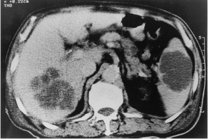
One patient had multi-foci abscess formation, liver and spleen abscesses (). As a result, there were 17 abscesses in 16 patients. Among the 17 extra-renal visceral abscess, 47.1% were in liver (8/17), 29.4% in lung (5/17), 5.9% in spleen (1/17), 5.9% in perianal fat tissue (1/17), 5.9% in psoas muscle (1/17) and 5.9% in prostate (1/17). Two patients had gas-forming liver abscess. Chest film revealed the air–fluid level in 4 patients with lung abscesses (80%). The results indicated the hepatic preference of the abscess formation.
Figure 1. Abdominal CT demonstrating hypodense densities in liver and spleen.
All patients presented with fever and chills. Other presenting symptoms included cough (4/16), chest pain (3/16), disturbance of consciousness (3/16), right upper quadrant pain (1/16), anal colonic protrusion (1/16), right flank pain (1/16) and suprapubic discomfort (1/16). It seemed that the presenting symptoms were non-specific to the disease, but more related to the location of abscess.
Laboratory data at the onset of extra-renal abscesses and the culture results are shown in . Laboratory studies revealed leukocytosis (> 10 000/mm3; 81.3%, 13/16), thrombocytopenia (<200 000/mm3; 68.8%, 11/16), and hypoalbuminemia (<3.5 g/dL; 81.3%, 13/16) in more than two third of the patients. The mean peripheral leukocyte count was 16 741 ± 7912/mm3 (range from 2400/mm3 to 34 400/mm3) and platelet count was 18.6 ± 15.4 × 10 000/mm3 (range from 3.8 × 10 000/mm3 to 52.1 × 10 000/mm3). The average serum albumin was 2.94 ± 0.55 g/dL (range 2.2–4.1 g/dL). Abscess pus culture grew out K. pneumoniae in 12 patients (75%), E. coli in 2 patients (12.5%), Proteus vulgaris in one patient (6.25%), and anaerobic bacteria in 1 patient (6.25%) including Bacteroides caccae, Prevotella melaninogenica, and Peptostreptococcus species. The yield of blood cultures was only 56.3% (9/16). Two third of the positive blood culture grew out K. pneumoniae as in the pus culture ().
Table 2. Laboratory Data and Culture Results of 16 Patients with Extra-renal Abscess
The nutritional status in the onset of abscess was analyzed. Serum albumin at the onset of the abscess formation was much lower (2.94 ± 0.55 mg/dL) than patients without abscess (4.05 ± 0.47 mg/dL, p < 0.001). The nPCR (normalized protein catabolism rate), which represent the short term nutrition status of the dialysis patient, was also lower (0.84 ± 0.11 gm/kg/day) than our patients without abscess (1.14 gm/kg/day, p < 0.001), The nPCR appeared to vary directly with kt/V, a measurement of dialysis adequacy. For this reason, we also analyzed the kt/V in these patients. There is no significant difference between abscess formation patients (1.31 ± 0.34, n = 16) and patients without extra-renal abscess formation (1.47 ± 0.36, n = 2152). The results indicated that the extra-renal abscess formation was associated with the poor nutritional status.
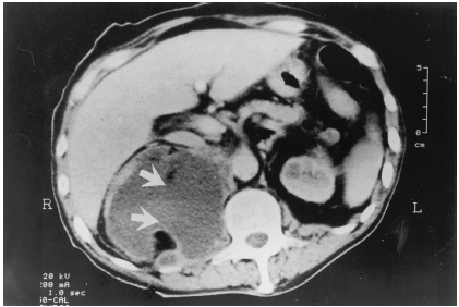
All 16 patients received parenteral antibiotic treatments. Three patients developed conscious disturbance due to severe systemic infection. Surgical drainage by laparotomy was performed in one patient (patient 2) with a gas-forming liver abscess due to poor response to antibiotic therapy for 12 days. He died of sepsis after the surgical intervention. One patient (patient 15) received open surgical drainage because of large abscess formation (18 × 9 × 7 cm) over right retroperitoneal space (). However, he died of ventricular arrhythmia 2 days after the surgical procedure. A total of six patients had needle aspiration and percutaneous drainage procedures in addition to the parenteral antibiotic therapy. Eight patients (50%, 8/16) received only medical treatment. In summary, twelve patients (75%, 12/16) recovered after intensive treatment for two to three weeks. The remaining four patients died of sepsis, ventricular arrhythmia, and upper gastrointestinal bleeding.
Figure 2. Abdominal CT demonstrating large loculated abnormal fluid collection right posterior para-renal region extended into the psoas muscle (arrows). The lesion size about 18 × 9 × 7 cm.
DISCUSSION
Infection is a common cause of morbidity and mortality in hemodialysis patients.Citation[[1]], Citation[[3]], Citation[[9]], Citation[[10]] Previous studies have disclosed that suppression of cell-mediated immunity is one of reasons.Citation[[11]], Citation[[12]], Citation[[13]], Citation[[14]] The incidence of infections in dialysis patients is around 13.7–36%.Citation[[9]], Citation[[10]], Citation[[15]] However, extra-renal abscess in chronic hemodialysis patients is very rare in previous reports.Citation[[16]], Citation[[17]], Citation[[18]], Citation[[19]], Citation[[20]] In our study, the incidence rate of extra-renal abscess was only 0.73% within a study period of 14 years. The majority patients were diabetes mellitus and there was male predominance.
There were no data of organ preference and microbiological characteristics of extra-renal abscess formation in hemodialysis patients in literature. The clinical features of extra-renal abscesses came from sporadic case reports.Citation[[9]], Citation[[16]], Citation[[17]], Citation[[18]], Citation[[19]], Citation[[20]] The results indicated that liver was the site of preference, followed by lung. There was no association of previous liver diseases with the occurrence of the liver abscess formation. Gram-negative bacilli were the major pathogens for extra-renal abscess, especially in diabetic patients. K. pneumoniae was the most common bacteria, and was found in 75% of these cases. E. coli, Proteus vulgaris and anaerobic were 12.5, 6.25, 6.25% respectively. It is very interested to find that there was no Staphylococcus aureus bacteremia in these patients. Staphylococcus is thought to be the most common pathogen in uremic infection. It is likely that Staphylococcus is not prone to extra-renal visceral abscess formation. The microbiological characteristics with the highest incidence rate of K. pneumoniae in pyogenic liver abscess are consistent with the previous reports in non-uremic patients in Taiwan.Citation[[21]], Citation[[22]] The results indicated that the microbiological characteristics of uremic extra-renal abscess were not different from general population. K. pneumoniae is a rare pathogen of septicemia comparing to the S. auras in hemodialysis patients.Citation[[9]] Our results suggested that the presence of K. pneumoniae infection might be a sign of extra-renal abscess formation and further surveillance for abscess might be indicated. The low yield of blood culture positive rate (56%, 9/16; and ) were similar to the previous study in non-uremic patients.Citation[[21]], Citation[[22]] The results indicated that the absence of the positive blood culture result did not exclude the diagnosis of the extra-renal abscess formation in hemodialysis patients.
Several studies have demonstrated that the functional integrity of the immune system is extremely dependent upon optimal nutrition.Citation[[8]], Citation[[23]], Citation[[24]] The serum albumin level and nPCR were much lower in our patients with abscess formation. The reduced nPCR was not associated with the decreased dialysis adequacy, as they had the same kt/V value. The poor might compromise the immunity. The infection itself might also worsen the nutritional status. However, the nutritional status and abscess formation is clearly associated.
We concluded that extra-renal abscess is uncommon in chronic hemodialysis patients. Liver abscess was the most common presentation. Diabetes mellitus and malnutrition were the risk factors. K. pneumoniae was the major pathogen. The prognosis is good with clinical alertness and intensive therapy.
REFERENCES
- Nsouli K.A., Lazarus M., Schoenbaum S.C. Bacteremic Infection in Hemodialysis. Arch. Intern. Med. 1979; 139: 1255–1258
- Wu M.S., Yu C.C., Yang C.W. Poor Pre-Dialysis Glycemic Control is a Predictor of Mortality in Type II Diabetic Patients on Maintenance Hemodialysis. Nephrol. Dial. Transplant 1997; 12: 2105–2110
- Mailloux L.U., Bellucci A.G., Wilkes B.M. Mortality in Dialysis Patients: Analysis of the Causes of Death. Am. J. Kidney Dis. 1991; 18: 326–335
- Sarnak M.J., Jaber B.L. Mortality Caused by Sepsis in Patients with End-Stage Renal Disease Compared with the General Population. Kidney Int. 2000; 58: 1758–1764
- Goldblum S.E., Reed W.P. Host Defenses and Immunologic Alterations Associated with Chronic Hemodialysis. Ann. Intern. Med. 1980; 93: 597–613
- Lees J.A., Falk R.M., Stone W.J. Pyocystis, Pyonephrosis and Perinephric Abscess in End Stage Renal Disease. J. Urol. 1985; 134: 716–719
- Fan P.Y., Schwab S.J. Vascular Access: Concepts for the 1990s. J. Am. Soc. Nephrol. 1992; 3: 1–11
- Wu C.H., Huang C.C., Wu M.S. Total Creatinine Appearance as Indicator of Risk of Infectious Complication in Peritoneal Dialysis. Adv. Perit. Dial. 2000; 16: 219–222
- Quarles L.D., Rutsky E.A., Rostand S.G. Staphylococcus aureus Bacteremia in Patients on Chronic Hemodialysis. Am. J. Kidney Dis. 1985; 6: 412–419
- Fernandez J.M., Carbonell M.E., Mazzuchi N. Simultaneous Analysis of Morbidity and Mortality Factors in Chronic Hemodialysis Patients. Kidney Int. 1992; 41: 1029–1034
- Churchill D.N., Taylor D.W., Cook R.J. Canadian Hemodialysis Morbidity Study. American Journal of Kidney Diseases 1992; 19: 214–234
- Kauffman C.A., Manzler A.D., Phair J.P. Cell-Mediated Immunity in Patients on Long-Term Hemodialysis. Clinical and Experimental Immunology 1975; 22: 54–61
- Lewis S.L., Van Epps D.E. Neutrophil and Monocyte Alterations in Chronic Dialysis Patients. American Journal of Kidney Diseases 1987; 9: 381–395
- Webel M.L., Ritts R.E.J., Briggs W.A. Lymphocyte Blastogenesis in Patients Receiving Hemodialysis. Archives of Internal Medicine 1976; 136: 682–687
- Keane W.F., Shapiro F.L., Raij L. Incidence and Type of Infections Occurring in 445 Chronic Hemodialysis Patients. Transactions – American Society for Artificial Internal Organ 1977; 23: 41–47
- Kolmos H.J. Spinal Epidural Abscess in Patients on Maintenance Hemodialysis (a Presentation of Two Cases). Int. Urol. Nephrol. 1979; 11: 249–253
- Tillman B.F., Gibson R.L., Stone W.J. Psoas Abscess in Chronic Dialysis Patients. J. Urol. 1987; 137: 489–490
- Fonseca V., Baillod R., Berger L. Splenic Abscess in Patients on Hemodialysis. Am. J. Kidney Dis. 1990; 15: 273–275
- Obrador G.T., Levenson D.J. Spinal Epidural Abscess in Hemodialysis Patients: Report of Three Cases and Review of the Literature. American Journal of Kidney Diseases 1996; 27: 75–83
- Witham M., Dittmer I., Williams A. Myocardial Abscess: An Unusual Complication of Long-Term Hemodialysis Line Presence. Clinical Nephrology 1999; 51: 193–194
- Yang C.C., Chen C.Y., Lin X.Z. Pyogenic Liver Abscess in Taiwan: Emphasis on Gas-Forming Liver Abscess in Diabetics. Am. J. Gastroenterol. 1993; 88: 1911–1915
- Lau Y.J., Hu B.S., Wu W.L. Identification of a Major Cluster of Klebsiella pneumoniae Isolates from Patients with Liver Abscess in Taiwan. J. Clin. Microbiol. 2000; 38: 412–414
- Kaminski M.V., Lowrie E.G., Rosenblatt S.G. Malnutrition is lethal, diagnosable, and treatable in ESRD patients. Transplant Proc. 1991; 23: 1810–1815
- Redmond H.P., Shou J., Kelly C.J. Immunosuppressive Mechanisms in Protein-Calorie Malnutrition. Surgery 1991; 110: 311–317