Abstract
Background. A recent report demonstrated that the presence of left ventricular hypertrophy was an independent predictor of mortality in patients with coronary artery bypass grafting (CABG) severely depressed left ventricular function. However, the impact of left ventricle (LV) mass index on the renal and patient outcomes in such patients with CABG has previously not been addressed. The present study thus considers this group of patient and uses LV mass index to assess renal and patient outcomes for these patients. Material and Method. All patients who arrived at the emergency room with severe cardiac dysfunction (EF<60%), triple vessel disease, and required CABG and LV hypertrophy (LVH) (LV mass index γ110 g/m2 in women, γ134 g/m2 in men) were admitted preoperatively to the intensive care unit (ICU) for supportive intervention from 01 1, 1998 to 01 1, 2001. Of all LVH patients, 44 underwent CABG, and were divided into two groups according to LV mass index. Results. Of all patients, 72.7% had severe echocardiographic LVH. The echocardiographic data of both dialysis and non-dialysis groups showed no difference with respect to echocardiographic findings. Histories of myocardial infarction were more frequent in the severe LVH group that in the mild LVH group. As for pre-operative systolic blood pressure and diastolic blood pressure, mean systolic and diastolic blood pressure values were significantly lower in the severe LVH group. Ejection fraction was also significantly lower in the severe LVH group than in the mild LVH group. The patients in the severe LVH group were significantly more likely to have received hemodialysis following CABG surgery (62.5% vs. 33.4%, p<0.05). Mortality was higher in the higher LV mass index group that in the lower LV mass index group (56.2% vs. 25%, p<0.05). Conclusion. Patients with a significantly higher LV mass index usually manifest lower pre-operative blood pressure and poor cardiac function. Consequently, these patients will have a poor renal outcome and higher mortality.
Introduction
Left ventricular dysfunction is considered a major risk factor for operative mortality after coronary artery bypass grafting (CABG).Citation[[1]] Establishing wall thickness and calculating ventricular mass by echocardiographic methods,Citation[[4]] seems important in patients with CABG and poor cardiac dysfunction since long-term arterial hypertension is a well-established risk factor for myocardial infarction and increases the likelihood that a patient needs to receive CABG.Citation[[2]] It is also known to be frequently associated with left ventricular hypertrophy.Citation[[3]] Moreover, echocardiography allows cardiac structure and function to be assessed non-invasively. Among patients with cardiovascular disease, echocardiographic left ventricular (LV) hypertrophy is a frequently observed abnormality that predicts mortality independently of age, diabetes, hypertension, hyperlipidemia, and smoking.Citation[[5]], Citation[[6]] Furthermore, several reports have shown that left ventricular hypertrophy is an independent and powerful predictor of future cardiovascular morbidity in various underlying diseases, including CABG.Citation[[7]], Citation[[8]], Citation[[9]] Among renal failure patients, LV hypertrophy is even more prevalent and is also thought to predict death independently.Citation[[10]]
A recent study indicated that the presence of left ventricular hypertrophy (LVH) is an independent predictor of mortality patients with CABG with severely depressed left ventricular function.Citation[[7]] However, the impact of LV mass index on the renal outcome and patient outcome of CABG has previously not been addressed in this subset of patients. The present study therefore firstly evaluate the relationship between echocardiographic findings and relevant hemodynamic parameters, according to the presence or absence of acute renal failure, and secondly, to use LV mass index to assess renal and patient outcomes.
Patients and Methods
Patients
This study included all patients who arrived at the emergency room with severe cardiac dysfunction (Ejection fraction <60%) and triple vessel disease; required coronary artery bypass grafting (CABG), and were admitted preoperatively to the intensive care unit (ICU) for supportive intervention from January 1, 1998 to January 1, 2001. Patients with only two vessel disease or left main coronary artery disease (CAD), who needed chronic hemodialysis prior to surgery and who were involved in concomitant valvular surgery in a single unit hospital were excluded from the analysis, leaving 44 patients as subjects of this study. All patients had a preoperative LV Ejection Fraction <60%, three vessel disease, and LV hypertrophy (LVH) (LV mass index γ110 g/m2 in women, mass γ134 g/m2 in men).Citation[[11]], Citation[[12]]
Methods
Demographic data were collected, concerning preoperative medical history and information, preoperative echocardiographic studies, preoperative blood pressure and renal outcome for patients with or without acute renal failure, and hemodialysis. For analytical purposes, acute renal failure was defined as a serum creatinine level that exceeded 3.2 mg/dL; as a two-fold creatinine rise in chronic renal failure, after correction for prerenal causes, or as an acute need for renal replacement therapyCitation[[13]], Citation[[14]] following cardiac surgery. Dialysis therapy was initiated by the attending base on the indication of dialysis including volume overload with inadequate control of pulmonary edema via diuretics, insufficient urinary output, and hyperkalemia. Echocardiographic examinations were performed immediately after admission and before operation. M-mode measurements were taken for all patients. The left ventricular internal diameter (LVID), the thickness of the interventricular septum (IVS), and the posterior left ventricular wall (PW) were determined by M-mode measurement, according to widely accepted criteria. These parameters were used in an anatomically validated formula to calculate the left ventricular (LV) mass:Citation[[15]] LV mass (g) = 1.04((IVSβLVIDβPW)3 − (LVID)3) − 13.6 g. Measurements of left ventricular mass were divided by body surface area to obtain the left ventricular mass index. The association between preoperative variables and accepting acute renal failure and hemodialysis was assessed. Preoperative variables included age, sex, presence of myocardial infarction, history of smoking, presence of diabetes, ejection fraction, presence of chronic obstructive pulmonary disease, echocardiographic data including LV, IVS, LV end diastolic diameter (LVEDD), LV end systolic diameter (LVESD), PWT, LV mass, and LV mass index. Of all the LVH patients, 44 patients who underwent CABG were divided into two groups according to LV mass index (+20 g/m2). One group with severe echocardiogenic LVH (LV mass index γ154 g/m2 in men, mass γ130 g/m2 in women) had a larger LV mass index and the other group with mild echocardiogenic LVH had a lower LV mass index (134φLV mass index <154 g/m2 in women, 110φ mass <130 g/m2 in women).
Statistical Analysis
Descriptive analyses are presented as mean ± SD. Continuous variables were compared using the student's t-test or Wilcoxon's rank-sum test, depending on the distribution of the values. Categorical variables were compared using the chi-square test. p less than 0.05 for two-side tests was considered significant.
Results
Baseline Characteristics
During the period of the study, 44 of 90 patients underwent CABG for triple vessel disease and were included in the analysis. Demographic characteristics in all triple vessel disease patients are as follows—mean age was 68.8 ± 0.6 years old, and the ratio of sexes (Male/Female) was 1.1:1 (23/21). Fortynine point five percent of the patients with severe cardiac dysfunction had diabetes; 28.6% had chronic obstructive pulmonary disease, and 71.2% had a history of myocardiac infarction. shows the baseline echocardiographic characteristics of the patients. Seventy two point seven percent of all patients showed severe echocardiographic LVH.
Table 1. Prevalence of echocardiographic abnormalities at baseline
Echocardiographic Findings vs. the Need for Dialysis
shows the echocardiographic data of dialysis and non-dialysis groups. The mean systolic BP and diastolic BP was the same for both groups. The two groups did not differ with respect to all echocardiographic findings, including LV, IVS, LVEDD, LVESD, PWT, aortic diameter (AD), LV mass (326.4 ± 7.4 vs. 327.5 ± 10.9), and LV mass index (170.3 ± 10.4 vs. 171.4 ± 11.6). Moreover, ejection fraction did not reach statistical significance.
Table 2. Comparisons of demographic and echocardiographic parameters in CABG patients between dialysis and non-dialysis groups
Parameters of Potential Relevance to Myocardial Mass
shows that the demographic characteristics do not differ significantly between the severe and mild LVH groups. However, histories of myocardial infarction were more common in the severe LVH group than in the mild LVH group. As for pre-operative systolic blood pressure and diastolic blood pressure, mean systolic and diastolic blood pressures were significantly lower in the severe LVH group than in the mild LVH group. Furthermore, ejection fraction was also noted to be significantly lower in the severe LVH group.
Table 3. The demographic and blood pressure characteristics between the severe and mild left ventricle hypertrophy (LVH) groups
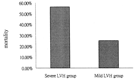
and plot the impact of LV mass index on patient and renal prognosis. In , the patients in the severe LVH group have a significantly higher incidence of receiving hemodialysis after CABG surgery (62.5% vs. 33.4%, p<0.05). reveals a higher mortality in the higher LV mass index group that in the lower mass index group (56.2% vs. 25%, p<0.05).
Figure 1. The severe LVH group has a significantly higher incidence of receiving hemodialysis (HD) after CABG surgery (62.5% vs. 33.4%, p<0.05).
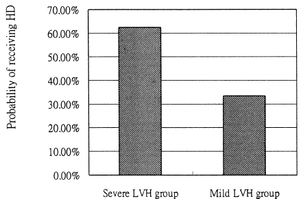
Figure 2. The higher LV mass index group has a higher mortality than the lower LV mass index group (56.2% vs. 25%, p<0.05).
Discussion
Echocardiographic abnormalities, including LV hypertrophy, LV dilation, and systolic dysfunction, are known to be highly prevalent in patients who undergo end stage renal disease therapy. In , the presence of dialysis therapy was used to categorize patients with LVH and CABG. Whether or not patients with CABG accepted hemodialysis, there is no significant difference in echocardiographic parameters included LV echocardiographic parameters included LV mass and LV mass index. However, in , the presence of prominent LVH was used to categorize these patients. Renal prognosis was clearly worse for those with severe LVH (LV mass index >154 g/m2) than for those with mild LVH. The results for this group were very similar to those observed in a population of essentially hypertensive patients and those who angiographically demonstrate coronary artery disease. Therefore, a high LV mass index in pre-CABG patients independently affects renal prognosis. For a given degree of hypertrophy, patients with a higher mass index tended to fare worse. Of the echocardiographic variables tested, LV mass index, especially when exceeding 154 g/m2, was strongly associated with an adverse prognosis.
has important implications. The severe LVH group of CABG patients had higher a percentage of histories of myocardial infarction and significantly lower blood pressure. An interesting study demonstrated that patients with severely depressed left ventricular function hypertensives exhibited lower hospital mortality and a lower incidence of postoperative low cardiac output than normotensive patients.Citation[[7]] This finding is an important message that pre-operative blood pressures are strongly associated with patient outcome. If so, patients in the high LVH group have a lower mean blood pressure and consequently a poorer prognostic outcome. The data presented here are consistent with this expectation.
The principal hypothesis of this study was that a high LV mass index has a poor prognostic effect in patients with CABG and LVH. LV geometry such as LV mass index seems to a major predictor of subsequent outcome, especially in those with CABG and poor LV function. Studies in the pre-operative phase are urgently needed because these abnormalities are already present in many patients at or before surgery. Moreover, a high LV mass index is known to be a major cardiovascular risk factor in patients with coronary artery disease.Citation[[16]] Cooper et al. has also demonstrated that left ventricular mass is a strong predictor of all causes of mortality, independently of the number of obstructed coronary arteries or the left ventricular ejection fraction.Citation[[17]] Of patients undergoing coronary artery bypass surgery, the left ventricular mass was 20% greater in those who died.Citation[[17]] Furthermore, demonstrates the presence of a high LV mass index was a risk factor for mortality after CABG in LVH patients. This result is compatible with much evidence to suggest that severe LV hypertrophy is a marker of poor prognosis in chronically ill patients.
In conclusion, this study has demonstrated that LV hypertrophy is common in patients with triple vessel coronary artery disease and undergoing CABG. Patients with a prominent higher LV mass index usually manifest lower pre-operative blood pressure and poor cardiac function. Consequently, these patients will have a poor renal outcome and higher mortality.
References
- Hammermeister K.E., DeRouen T.A., Dodge H.T. Variables predictive of survival in patients with coronary disease. Selection by univariate and multivariate analyses from the clinical, electrocardiographic, exercise, arteriographic, and quantitative angiographic evaluations. Circulation 1979; 59(3)421–430
- Devereux R.B., Roman M.J. Inter-relationships between hypertension, left ventricular hypertrophy, and coronary heart disease. J. Hypertens. Suppl. 1993; 11(4)S3–S9
- Tingleff J., Munch M., Jakobsen T.J., Torp-Pedersen C., Olsen M.E., Jensen K.H., Jorgensen T., Kirchoff M. Prevalence of left ventricular hypertrophy in a hypertensive population. Eur. Heart J. 1996; 17(1)143–149
- Devereux R.B. Detection of left ventricular hypertrophy by M-mode echocardiography. Anatomic validation, standardization, and comparison to other methods. Hypertension 1987; 9: II9–II26, 2 Pt 2
- Levy D., Garrison R.J., Savage D.D., Kannel W.B., Castelli W.P. Prognostic implications of echocardiographically determined left ventricular mass in the Framingham Heart Study. N. Engl. J. Med. 1990; 322(22)1561–1566
- Lauer M.S., Evans J.C., Levy D. Prognostic implications of subclinical left ventricular dilatation and systolic dysfunction in men free of overt cardiovascular disease (the Framingham Heart Study). Am. J. Cardiol. 1992; 70(13)1180–1184
- Christenson J.T., Simonet F., Schmuziger M. The impact of arterial hypertension on the results of coronary artery bypass grafting. Thorac. Cardiovasc. Surg. 1996; 44(3)126–131
- Levy D., Garrison R.J., Savage D.D., Kannel W.B., Castelli W.P. Prognostic implications of echocardiographically determined left ventricular mass in the Framingham Heart Study. N. Engl. J. Med. 1990; 322(22)1561–1566
- Sullivan J.M., Vander Zwaag R.V., el-Zeky F., Ramanathan K.B., Mirvis D.M. Left ventricular hypertrophy: effect on survival. J. Am. Coll. Cardiol. 1993; 22(2)508–513
- Silberberg J.S., Barre P.E., Prichard S.S., Sniderman A.D. Impact of left ventricular hypertrophy on survival in end-stage renal disease. Kidney Int. 1989; 36(2)286–290
- Ganau A., Devereux R.B., Roman M.J., de Simone G., Pickering T.G., Saba P.S., Vargiu P., Simongini I., Laragh J.H. Patterns of left ventricular hypertrophy and geometric remodeling in essential hypertension. J. Am. Coll. Cardiol. 1992; 19(7)1550–1558
- Hammond I.W., Devereux R.B., Alderman M.H., Lutas E.M., Spitzer M.C., Crowley J.S., Laragh J.H. The prevalence and correlates of echocardiographic left ventricular hypertrophy among employed patients with uncomplicated hypertension. J. Am. Coll. Cardiol. 1986; 7(3)639–650
- Tran D.D., Groeneveld A.B., van der Meulen J., Nauta J.J., Strack van Schijndel R.J., Thijs L.G. Age, chronic disease, sepsis, organ system failure, and mortality in a medical intensive care unit. Crit. Care Med. 1990; 18(5)474–479
- Cioffi W.G., Ashikaga T., Gamelli R.L. Probability of surviving postoperative acute renal failure. Development of a prognostic index. Ann. Surg. 1984; 200(2)205–211
- Devereux R.B., Reichek N. Echocardiographic determination of left ventricular mass in man. Anatomic validation of the method. Circulation 1977; 55(4)613–618
- Ghali J.K., Liao Y., Simmons B., Castaner A., Cao G., Cooper R.S. The prognostic role of left ventricular hypertrophy in patients with or without coronary artery disease. Ann. Intern. Med. 1992; 117(10)831–836
- Cooper R.S., Simmons B.E., Castaner A., Santhanam V., Ghali J., Mar M. Left ventricular hypertrophy is associated with worse survival independent of ventricular function and number of coronary arteries severely narrowed. Am. J. Cardiol. 1990; 65(7)441–445