Abstract
Acute renal failure associated with scrub typhus infection is not rare as previously thought. The possibility of scrub typhus should be borne in mind when patients present with fever and varying degrees of acute renal failure, particularly if an eschar exists, along with a history of environmental exposure in an area like Taiwan, where scrub typhus is endemic. Prompt diagnosis and the use of appropriate antibiotics can rapidly alter the clinical course of the disease and prevent the development of serious or fatal complications. To illustrate the above point, this study reports 3 cases of scrub typhus associated with acute renal failure. They were seen at Chang Gung Memorial Hospital in a 2-year interval. Case 1 was referred from district hospital with clinical features of multiple organ dysfunctions, including shock, fever, acute respiratory failure, acute renal failure, and acute hepatitis. Case 2 was admitted with the chief problems of shock, fever, acute renal failure, and DIC. Case 3 visited our outpatient clinic due to fever, maculopapular rash and acute renal failure. In all these patients, the diagnosis was confirmed using immunofluorescence techniques, which showed that Orientia tsutsugamushi had an IgM titer of 1:80 or greater. Notably, despite having varying degrees of acute renal deterioration, the patients responded very well to doxycycline therapy and recovered completely. Additionally, a total of 4 similar cases of scrub typhus associated with acute renal failure were reviewed from th past literature.
Introduction
Scrub typhus, also called tsutsugamushi disease or chigger-borne typhus, is an acute febrile illness caused by Orientia tsutsugamushi, a gram-negative obligate intracellular organism that had, until recently, been classified within the genus Rickettsia. Orientia tsutsugamushi is transmitted to humans by the bite of the larval stage of the trombiculid mite, belonging to the genus Leptotrombidium. The larval mites, also known as chiggers, maintain the infection through successive generations via transovarial transmission. Certain species of rats, tree shrews, birds, and monkeys form the major reservoirs of this organism. Human infection arises from accidents attributable to close contact, placing those involved agricultural activities on a daily basis at high risk.Citation[[1]], Citation[[2]], Citation[[3]]
The clinical spectrum of scrub typhus is wide, ranging from mild and probably subclinical to severe and prostrating courses. Most of the infected patients have a benign course.Citation[[2]], Citation[[3]] Fever, headaches and myalgia begin about 1–3 weeks after a bite by an infected mite. An eschar develops in over half of patients at the site of the original bite, usually located at pressure points and in moist skin folds.Citation[[2]], Citation[[3]] The bite of the chigger forms an erythematous papule, which soon develops into a small bulla, usually overlying a pressure point. The blister eventually sloughs leaving an ulcer. A black eschar then develops and is surrounded by a 1–2 cm ring of red tissue.Citation[[2]], Citation[[3]] Additionally, a non-pruritic maculopapular rash, first appearing on the trunk and then radiating outwards, occurs in approximately half of patients, along with the constitutional symptoms. General and regional lymphadenopathy follows, and the nodes draining the primary lesion may become tender and eventually suppurate.Citation[[2]], Citation[[3]] Occasionally, patients suffer serious complications resulting from rickettsemia, where the invasion of Orientia tsutsugamushi into the endothelial cells causes disseminated multi-organ vasculitis. Depending on the organ involved, patients might present with pneumonitis,Citation[[4]] acute respiratory distress syndrome (ARDS),Citation[[5]] hemophagocytic syndrome,Citation[[6]], Citation[[7]] meningitis, encephalitis,Citation[[8]], Citation[[9]] myocarditis,Citation[[10]] acute pulmonary edema,Citation[[11]] pericarditis,Citation[[12]] hepatitis,Citation[[13]] acute renal failure,Citation[[14]], Citation[[15]], Citation[[16]], Citation[[17]] or even multiple organ failure.Citation[[15]], Citation[[16]], Citation[[17]] Respiratory distress and encephalitis are the principal cause of death in patients with severe disease.Citation[[15]]
The disease has been reported from various parts of Southeast Asia, Australia, Japan, and certain Southwest Pacific islands.Citation[[1]], Citation[[2]], Citation[[3]] Taiwan was once considered to be an area of endemic scrub typhus, although the incidence of this disease has declined with industrialization. However, scrub typhus is still under-diagnosed in Taiwan today. Since the medical literature states that kidney involvement from scrub typhus is rare, Taiwanese physicians seldom make a correct diagnosis even when they do come across the disease. Recently we encountered 3 patients who presented with acute renal failure after scrub typhus infection, and this has prompted us to review literature of renal manifestation of the disease.
Patients and Methods
Patients
Between January 2000 and December 2001, 39 patients at Lin-Kou Medical Center of Chang Gung Memorial Hospital had a diagnosis of scrub typhus confirmed serologically. Three of the patients were presented with predominately renal manifestations and were treated by our nephrologists, but all exhibited various degrees of renal function deterioration. These three patients denied any previous renal disease previously. Lin-Kou Medical Center is situated in Taoyuan, about 15 km from Taipei city and is a referral center for many district hospitals. Hospital records show that cases of florid scrub typhus have become very rare since Taoyuan is now a well-developed city. Meanwhile, patients presenting with scrub typhus with minor complications were easily treated at district hospitals, and thus were seldom been referred to Chang Gung Memorial Hospital.
A total of 4 cases of scrub typhus associated with acute renal failure were found from Medline from 1980 to 2001.Citation[[14]], Citation[[15]], Citation[[16]], Citation[[17]] Similarly, all patients exhibited various degrees of renal function deterioration after Orientia tsutsugamushi infection. They also did not have a renal disease in the past time.
Methods
For the 3 patients studied herein, all serum hematological and biochemical tests were conducted in the clinical pathology laboratories of Chang Gung Memorial Hospital using routine automated techniques. To confirm the diagnosis of scrub typhus, serum samples were also sent to the Center for Disease Control at the Taiwanese Ministry of Health, for testing for immunoglobulin M (Ig M) antibody to Orientia tsutsugamushi, strains Kato, Karp and Gilliam using micro-immunofluorescence. The criteria for a diagnosis of scrub typhus was an Ig M titer for orientia tsutsugamushi of 1:80 or greater. Renal biopsy was not performed in these 3 patients. Instead, skin biopsy was performed in Case 3 and the specimens were submitted for light microscopic study.
Results
Report of Case 1
A 72-year-old, healthy man had been working as a laborer in a forested mountainous area since his youth. Seven days before presenting at the district clinic he began suffering from fever, headaches, and myalgia. These initially symptoms were followed a day later by vague abdominal pain and jaundice discoloration. Urine output was decreasing day by day, and his daughter told us that it was around 500 mL. Symptoms of respiratory distress gradually developed and the man finally became confused. Upon presentation at a district clinic, the patient was immediately sent to our emergency department. On arrival, blood pressure was 85/55 mmHg, and a high body temperature of up to 39.4°C was noted. The patient displayed evident of respiratory distress, with a respiratory rate of up to 30/min. Physical examination found an ill-looking and confused man. An ecchymotic 3-cm plaque with a 1-cm black eschar was found over the right lower back. There were no palpable lymph nodes, and the abdomen was found to be soft and flat, with mild tenderness on light pressure over the epigastric area. No other physical abnormalities were found. Arterial blood gas while breathing from an oxygen mask with FIO2 50% revealed PH: 7.010, PCO2: 45.3 mmHg, PO2: 68 mmHg, HCO3: 11.8 mmHg. Endotracheal intubation was performed immediately, and the patient was then transferred to the medical intensive care unit (MICU) for further care.
Laboratory tests from MICU revealed the following results. A hemogram showed hemoglobin (HB) 13.0 g/dL; platelet (PLT) 165,000/µL; white blood cell (WBC) 13,400/µL; 92% segment form neutrophil. Blood biochemistry showed: blood urea nitrogen (BUN) 74 mg/dL (normal range 6–21 mg/dL); creatinine (CRE) 7.1 mg/dL (normal range 0.4–1.2 mg/dL); aspartate aminotransferase (AST) 49 IU/L (normal range 0–34 IU/L); alanine aminotransferase (ALT) 48 IU/L (normal range 0–36 IU/L); bilirubin total/direct (BIL-T/D) 6.7/4.0 mg/dL (normal range 0–1.3 mg/dL); alkaline phosphatase (ALP) 310 mg/dL (normal range 28–94 mg/dL); gamma-glutamyl transferase (GGT) 118 mg/dL (normal range 0–26 mg/dL); albumin (ALB) 2.7 g/mg/dL (normal range 3.5–5.5 g/dL); total protein (TP) 5.3 g/dL (normal range 6.3–8.0 g/dL). Meanwhile, urinalysis showed protein 1+, blood −, bilirubin 1+, 2–4 RBC cells/HPF, and 1–2 WBC cells/HPF. The chest X-ray was normal. Renal ultrasound revealed normal-sized kidneys with increased echogenicity. Accordingly, the patient was found to have multiple organ dysfunctions, including shock, fever, acute respiratory failure, acute renal failure, and acute hepatitis. The patient was treated aggressively with a saline infusion, dopamine infusion, and broad-spectrum antibiotic injections as well as other supportive medications. The blood pressure well again later and the dopamine was tapered off successfully on the 2nd day. At the MICU, his urine output was around 600–700 mL with intravenous furosemide.
The fever persisted. Scrub typhus was highly suspected following consultations with an infection specialist and dermatologist, and the man was prescribed doxycycline 100 mg bid from his 3rd hospital day. The patient responded dramatically to this treatment, and the fever vanished after 1 day of doxycycline treatment. On the 5th hospital day, the endotracheal tube was extubated successfully, and the next day saw the man transferred to an ordinary ward. The patient recovered smoothly from then on with acute hepatitis being resolved by supportive therapy. Urine output increasing day by day, and BUN was 12 mg/dL and CRE was 0.9 mg/dL on the 13th hospital day. Notably, the micro-immunofluorescence IgM antibody against rickettsia tsutsugamushi was found to be positive with a titer of ≥1:80 after 2 week. The man was discharged 3 weeks after admission.
Report of Case 2
A 37 year-old, healthy housewife had visited her hometown in a rural area 3 weeks previously and had helped on the family farm. The woman began to suffer from fever, myalgia, dry cough and watery diarrhea from 10 days after her visit, and visited our emergency department on a winter evening. Of note, she denied any decrement in her urine output. On arrival, the patient appeared lethargic and emaciated. Blood pressure was 90/50 mmHg, and body temperature was 39.0°C. A thorough examination found no eschar on her skin, but a 1-cm lymph node was found over the right neck area. Basal rales were audible over both lower lung fields, and heart sounds were normal. A soft and protruded abdomen was noted, with shifting dullness on percussion. No lower leg edema was found. The patient was thus admitted to our nephrology ward for further evaluation and treatment.
Laboratory investigations revealed the following results. A hemogram showed HB 12.4 g/dL; PLT 35,000/µL; WBC 10,200/µL; 72% segment neutrophil. Meanwhile, DIC profiles revealed a prothrombin time of 16.4 s (12.9 s of control); activated partial thromboplastin time of 30.1 s (26.0 s of control); thrombin time of 19.0 s (17.0 s of control); fibrinogen 7.03 g/L; fibrin degradation product >40 mg/L and D-dimer >1000 g/L. Blood biochemistry showed BUN 56 mg/dL; CRE 2.1 mg/dL; AST 12 IU/L; ALT 13 IU/L; BIL-T/D 1.1/0.3 mg/dL; ALP 31 mg/dL; GGT 18 mg/dL; ALB 2.0 g/dL, and TP 3.3 g/dL. Furthermore, urinalysis showed protein 1+, blood +, bilirubin −, 10–12 RBC cells/HPF, and 1–2 WBC cells/HPF. Chest X-ray displayed minimal pleural effusion over both sides. Renal ultrasound revealed normal-sized kidneys with increased echogenicity as well as moderate ascites.
The preliminary diagnosis was shock, fever, acute renal failure and DIC, and the patient was treated with saline supplementation, dopamine infusion and broad- spectrum antibiotics. The blood pressure well again later and the dopamine was tapered off successfully on the 2nd day. Ascites and pleural effusion studies revealed transudate findings mainly comprised of reactive mesothelial cells on cytologic examination. Biopsy of the lymph node over the right neck discovered dermatopathic lymphadenopathy. Bone marrow biopsy was also performed, revealing megakaryocytic hypercellularity and some plasma cells infiltration.
Meanwhile, the fever persisted. The patient was prescribed a trial of doxycycline 100 mg bid from the 7th hospital day, and responded very well to this treatment, with the fever resolving itself after 2 days. Renal function returned to normal, and her BUN was 1.0 mg/dL and CRE was 0.7 mg/dL on the 11th hospital day. Follow studies found normal DIC profiles. Notably, the micro-immunofluorescence IgM antibody against rickettsia tsutsugamushi was positive at a titer of ≥1:80 on the 21st hospital day. The woman was discharged 4 weeks after admission.
Report of Case 3
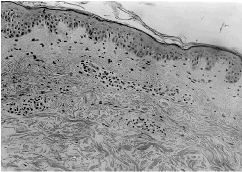
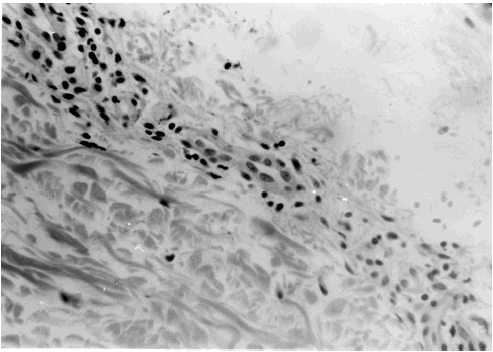
A 69 year-old male patient had been mountain climbing the week before presenting with intermittent fever and chills, accompanied by headaches and general myalgia, developing over the previous 4 days. The man had a history of hypertension and angina pectoris dating back 4 years ago, but his general medical condition had been stable to date. He denied any decrement in urine output so far. On arrival at our clinic, physical examination revealed a well-nourished old man with clear consciousness. Blood pressure was 140/90 mmHg and body temperature was 39.5°C. Notably, an ecchymotic 3-cm plaque with a 2-cm black eschar was found over the left chest wall. A maculopapular rash was found on her trunk. No lymphadenopathy was palpated. No other physical abnormalities were found. Laboratory tests revealed the following results. A hemogram showed HB 14.4 g/dL; PLT 180,000/µL; WBC count 4700/µL; 68% segment form neutrophil. Blood biochemistry found: BUN 30 mg/dL; CRE 1.8 mg/dL; AST 228 IU/L; ALT 238 IU/L; BIL-T/D 0.5/0.1 mg/dL; ALP 160 mg/dL; GGT 18 mg/dL; ALB 3.7 g/dL; and TP 7.0 g/dL. Urinalysis showed protein 1+, blood −, bilirubin −, 10–12 RBC cells/HPF, and 2–4 WBC cells/HPF. The chest X-ray was normal. Renal ultrasound found normal-sized kidneys with normal echogenicity. After consultation with infection specialists and dermatologists, scrub typhus was impressed and the patient was given doxycycline 100 mg bid. Meanwhile, a skin biopsy of the maculopapular rash was performed for pathology examination. The fever disappeared after 2 days of treatment. The patient returned to our clinic 2 weeks after treatment, which time his BUN was 14 mg/dL, and CRE was 1.1 mg/dL. As shown in and , the pathological report from the skin biopsy revealed perivascular mononuclear cells infiltration. Finally, the micro-immunofluorescence IgM antibody against rickettsia tsutsugamushi was positive at a titer of ≥1:80, further confirming the diagnosis of scrub typhus.
Report of Case 4–Case 7
Hsu et al.Citation[[14]] reported a 25 year-old male who had scrub typhus with the unusual complication of acute renal failure. The renal deterioration was described as acute, severe and oliguric. The patient had a complete recovery after tetracycline therapy. Watt and StrickmanCitation[[15]] reported a 51-year-old American traveler who developed multi-organ failure including ARDS that required prolonged ventilation and tracheostomy, renal failure that required dialysis, meningoencephalitis with extended coma, bilaterally decreased vision and hearing as well as hyperbilirubinemia after returning from Thailand. The correct diagnosis was made retrospectively and the antirickettsia agent was not administrated during hospitalization. Additionally, Chi et al.Citation[[16]] reported a 48 year-old male farmer who suffered scrub typhus with unusual and serious multiple organ involvement, including tubulointerstitial nephritis with acute renal failure, interstitial pneumonitis with ARDS, disseminated intravascular coagulation, liver function impairment, upper gastrointestinal bleeding, prolonged hyperamylasemia and hyperlipasemia. This patient received three sessions of hemodialysis therapy. The administration of chloramphenicol rapidly altered the clinical course of the patient's disease, but renal impairment and prolonged hyperamylasemia and hyperlipasemia persisted for 10 months. Finally, Cracco et al.Citation[[17]] reported a 32 year-old woman in whom probable scrub typhus was complicated by life-threatening ARDS with multiple organ failure. The acute renal failure was especially notable, and required continuous venovenous hemodiafiltration for life support. This patient also recovered fully after ofloxacin therapy.
Summary of Results
summarizes the demographic data and clinical courses of the 3 patients studied herein as well as the 4 similar patients from past literature.Citation[[14]], Citation[[15]], Citation[[16]], Citation[[17]] As listed in , all of them exhibited acute renal failure, with clinical symptoms onset between 3 to 21 days after orientia tsutsugamushi infection. Constitutional symptoms, for example fever, headache and myalgia were presented in all patients (100%). Eschar was presented in 4 out of 7 patients (57.1%). Maculopapular rash was presented in 2 out of 7 patients (28.6%). Depending on the organs involved, patients might present with acute renal failure alone or multi-organ failure that including renal failure, respiratory failure (42.9%), shock (42.9%), central nervous system infection (14.3%), hepatitis (71.4%), pancreatitis (14.3%), or DIC (28.6%). The duration of renal failure lasted 11–300 days with peak serum creatinine ranged from 1.8 mg/dL to 8.6 mg/dL. Sometimes, the renal failure was severe and oliguric, necessitating hemodialysis for life support in 3 out of 7 patients (42.9%). Urinary analysis revealed either mild proteinuria or microscopic hematuria. Meanwhile, renal ultrasound generally found normal-sized kidneys with either normal or increased echogenicity. Percutaneous renal biopsies were performed in 2 patients, and the pathology reports showed minimal mesangial hyperplasia and tubulointerstitial nephritis. Renal biopsy was not performed in our 3 patients studied herein since they hesitated to receive such an invasive procedure. Biopsy of maculopapular rash in Case 3 revealed perivascular mononuclear cells infiltration after H & E stain. Importantly, although all of the patients suffered varying degrees of acute renal deterioration, they responded dramatically to anti-rickettsia therapy once a correct diagnosis was made, and all recovered full or partial renal function. Case 4 was diagnosed retrospectively and the appropriate antibiotic was not prescribed at that time. The fate of his renal function, however, was not mentioned in the article.
Table 1. The demographic data and clinical courses of the 3 patients studied herein as well as 4 patients from past literature
Discussion
Our data showed that acute renal failure associated with scrub typhus is not rare as previously thought. As you can see, 3 out of 39 patients (7.7%) studied herein had acute renal failure after Orientia tsutsugamushi infection. Though smaller in case's number, literatureCitation[[14]], Citation[[15]], Citation[[16]], Citation[[17]] had reported the occurrence of this renal complication. Importantly, the renal failure was severe and oliguric, necessitating hemodialysis for life support in 3 out of 7 patients (42.9%). Although all of the patients suffered varying degrees of acute renal deterioration, they responded very well to anti- rickettsia therapy once a correct diagnosis was made, and all recovered full or partial renal functions. Renal complications may cause prolonged morbidity or even mortality if diagnosis is delayed. Therefore, it's paramount important to have a correct diagnosis to enable initiation of anti-rickettsia therapy.
Constitutional symptoms were presented in all 7 patients (100%). Eschar was presented in 4 out of 7 patients (57.1%) while the maculopapular rash was presented in 2 out of 7 patients (28.6%). Though small in case number, it seem that the incidence of constitutional symptoms and eschar in patients with renal failure are not much different from those without renal failure. On the contrary, a lower incidence of maculopapular rash is noted in patients with renal failure.
Urinary analysis revealed either mild proteinuria or microscopic hematuria. Renal ultrasound found normal-sized kidneys with either normal or increased echogenicity. Hence, the accurate diagnosis of this disease is depending on other relevant clinical features. In this regard, the clinical manifestations, the history of recent exposure in an area or country with sporadic occurrence of tsutsugamushi disease, the finding of a bite site, and serological tests are extremely helpful in diagnosing this disease. The 3 patients studied herein and 4 patients from literature are considered high risk for scrub typhus because of their involvement in occupational or recreational behavior that brought them in contact with mite-infested grassy and brushy habitats in rural areas of TaiwanCitation[[14]], Citation[[16]] and Thailand.Citation[[15]], Citation[[17]]
The basic pathologic lesions are characterized by focal or disseminated multiorgan vasculitis of the small blood vessels. Because the rickettsiae invade endothelial and possibly smooth muscle cells, a vasculitis is seen histologically.Citation[[3]] The initial target is the microcirculation (i.e., capillaries and venules), but with time and disease spread, the contiguous arteries and veins are affected. Early lesions contain swelling of the endothelial cells and vessels and accumulation of mononuclear inflammatory cells. As vascular cells become necrotic, macrophages and extravasated erythrocytes are found. Finally, fibrin thrombi occlude the vessels leading to the clinical findings of infarction and gangrene if the involved vessel is large enough.Citation[[3]] Systemic vasculitic lesions are also found and are essentially identical to those of the skin.Citation[[3]] Biopsy of maculopapular rash in Case 3 revealed perivascular mononuclear cells infiltration. This is compatible with invasion of Orientia tsutsugamushi into the endothelial cells of skin that causes perivasculitis.
There are several possible mechanisms that may lead to acute renal failure after Orientia tsutsugamushi infection. First, of course is vasculitis. As the vasculitis comprises the basic pathologic changes, one may try to look for vasculitic changes in renal biopsy specimens. However, no evidence of vasculitic lesions is found in Case 4 and Case 6 who underwent renal biopsies during their hospitalization. Instead, the results revealed minimal mesangial hyperplasia and tubulointerstitial nephritis. Either these changes are due to a direct invasion of the renal parenchyma by the microorganism, or to a reactive response of the kidney to a systemic bacteria infection is not known at the present time. Lack of such vasculitic lesions in Case 4 and Case 6, however, did not exclude the possibility since there are only 2 patients who had underwent renal biopsy in this study. Second is shock. As Case 1, Case 2 and Case 7 had septic shock in their initial presentations; this may contribute to acute renal failure after compromising renal perfusions. Third is DIC. DIC may play a role in developing acute renal failure as Case 2 and Case 6 developed DIC during active Orientia tsutsugamushi infection. In DIC, elevated fibrin-split products are known to be associated with fibrin deposition in the microcirculation. Therefore, thrombosis and coagulation will induce microangiopathy in multiple organs and lead to multiple organ injuries. Fourth is volume depletion. Volume depletion can occur if the Orientia tsutsugamushi-induced diarrhea is severe or patients are unable to take enough water after the insults. Volume depletion, by compromising renal perfusions, may also contribute to acute renal failure. Case 4 had marked dehydration on arrival.
Confirmation of Orientia tsutsugamushi infection in 3 patients studied herein was by serology test. Serological diagnosis of scrub typhus depends on demonstrating an antibody elevation to Orientia tsutsugamushi antigens, with a specific IgM titer of the indirect immunofluorescent test of greater than 1:50 being recognized as significant.Citation[[18]] Alternatively, the diagnosis is achieved by demonstrating either (1) a fourfold or greater rise in indirect immunofluorescent or indirect immunoperoxidase antibody titers between paired serum samples to at least 1:200, or (2) a single or stable indirect immunofluorescent or indirect immunoperoxidase antibody titer of 1:400 or greater.Citation[[19]] Though relatively simple to use, the Weil–Felix test has been demonstrated to be neither sensitive nor specific in the serodiagnosis of scrub typhus, and thus is no longer used in most countries. In Taiwan, the criteria for diagnosis of scrub typhus is an Ig M titer for orientia tsutsugamushi of 1:80 or greater.
Immunofluorescence and immunoperoxidase techniques are commonly used to diagnose scrub typhus.Citation[[18]], Citation[[19]] However, diagnosis is sometimes difficult in the early stage of the illness, when the antibody titers are too low to be detected. Polymerase chain reaction (PCR) method is especially helpful when applying immunological techniques to diagnose early rickettsia disease is difficult. For example, PCR can reveal rickettsial infection during the acute rickettsemia phase, which occurs before the antibody titer increases,Citation[[20]], Citation[[21]] allowing diagnosis during the early phase of the illness. However, in Taiwan the PCR method is currently only available in certain selected research laboratories. Furthermore, this method is expensive and not covered by Taiwan's National Insurance system.
Generally, prompt anti-rickettsial chemotherapy causes almost immediate defervescence, usually within 24–48 h, and rapid resolution of the disease process, thus virtually eliminating mortality. The drugs of choice are chloramphenicol and tetracycline,Citation[[22]] with tetracycline eliminating the fever and other symptoms faster than chloramphenicol.Citation[[22]] Of the tetracycline congeners, doxycycline and monocycline are considered the most active anti-rickettsia agents. Both drugs also enjoy a long life in the serum and, because of their high degree of lipid solubility, penetrate numerous body tissues. However, the safety of minocycline has been questioned owing to reports of vestibular toxicity.Citation[[23]] In contrast, doxycycline is relatively free of deleterious effects, and has proven useful in treating rickettsial infection.Citation[[22]] Some reports have also proven ciprofloxacin to be effective.Citation[[17]], Citation[[24]] As displays, although these 7 patients exhibited varying degrees of acute renal deterioration, they responded very well to these anti-rickettsia therapy and recovered either fully or partially.
Scrub typhus, including the acute renal failure sometimes associated with it, is currently under-diagnosed in Taiwan and the other countries. Several important reasons for the correct diagnosis are easily missed. First, physicians are unfamiliar and unaware of the disease entity, and know little about the natural course of the disease. Second, the disease has protean manifestations, which affect the lung, brain, heart, liver, kidney, and other organs, thus opening up a wide range of alternative diagnosis. Third, the literature has rarely described reports of acute renal failure related to scrub typhus infection, making physicians less likely to consider the possibility of it inducing renal deterioration of their patients. Fourth, IFA testing is not usually available, an inconvenience that prevents physicians from send suspicious blood samples for IFA testing. At present, there is no rapid diagnostic kit commercially available in Taiwan.
In conclusion, acute renal failure associated with scrub typhus is not rare as previously thought. The possibility of scrub typhus should be borne in mind when diagnosing patients presenting with fever and varying degrees of acute renal failure, particularly if there is an eschar and a history of environmental exposure in a scrub typhus endemic area like Taiwan. In Taiwan, the disease can be serologically confirmed only at the Center for Disease Control of Ministry of Health and it take 2–3 weeks for the result to come out. Treatment therefore must be presumptive, but the benefits of avoiding severe scrub typhus by early administration of antibiotics generally far outweigh the risks of 1-week course of doxycycline.
Acknowledgment
The kind staff of the Infection Control Committee of Chang Gung Memorial Hospital is thanked for their excellent help in data collection.
References
- Rapmund G. Rickettsia disease of the Far East: new perspectives. J. Infect. Dis. 1984; 149: 330–338
- McDonald J.C., MacLean J.D., McDade J.E. Imported rickettsia disease: clinical and epidemiologic features. Am. J. Med. 1988; 85: 799–805
- Boyd A.S., Neldner K.H. Typhus disease group. Int. J. Dermatol. 1992; 31: 823–832
- Chayakul P., Panich V., Silpapojakul K. Scrub typhus pneumonitis: an entity which is frequently missed. Q. J. Med. 1988; 68: 595–602
- Lee W.S., Wang F.U., Wang L.S., Wong W.W., Young D., Fung C.P., Liu C.Y. Scrub typhus complicating acute respiratory distress syndrome: a report of two cases. Chin. Med. J. (Taipei) 1995; 56: 205–210
- Iwasaki H., Hashimoto K., Takada N., Nakayama T., Ueda T., Nakamura T. Fulminant rickettsia tsutsugamushi infection associated with hemophagocytic syndrome. Lancet 1994; 343: 1236
- Chen Y.C., Chao T.Y., Chin J.C. Scrub typhus associated hemophagocytic syndrome. Infection 2000; 28: 178–179
- Silpapojakul K., Ukkachoke C., Krisanapan S., Silpapojakul K. Rickettsia meningitis and encephalitis. Arch. Intern. Med. 1991; 151: 1753–1757
- Pai H., Sohn S., Seong Y., Kee S., Chang W.H., Choe K.W. Central nervous involvement in patients with scrub typhus. Clin. Infect. Dis. 1997; 24: 436–440
- Yotsukura M., Aoki N., Fukuzumi N., Ishikawa K. Review of a case of tsutsugamushi disease showing myocarditis and confirmation of rickettsia by endomyocardial biopsy. Japanese Circ. J. 1991; 55: 149–153
- Walker D.H., Mattern W.D. Rickettsia vasculitis. Am. Heart J. 1980; 100: 896–906
- Chang J.H., Ju M.S., Chang J.E., Park Y.S., Han W.S., Kim I.S., Chang W.H. Pericarditis due to tsutsugamushi disease. Scand. J. Infect. Dis. 2000; 32: 101
- Chien R.N., Liu N.J., Lin P.Y., Liaw Y.F. Granulomatous hepatitis associated with scrub typhus. J. Gastroenterol Hepatol. 1995; 10: 484–487
- Hsu G.J., Young T.G., Peng M.Y., Chang F.Y., Chou M.Y., Sheu L.F. Acute renal failure associated with scrub typhus: report of a case. J. Formos. Med. Assoc. 1993; 92: 475–477
- Watt G., Strickman D. Life-threatening scrub typhus in a traveler returning from Thailand. Clin. Infect. Dis. 1994; 18: 624–626
- Chi W.C., Huang J.J., Sung J.M., Lan R.R., Ko W.C., Chen F.F. Scrub typhus associated with multiorgan failure: a case report. Scand. J. Infect. Dis. 1997; 29: 634–635
- Cracco C., Delafosse C., Baril L., Lefort Y., Morelot C., Derenne J.P., Bricaire F., Similowski T. Multiple organ failure complicating probable scrub typhus. Clin. Infect. Dis. 2000; 31: 191–192
- Bozman F.M., Elishbeng B.L. Serological diagnosis of scrub typhus by indirect immunofluorescence. Proc. Soc. Exp. Bio. Med. 1963; 112: 568–673
- Brown G.W., Shirai A., Rogers C., Groves M.G. Diagnostic criteria for scrub typhus: probability values for immunofluorescent antibody and Proteus OXK agglutinin titers. Am. J. Trop. Med. Hyg. 1983; 32: 1101–1107
- Furuya Y., Yoshida Y., Katayama T., Kawamori F., Yamamoto S., Ohashi N., Tamura A., Kawamura A. Specific application of rickettsia tsutsugamushi DNA from clinical specimens by polymerase chain reaction. J. Clin. Microbiol. 1991; 29: 2628–2630
- Sugita Y., Nagatani T., Yoshida Y., Nakajima H. Diagnosis of typhus infection with rickettsia tsutsugamushi by polymerase chain reaction. J. Med. Microbiol. 1992; 37: 357–360
- Sheehy T.W., Hazlett D., Turk R.E. Srub typhus: a comparison of chloramphenicol and tetracycline in its treatment. Arch. Intern. Med. 1973; 32: 77–80
- Fanning W.L., Gump D.W., Sofferman R.A. Side effects of minocycline: a double blind study. Antimicrob. Agents Chemother. 1977; 11: 712–717
- Eaton M., Cohen M.T., Shlim D.R. Ciprofloxacin treatment of typhus. JAMA 1989; 262: 772–773

