Abstract
Background. Vascular access failure is a severe and common complication for hemodialysis patients. The possible vascular access sites are limited in dialysis patients. Axillary artery to contralateral axillary vein arteriovenous fistula (AVF) is one of the possibilities. However, the clinical outcome of this procedure is still un-defined. Object. The purpose of this study is to review the clinical outcome of axillary artery to contralateral axillary vein AVF as a hemodialysis vascular access. Patients and Methods. We retrospectively reviewed native or graft arteriovenous fistula records for chronic hemodialysis patients at Chang Gung Memorial Hospital in Kaohsiung, Taiwan, from 01 1986 to 03 2001. Records were reviewed for all chronic hemodialysis patients, with more than 2000 individuals receiving more than 10,000 fistulas. Eight patients received axillary artery to contralateral axillary vein AVF. Results. The mean age for these patients was 61.7 ± 16.3 year-old at time of surgery. All patients had received multiple native or graft arteriovenous fistula creation. The 2-year and 4-year AVF graft survival is 87.5% and 43.8% respectively. One patients developed brachial plexopathy after operation. Another patient had venous hypertension distal to the AVF site. Both patients were managed conservatively. There is no AVF-related mortality in these patients. Conclusion. We conclude that axillary artery to contralateral axillary vein graft fistula may be a feasible alternative choice for chronic hemodialysis access.
Introduction
Fistula dysfunction causes a severe problem for chronic hemodialysis patients despite advances in surgical techniques and progressive development of graft materials. The common finding in failed arteriovenous fistula (AVF) is the irreversible endothelial intimal hyperplasia,Citation[[1]], Citation[[2]], Citation[[3]] which limits the repeated AVF creation on the same site. Upper extremities are the preferred sites for the AVF. The major drawback of the upper extremities' AVF is the limited sites for repeated AVF creation. Wrist, elbow, and axillary area are the sites of usual AVF creation. However, some patients with poor AVF function need more AVF sites than these areas. Axillary artery to contralateral axillary vein AVF creationCitation[[4]], Citation[[5]] is one of the options for further AVF creation sites. However, the clinical outcome of this procedure is still unclear. We intended to review our cases with axillary-contralateral axillary AVF and to understand the clinical outcome of this specific procedure.
Patients and Methods
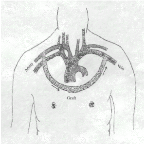
We reviewed medical records for more than 2000 chronic hemodialysis patients, receiving more than 10,000 fistulas from January 1986 to March 2001 in our hospital. We report outcomes of eight cases of axillary-contralateral axillary AVF from these chronic hemodialysis patients (). All eight cases received surgical procedure from subclavicular or infraclavicular skin incisions. After splitting the fibers of the pectoralis major muscle the axillary artery and the contralateral axillary vein were prepared, carefully sparing the brachial nerve plexus. The material of Gore-Tex is polytetrafluoroethylene. Cases 1–4 and 8 used 8 mm graft and Cases 5–7 used 6 mm graft. The underlying disease, duration of dialysis, operation, clinical presentations, radiology, treatment, and outcomes of the patients were recorded. Graft survival curve was derived from Kaplan–Meier analysis. Patient death was treated as a censored end-point in AVF graft survival analysis. The major statistics were calculated on a personal computer using Stat View 5.0 with Survival Tool (Abacus Concept Inc., Berkeley, CA, USA).
Figure 1. Anterior chest wall axillary artery to contralateral axillary vein bridge vascular access for hemodialysis.

All data are presented as mean ± SD. The patients' history was summarized as below.
Case 1. A 65-year-old woman with long-standing diabetes mellitus began hemodialysis in December 1991. She received three native arteriovenous fistulas, two graft revisions on both hands and three femoral double lumens from 1991 to 1992. In addition, she received a left femora-great saphenous vein graft in July 1993 and a left axillary artery to right axillary vein graft in January 1994. In April of 1995, progressive swelling of the right hand was noted, with venous hypertension of the right arm diagnosed after a series of examinations. Conservative treatment was given and patient recovered after cooling and limb elevation. She had been under regular dialysis by the AVF until she died of pneumonia 47 months after axillary-contralateral axillary AVF creation at age of 69.
Case 2. A 63-year-old man with diabetic nephropathy commenced hemodialysis in October 1991. He received one native arteriovenous fistula and nine graft revisions on both hands from 1992 to 1996. During this period, he underwent four neck and two femoral double-lumen insertions. In addition, he received left and right femora-great saphenous vein grafts in October 1994 and June 1996, respectively, and a right axillary artery to left axillary vein graft in June 1996. Although no problems were noted during surgery, graft failed due to thrombosis 25 months later. Finally, the patient was switched to continuous ambulatory peritoneal dialysis.
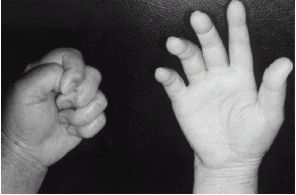
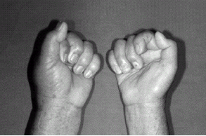
Case 3. A 61-year-old woman with end-stage renal disease as a result of chronic glomerulonephritis commenced hemodialysis in January 1992. She received three native arteriovenous fistulas and five graft revisions on both hands from 1992 to 1998. During the same period, she received three neck and four femoral double-lumen catheter insertions due to fistula dysfunction. In addition, she received a right femora-great saphenous vein graft in August 1998, and a right axillary artery to left axillary vein graft in September 1998 due to multiple graft occlusion. Unfortunately, paralysis with weakness of right hand was discovered immediately after the operation, with brachial plexopathy, as a result of operation sequelae diagnosed. A rehabilitation program and neurological examinations were arranged. The electromyography report revealed right plexopathy with active denervation superimposed on axonal polyneuropathy. After a six-month rehabilitation program, the patient was able to grasp objects and feed herself ( and ). The graft is still in use at present for 50 months after the AVF creation.
Figure 2. One week after the operation, left hand was normal and right hand could neither be flexed nor extended.

Figure 3. After 18 months, left hand was normal with complete flexion and right hand can extend partially.

Case 4. A 69-year-old male with end-stage renal disease owing to chronic glomerulonephritis commenced hemodialysis in March 1997. He received two native arteriovenous fistula, three graft fistulas and one great-great saphenous vein graft during the 1997–1998 period. During this time, he underwent two femoral double-lumen insertions, followed by a right axillary artery to left axillary vein graft in April 1999. Although no problems were noted during surgery, graft failure was detected 8 months later. He continues his hemodialysis by the access of intrajugular vein catheter.
Case 5. An 81-year-old female with end-stage renal disease due to chronic glomerulonephritis commenced hemodialysis in January 1994. She received two native arteriovenous fistulas and six graft revisions during the 1994–2000 period. During this period, she underwent three neck and six femoral double-lumen insertions. In addition, she received a right axillary artery to left axillary vein graft in October 2000. She continues to have a regular hemodialysis with the AVF after 26 months.
Case 6. A 70-year-old female with end-stage renal disease owing to chronic glomerulonephritis commenced hemodialysis in June 1993. He received two native arteriovenous fistulas and four graft revisions the 1993–2000 period. During this period, he underwent four femoral double-lumen insertions. In addition, he explored bilateral inguinal femoral vessels but unable to do thigh loop graft, and simultaneous a right axillary artery to left axillary vein graft in November 2000. She is under regular hemodialysis with this AVF 25 months after the AVF creation.
Case 7. A 19-year-old male with end-stage renal disease owing to chronic glomerulonephritis commenced continuous ambulatory peritoneal dialysis in December 1992. He received continuous ambulatory peritoneal dialysis for 8 years then shifted to hemodialysis due to severe peritonitis with Escherichia coli sepsis in November 2000. During this period, he underwent one neck and one femoral double-lumen insertions. In addition, he received a right axillary artery to left axillary vein graft due to congenital bilateral upper limb atrophy. The AVF is still in use with a period of 25 months.
Case 8. A 66-year-old male with end-stage renal disease owing to chronic glomerulonephritis commenced hemodialysis in September 1996. He received two native arteriovenous fistuli and six graft revisions the 1996–2000 period. During the propertied, he went one neck and one femoral double-lumen insertions. In addition, he received a right axillary artery to left axillary vein graft in February 2001. The AVF continues to provide permanent access after 22 months of service.
Results
Eight patients were reviewed in the study. The mean age of the patients was 61.7 ± 16.25 year-old at the time of surgery. The underlying causes for the end-stage renal diseases were chronic glomerulonephritis in six patients (75%, 6/8) and diabetic nephropathy in two patients (25%, 2/8). There was no severe peripheral vascular disease, which leads to amputation in these eight patients. All patients had undergone multiple native or graft arteriovenous fistula operations on both hands during the course of hemodialysis. Four patients had a history of intrajugular vein double-lumen catheters for temporary vascular access. All patients had a history of femoral double-lumen access. A preoperative evaluation of the axillary or femoral vein had not been arranged for all patients.
The axillary-contralateral axillary AVF failed in two patients at the graft age of 47 and 8 months. The 2-year and 4-year graft survival were 87.5% and 43.8% respectively. One of the graft failure patients was diabetes. Six patients had chronic hypotension during the period of hemodialysis. The diabetes is not a risk factor for AVF graft failure in our small population observation. AVF thrombosis was the unique cause of the AVF graft failure. One patient developed venous hypertension with swollen hands. One patient developed more severe morbid condition of brachial plexopathy. The complications can be managed conservatively.
Discussion
Fistula dysfunction is very common for chronic hemodialysis patients. The native arteriovenous fistula is the first choice for permanent initial access, with hand or femoral graft placement providing alternative options. Axillary to contralateral axillary AVF is a rare and more complicated procedure for patients without further vascular access for hemodialysis. There are few reports for axillary artery to contralateral axillary vein grafts.Citation[[4]], Citation[[5]] There are only eight patients who received this procedure in our 10,000 AVF created.
Hypotension occurring during dialysis is associated with a number of factors including baroreflex impairment, alteration of heart rate response and decreased vascular resistance.Citation[[6]] The patient with chronic hypotension on hemodialysis has a higher prevalence of multiple graft occlusion than the patient with normotension. In our patients, six patients exhibited chronic hypotension during hemodialysis.
Venous hypertension is an infrequent complication of fistula surgery. Central venous stenosis or obstruction is the common cause of venous hypertension in chronic hemodialysis patients. Venous occlusions or stenosis may sometimes be revealed by angiography. Rarely, such occlusion may be improved using percutaneous transluminal angioplasty or surgical bypass.Citation[[7]], Citation[[8]], Citation[[9]] Case 1 exhibited progressive swelling of the right hand 16 months after the procedure. Conservative treatment was given for the management. She recovered and continued her dialysis with the AVF.
Complications resulting from permanent hemodialysis access include thrombosis, stenosis, aneurysm, pseudoaneurysm, and ischemic symptoms.Citation[[10]], Citation[[11]] Brachial plexus palsy has previously been reported for patients with gunshot wounds, clavicular fracture, IV cocaine overdose or internal jugular vein catheter cannulationCitation[[12]], Citation[[13]], Citation[[14]], Citation[[15]], Citation[[16]] and has associated with a poor prognosis.Citation[[12]], Citation[[13]], Citation[[15]] In our observation, Case 3 developed permanent brachial plexopathy, even after vigorous rehabilitation. The possible cause of brachial plexopathy was surgical injury. No ischemic sign or cyanosis of right hand was found after the operation.
In previous studies, a preoperative venogram was performed before surgery.Citation[[4]], Citation[[17]] In our patients, four of them received previous intrajugular vein double-lumen catheter insertion for temporary access. Pre-existing intra-venous lesion is likely present in these patients. It seems reasonable to suggest that the preoperative venogram may be helpful especially where there is a history of previous double-lumen access.
No AVF-related mortality was found in our eight patients. The 2-year AVF graft survival was 87.5% and 4-year AVF graft survival was 43.8%. Our observation indicated that axillary to contralateral axillary AVF is a safe and effective procedure for patients with difficulty in vascular access.
We conclude that axillary artery to contralateral axillary vein graft fistula may be an alternative choice for vascular access in patients having difficult to find an easy vascular access.
References
- Alber F. Causes of hemodialysis access failure. Adv. Renal Repl. Ther. 1994; 1: 107–118
- Choudhurg D., Lee J., Elivera H.S., Ball D., Roberts A.B., Ahmed Z. Correlation of venography, venous pressure and hemoaccess function. Am. J. Kidney Dis. 1995; 25: 269–275
- Sukhatme V.P. Vascular access stenosis: prospects for prevention and therapy. Kidney Int. 1996; 49: 1161–1174
- Ono K., Muto Y., Yano K., Yukizane T. Anterior chest wall axillary artery to contralateral axillary vein graft for vascular access in hemodialysis. Artif. Organs 1995; 19: 1233–1236
- Debing E. Pierre Van den Brande. Axillo-axillary arteriovenous fistula as a suitable surgical alternative for chronic hemodialysis access. Nephrol. Dial. Transplant. 1999; 14: 1252–1253
- Daugirdas J.G. Dialysis hypotension: a hemodynamic analysis. Kidney Int. 1991; 3: 233–246
- Duncan J.M., Baldwin R.T., Caralis J.P., Cooley D.A. Subclavian vein-to-right atrial bypass for symptomatic venous hypertension. Ann. Thoracic. Surg. 1991; 52: 1342–1343
- Puskas J.D., Gertler J.P. Internal jugular to axillary vein bypass for subclavian vein thrombosis in the setting of brachial arteriovenous fistula. J. Vasc. Surg. 1994; 19: 939–942
- Kalman P.G., Lindsay T.F., Clarke K., Sniderman K.W., Vanderburgh L. Management of upper extremity central venous obstruction using interventional radiology. Ann. Vas. Surg. 1998; 12,: 202–206
- Palder S.B., Kirkman R.L., Whittemore A.D., Hakim R.M., Lazarus J.M., Tilney N.L. Vascular access for hemodialysis. Ann. Surg. 1985; 202: 235–239
- Duncan H., Ferguson L., Faris I. Incidence of the radial steal syndrome in patients with Brescia fistula for hemodialysis: its clinical significance. J. Vasc. Surg. 1986; 4: 144–147
- Raju S., Carner D.V. Brachial plexus compression: complication of delayed recognition of arterial injuries of the shoulder girdle. Arch. Surg. 1981; 116: 175–178
- O'Leary M.R. Subclavian artery false aneurysm associated with brachial plexus palsy: a complication of parenteral drug addiction. Am. J. Emerg. Med. 1990; 8: 129–133
- Donohoe C.D., McGuire T.J. Delayed weakness following a gunshot wound. Postgrad. Med. 1991; 90: 219–222
- Hansky B., Murray E., Körfer M.R. Delayed brachial plexus paralysis due to subclavian pseudoaneurysm after clavicular fracture. Eur. J. Cardiothorac. Surg. 1993; 7: 497–498
- Tarng D.C., Huang T.P., Lin K.P. Brachial plexus compression due to subclavian pseudoaneurysm from cannulation of jugular vein hemodialysis catheter. Am. J. Kidney Dis. 1998; 31: 694–697
- Surratt R.S., Picus D., Hicks M.E., Darcy M.D., Kleinhoffer M., Jendrisak M. The importance of preoperative evaluation of the subclavian vein in dialysis access planning. Am. J. Roentgenol. 1991; 156: 623–662