Abstract
Mg alloys with high Al contents have superior corrosion resistance in aqueous environments, but poor cytocompatibility compared to that of pure Mg. We have silanized the cast AZ91 alloy to improve its cytocompatibility using five different silanes: ethyltriethoxysilane (S1), 3-aminopropyltriethoxysilane (S2), 3-isocyanatopyltriethoxysilane (S3), phenyltriethoxysilane (S4) and octadecyltriethoxysilane (S5). The surface hydrophilicity/hydrophobicity was evaluated by water contact angle measurements. X-ray photoelectron analysis was performed to investigate the changes in surface states and chemical composition. All silane reagents increased adsorption of the albumin to the modified surface. In vitro cytocompatibility evaluation revealed that silanization improved cell growth on AZ91 modified by silane S1. Measurement of the concentration of Mg2+ ions released during the cell culture indicated that silanization does not affect substrate degradation.
1. Introduction
Magnesium (Mg) and its alloys have been investigated as promising bioresorbable metallic materials for biomedical applications, especially for bone implants and cardiovascular stents [Citation1]. One of the reasons for this interest is their excellent mechanical properties. In particular, these materials have elastic moduli close to that of human bone and thus can support tissue regeneration during the healing process. Other reasons are biocompatibility [Citation1, Citation2] and biodegradability [Citation3, Citation4] that can prevent a second operation after successful recovery of damaged tissue. However, Mg and its alloys have high biodegradation/corrosion rates, especially at the early stage of implantation in the human body. The rapid corrosion causes formation of hydrogen bubbles in the tissue surrounding the implant, which may delay healing or even induce necrosis of tissue [Citation1, Citation5]. OH− anions generated in the corrosion reaction increase the pH of the fluid around Mg surfaces, which influences surrounding cellular functions and decreases cytocompatibility.
One of the methods to increase the corrosion resistance of Mg is alloying. Adding 9% Al to a Mg–Zn alloy yields the AZ91 alloy that has a higher corrosion resistance than Al-free Mg alloys and pure Mg [Citation6–Citation8], probably owing to the β-Mg17Al12 phase. However, Mg alloys with relatively high Al content suppress cell growth on their surface [Citation9]. Based on this phenomenon, AZ91 alloy is employed to demonstrate the effectiveness of surface modification, which can improve cytocompatibility. It is well known that surface properties such as hydrophilicity play a significant role in cell ability and proliferation. For example, adhesion of murine fibroblast L929 depends on the surface hydrophilicity/hydrophobicity, with the optimum water contact angle of about 70° [Citation10]. Aiming for biomedical applications, many kinds of surface modification techniques are applied to Mg alloys, such as various Ca–P coatings [Citation11–Citation14], micro-oxidation coatings [Citation15], fluoride coatings [Citation16], biodegradable polymer coatings [Citation17, Citation18], silanization [Citation19] or a combination of anodization and silanization [Citation20]; their main purpose is to improve corrosion resistance of Mg alloys.
The aim of this study is to apply different silanes on the surface of magnesium alloy AZ91 to improve its cytocompatibility. Silanes are silicon-containing chemical compounds possessing a hydrolytically sensitive center that can react with inorganic substrates to form covalent bonds. They have an organic substitution that can change the physical interactions of treated substrates. This process can easily modify properties such as absorption, electrical conduction, hydrophobicity/hydrophilicity of the surface [Citation21], and in the same way it will influence cellular response. Silanization can be applied to a wide range of materials, especially polymeric [Citation22] or ceramic materials [Citation23]. In this study, we prepared different silane layers on the magnesium alloy AZ91. Protein adsorption, cell proliferation and substrate degradation of modified samples were examined.
2. Materials and methods
2.1. Sample preparation
We used a cast AZ91 magnesium alloy (AGH, Krakow) with main alloying elements being 9.0 ± 0.5 wt% Al and 1.0 ± 0.5 wt% Zn. Its slabs were cut into disks 6 mm in diameter and 1.8 mm in thickness, which were polished with a 600 grit (14 μm) SiC paper. For x-ray photoelectron spectroscopy (XPS) analysis, these samples were further polished with a diamond paste (0.25 μm). Finally, all samples were ultrasonically washed with acetone for 15 min.
Such prepared surfaces were modified by five different silanes as listed in table . Silane S1 was chosen to introduce a small and simple organic moiety to the substrate surface. S2 and S3 were selected to introduce positive and negative charges on the AZ91 surface. S4, which contains phenyl group, was selected to examine the effect of π electrons on the surface. S5 was chosen as it has a long alkyl chain and thus was expected to increase surface hydrophobicity.
Table 1. List of used silane solutions. MW stands for molecular weight.
The silane reagent was spin-coated on both sides of the sample disks. Then, the samples were dried at 100 °C for 1 h in a Teflon vessel and ultrasonically washed in absolute ethanol for 15 min. Additionally, unmodified and silanized (S1–S5) specimens for the study of protein absorption and cell culture were sterilized in ethylene oxide gas (EOG) for 23 h at 44 °C.
2.2. Surface characterization after silanization
Wettability of unmodified and silanized AZ91 surfaces was measured by a contact angle meter (DM 700, Kyowa Interface Science Co. Ltd, Japan) via the shape of a distilled water droplet in static condition. Measurements were repeated 3 times for each sample. For every measurement, 21 readings were taken with an interval of 1000 ms at room temperature.
Changes in surface states and chemical composition due to silane modification were also investigated by XPS (Theta Probe, Thermo Electron Co.). The XPS measurements were performed using x-ray Gun 400 μm, Al Kα (1486.6 eV) at take-off angle 50° and in vacuum 2 × 10⁻⁵ Pa. All binding energies were corrected using the binding energy of C1s peak at 285 eV. We analyzed the unmodified sample and silanized samples S1 and S5.
2.3. Albumin absorption
To demonstrate the effect of surface modification on protein absorption, albumin adsorption was examined. Unmodified and silanized AZ91 samples were placed into a 96-well microplate, immersed in 310 μl of bovine serum albumin solution with a concentration of 20 μg ml−1 in phosphate buffered saline (PBS) for 3 h at 37 °C. Then, 150 μl of supernatant was collected from each well to quantify albumin concentration using a protein assay kit (Coomassie Plus Assay Kit, Pierce Biotechnology, USA) following the instruction supplied with the kit. The amount of adsorbed albumin (Aabs) was calculated as follows:
where Co and Cs are the initial and supernatant (after immersion) concentrations of albumin in PBS(−), and So and Ss indicate the top and lateral surface areas of the sample, respectively.
2.4. Cell culture
To examine cell response to unmodified and silanized (S1–S5) AZ91 samples, tests with human osteosarcoma cell line (SaOS-2) were performed. Samples were separately placed into a glass deep dish, and SaOS-2 were inoculated at a density of 10 000 cells ml−1 in 27.5 ml of Dulbecco's modified Eagle minimum essential medium (D-MEM) supplemented with 10% (v/v) fetal bovine serum (FBS). Then, the cells were incubated under cell culture condition (37 °C, 5% CO2) for 1, 4 and 7 days. After the applied period, 1 ml portion of the supernatant was poured into a well of the 24-well microplate, and the same amount into another well as control. Then, 110 μl of the mixed solution of 1 mM WST-1 and 0.2 mM 1-methoxy-5-methylphenazinium methylsulfate (1-methoxy PMS) in PBS(−) was added into each of the wells, which contained a sample moved from the deep dish. The microplate was placed into a CO2 incubator for 4 h, and the absorbance of the supernatant was measured at 450 nm by a Multiskan FC Microplate Photometer (Thermo Scientific). The relative viability of cells (RVC) was calculated as where As and Ab are the absorbances of the supernatant in the sample and blank wells, respectively, and So is the top surface area of the samples.
Then, the specimens were fixed with 25% glutaraldehyde solution for 10 min and stained by 10% (v/v) Giemsa's staining solution for 15 min. Air-dried samples were observed using an optical microscope (Axiotech 100, Carl Zeiss AG, Germany), and digital images were recorded by a CCD camera (DS-5M, Nikon Co. Ltd, Tokyo, Japan). Cell culture experiments were performed in triplicate.
2.5. Quantification of the Mg2+ ion
Degradation of the substrate during cell culture experiment was examined by quantification of released Mg2+ ions. Portions of supernatant from each of the deep dishes were collected after 1, 4 and 7 days of incubation. The quantification of released Mg2+ ions was performed using Magnesium B test (Wako Pure Chemical Industries, Ltd, Osaka, Japan) following the instruction supplied with the kit.
3. Results
3.1. Water contact angle
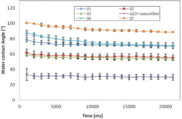
Water contact angle was measured shortly after sample preparation procedure. Figure shows values obtained for unmodified and silanized (S1–S5) AZ91 samples, which reveal that silanization affected wettability. Whereas the contact angle for unmodified surface is ∼ 30°, the value for S1-treated sample is ∼ 72°, which is favorable for cell proliferation [Citation10]. The angles for S2, S3, S4 and S5 treatment are approximately 57°, 54°, 71°–88° and 89°–100°, respectively. The values were stable in time, except those for silanes S4 and S5.
Figure 1 Time evolution of water contact angles of unmodified and silanized AZ91.
3.2. XPS
Table shows XPS binding energies for unmodified and silanized (S1, S5) samples. For unmodified specimen, double peaks for Mg2p, Al2p, O1s and C1s and single peaks for Si2p and F1s were observed. For the silanized samples, the same photoelectron spectra with comparable binding energies were obtained beside Si2p. Table shows atomic concentrations of Si, C, O, Mg, Al and F for unmodified and silanized (S1, S5) samples. Based on the amount of Si on the surface, we can confirm that the silanized layer was not removed by the ultrasonic rinsing with absolute ethanol. The F1s peak originates from the Teflon vessel, which was used to avoid contamination of Si by a glass vessel.
Table 2. Binding energies (BE, in eV) for unmodified and silanized (S1, S5) samples.
Table 3. Chemical composition of AZ91 magnesium alloy before and after silanization (S1, S5).
3.3. Albumin absorption
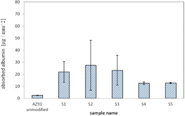
Figure illustrates the effect of silanization on protein absorption. The quantity of absorbed protein differs between unmodified and silanized (S1–S5) AZ91 samples. The lowest amount was measured on unmodified samples, ∼ 2.4 μg mm−2. Absorbed amounts of albumin were higher for S1, S2 and S3 than S4 or S5 silanes.
Figure 2 Amounts of albumin adsorbed onto unmodified and silanized AZ91.
3.4. Cell experiments
3.4.1. Cell viability.
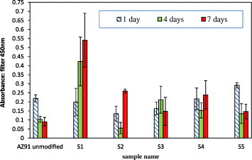
Cell viability on unmodified and silanized (S1–S5) AZ91 magnesium samples is shown in figure . There were no significant differences among specimens after 1 day of incubation in D-MEM + 10% FBS. At day 4, a slight decrease in the number of cells was observed for unmodified, S2, S4 and S5 samples, whereas a small growth was noted for S3 sample. After 7 days of incubation cell growth was observed only for S1-treated AZ91 alloy, whereas other silanes (S2–S5) and unmodified AZ91 seem to suppress cell growth.
Figure 3 Evaluation of cell viability by WST-1 assay after 1, 4 and 7 days of incubation on unmodified and silanized AZ91 magnesium alloy samples.
3.4.2. Morphology of cells and Mg substrate.
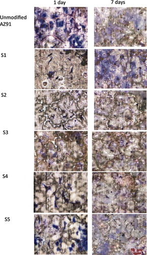
Microscopy images of unmodified and silanized (S1–S5) AZ91 samples after cell culture for 1 and 7 days are shown in figure , illustrating different responses of the cells to the unmodified and silanized surfaces. After 1 day of incubation, elongated and spread cells were observed on all surfaces. The microstructure of the AZ91 substrate is clearly observed on some surfaces, especially S2. After 7 days, almost no cells were left, except on S1-treated surface. White corrosion products are observed on all silanized surfaces.
Figure 4 Cell morphology of SaOS-2 cultured on unmodified and silanized AZ91.
3.4.3. Mg2+ release.
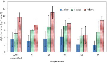
The Mg2+ release from samples during cell culture is presented in figure . In every sample, ion release was observed even after 1 day of cell culture, however, no significant difference was observed among specimens including unmodified AZ91. Along the incubation period, the amount of released Mg2+ increased for all samples.
Figure 5 Surface density of Mg2+ ions released from unmodified and silanized AZ91 during cell culture.
4. Discussion
Wettability of samples can be easily changed by surface modification. Liu et al [Citation24] fabricated a superhydrophobic surface on a Mg–Li alloy using fluoroalkylsilane (FAS) molecules. Magnesium alloy AZ31 is hydrophilic with a water contact angle (θ) of ∼ 45° because of its surface oxide layer, but it became superhydrophilic with θ ∼ 150° after immersion in ultrapure water followed by the silanization with n-octadecyltrimethoxysilane [Citation21]. In this study, 5 different silanes were used to obtain various water contact angles. The water contact angle increased after treatment with any silane reagent. For S1, S4 and S5, which have no electrically charged functional groups, the water contact angle increased from 72° to 100° along with the increase in molecule size, suggesting that surface wettability decreases with the increasing amount of CH2 groups at the surface. In case of S4 and S5, water contact angle decreased with time. This may be attributed to the larger molecules of S4 and S5 comparing to S1, which results in lower silanization reaction efficiency at the substrate surface. Concerning S2 and S3, which have electrically positive and negative functional groups respectively, the water contact angles were lower compared to S1. The result is reasonable because polarization of a molecule strengthens its interaction with water.
Proteins play an important role in mediating cell attachment onto a material. The culture medium supplemented with FBS contains various kinds of proteins, however albumin is the most common one and its adsorption onto material surface is well studied. Albumin shows greater adsorption to hydrophobic surface with a contact angle ∼ 80° [Citation25]. In this study, albumin adsorption test revealed that silanization decreases surface wettability and increases albumin adsorption onto AZ91 surface. Albumin adsorption was relatively strong on surfaces treated with S1, S2 or S3, which are relatively small molecules, and these surfaces had water contact angles of 60°–70°. As described above, molecular polarization will increase the adsorption of some proteins because they have charged functional groups. In case of S1, which has the smallest number of CH2 groups per molecule at the substrate surface, the water contact angle was lower than those of S4 and S5. This lower water contact angle suggests that S1 surface can have slightly higher electrostatic interaction, which may enhance its albumin adsorption as compared to S4 and S5 surfaces.
Results for SaOS-2 culture on these silanized surfaces revealed that only S1 can improve cell growth during 7 days of incubation. Based on the Mg2+ quantification, silanization does not influence the AZ91 substrate degradation under cell culture condition. This result may be attributed to the thickness of silane layer prepared on the substrate surface. According to the XPS analysis, the S1 layer was too thin (1–1.5 nm) to stop water molecules from penetrating through and reacting with the Mg substrate. However, the released amount of Mg2+ is similar to those of pure Mg or AZ31 alloy, on which cells can grow well [Citation9]. This result suggests that released Mg2+ or OH− ions are not the main cause of the inhibition of cell growth. Therefore we conclude that protein adsorption behavior may be an important factor controlling cellular behavior on unmodified and silanized AZ91 surfaces.
As shown in figure , more elongated and spread cells were observed on unmodified and S5 surfaces after 1 day of incubation. These surfaces exhibit water contact angles lower and higher than 70°, respectively, as well as relatively low albumin adsorption. Albumin is a major protein in blood plasma, but it does not mediate cellular adhesion onto material surface. Fibronectin, which is well known to influence cellular adhesion and growth, is reported to preferentially adsorb on hydrophilic surfaces (contact angle <60°) than hydrophobic surfaces (contact angle of 100°) [Citation26]. These facts suggest that silanization changes water contact angle and thus the protein adsorption. As shown in figure , cell growth after 4 and 7 days of incubation is inefficient for all surfaces except that treated with S1, suggesting that spread and elongated cells observed at day 1 could not grow further. While the cause of this inhibition of cell growth remains unclear, the results of S1 treatment demonstrate that silanization of AZ91 alloy surface can improve cytocompatibility while maintaining the degradation profiles.
5. Conclusions
Silanization was applied via spin-coating to cast AZ91 magnesium alloy to investigate its influence on cytocompatibility of the alloy. Water contact angle measurements suggest that the silanization changed the surface wettability. XPS revealed an additional Si2p peak on the modified surface, which might originate from the silane layer. Silanization enhanced absorption of albumin onto the modified surface. Cell culture experiments show that only silane S1 improved cell growth during 7 days of incubation. The applied surface modifications do affect cytocompatibility, but not the degradation of AZ91 substrate during cell culture.
References
- StaigerMPietakAHuadmaiJ 2006 Biomaterials 27 1728 10.1016/j.biomaterials.2005.10.003
- MaguireMCowanJ 2002 Biomaterials 15 203
- WitteFKaeseVHaferkampHSwitzerEMeyer-LindenbergAWirthC 2005 Biomaterials 26 3557 10.1016/j.biomaterials.2004.09.049
- XuLYuGZhangEPanFYangK 2007 J. Biomed. Mater. Res. A 83 703-
- SongG 2007 Corros. Sci. 49 1696 10.1016/j.corsci.2007.01.001
- PietakAMahoneyPDiasGStaigerM 2008 J. Mater. Sci.: Mater. Med. 19 407 10.1007/s10856-007-3172-9
- HeubleinBRohdeRKaeseVNiemeyerMHartungWHaverichA 2003 Br. Med. J. 89 651
- MullerWNascimentoMZeddiesMCorsicoMGassaLMeleM 2007 Mater. Res. 10 5
- YamamotoAKohayamaY 2009 22nd Eur. Conf. Biomater. T97
- TamadaYIkadaY 1993 Polymer 34 2208 10.1016/0032-3861(93)90752-V
- HiromotoSShishidoTYamamotoAMaruyamaNSomekawaHMukaiT 2008 Corros. Sci. 50 2906 10.1016/j.corsci.2008.08.013
- XuLPanFYuGYangLZhangEYangK 2009 Biomaterials 30 1512 10.1016/j.biomaterials.2008.12.001
- SongYZhangSLiJZhaoCZhangX 2010 Acta Biomater. 6 1736 10.1016/j.actbio.2009.12.020
- XuLZhangEYangK 2009 J. Mater. Sci.: Mater. Med. 20 859 10.1007/s10856-008-3648-2
- ChenJZengR C MHuangW JZhengZ QWangZ LWangJ 2008 Trans. Nonferr. Metal Soc. 18 361 10.1016/S1003-6326(10)60232-4
- YanTTanLXiongDLiuXZhangBYangK 2010 Mater. Sci. Eng. C 30 740 10.1016/j.msec.2010.03.007
- ManoJSouseRBoeselLNevesNReisR 2004 Compos. Sci. Technol. 64 789 10.1016/j.compscitech.2003.09.001
- WongH M 2010 Biomaterials 31 2084 10.1016/j.biomaterials.2009.11.111
- ZomorodianABrusciottiFFemandesACarmezimM JMoura e SilvaTFernandesJ C SMontemorM F 2012 Surf. Coat. Technol. 206 4368 10.1016/j.surfcoat.2012.04.061
- PintoRCarmezimM JFerreiraM G SMontemorM F 2010 Prog. Org. Coat. 69 143 10.1016/j.porgcoat.2010.04.014
- IshizakiTSakamotoM 2011 Langmuir 27 2375 10.1021/la1051029
- BùiL NThompsonMMcKeownN BRomaschinA DKalmanP G 1993 Analys 118 463 10.1039/an9931800463
- SchuesseleAMayrHTessmarJGoepferichA 2009 J. Biomed. Mater. Res. A 90 959
- LiuKZhangMZhaiJWangJJiangL 2008 Appl. Phys. Lett. 92 183103 10.1063/1.2917463
- WeiJIgarashiTOkumoriNIgarashiTMaetaniTLiuBYoshinariM 2009 Biomed. Mater. 4 045002 10.1088/1748-6041/4/4/045002
- DekkerAReitsmaKBeugelingTBantjesAFeijenJVanakenW G 1991 Biomaterials 12 130 10.1016/0142-9612(91)90191-C