
ABSTRACT:
Iatrogenic problems resulting from inappropriate bandaging techniques are very common. Owners should be warned that there may be consequences, particularly with long-term dressings. However, as this article will show, many of these issues are avoidable. Measures can be taken to prevent the commonest problems and to ensure that we, as nurses, are providing the best possible standard of care for our patients. This article discusses the techniques recommended and the theory behind them, to help us achieve gold-standard care. The foundation for gold-standard care is applying sound evidence-based medicine to practice.
The Veterinary Wound Library, www.vetwoundlibrary.com, has set up a new CPD initiative that aims to educate veterinary staff on gold-standard bandaging techniques. The founder, Georgie Hollis, was prompted to set up the ‘Bandaging Angels’ in response to an increasing number of wound cases caused by inappropriate bandaging techniques submitted to the library. It highlighted the need for increased knowledge and education within veterinary practices.
The aim of the group is to improve animal welfare and reduce iatrogenic injury from bandaging. The ‘Angels’ will teach a range of gold-standard techniques that have been developed by working with specialists in the field. As very little evidence is available on bandaging techniques, where standards are lacking, the ‘Angels’ will work with the specialists to provide an evidence-based solution.
This knowledge will be used to help and educate vets and nurses to resolve issues and improve standards. The training will be delivered as an in-house CPD theoretical and practical session involving the whole team.
There are several types of problem that can occur whilst bandaging. Some are within our control, whilst others result from external factors that come into play when the patient leaves the surgery and is then under the care of the owner. The focus of this article will be on limb dressings, describing the complications that can occur, and the steps we can take to try to prevent them. The importance of good client communication and compliance is also highlighted.
Functions of a bandage
Why do we dress a limb? There are many functions of a bandage (Williams & Moores) (). All these functions positively promote healing. When a dressing bandage malfunctions, the bandage itself can actively cause further injury to the patient.
Table 1. Functions of a bandage (from CitationWilliams & Moores, 2009)/
Why do things go wrong?
There are many reasons why a patient may develop further injuries following bandage application. The two main causes can be classified into Primary and Secondary ischaemic injury.
Primary injury occurs whilst the bandage is still in place. There may be an interruption – or reduced blood flow – to the tissues in the limb, which causes direct pressure necrosis.
Secondary injury and reperfusion injury occur 24–48 hours after the removal of a bandage. It is caused by the return of blood into tissue that has suffered a period of ischaemia or lack of oxygen. As blood flows back into the tissue, an inflammatory reaction is created which causes deterioration even after the bandage is removed (CitationNelissen, 2013).
Causes of injury arising from inappropriate bandage application include:
| • | bandage too tight, either on initial application or if it later constricts because the limb swells or the dressing gets wet | ||||
| • | bandage too loose – this can cause problems if the bandage then slips, causing friction and soft tissue abrasions. Further problems will arise where the slipped bandage ceases to provide the intentioned immobilisation as the area will cease to be effectively supported | ||||
| • | inadequate padding | ||||
| • | inadequate protection of vulnerable areas, such as joints | ||||
| • | skin damage from the use of adhesive tape/stirrups. | ||||
Causes of injury relating to owner compliance include:
| • | allowing the bandage to become wet, which would then cause it to tighten | ||||
| • | leaving the bandage on for too long after failure to return for a planned appointment or reluctance to return for a dressing change owing to costs involved | ||||
| • | over-exercise whilst the bandage is in place. (CitationMcKeating, 2013 & CitationNelissen, 2013). | ||||
If any one of these factors is present, it can lead to complications.
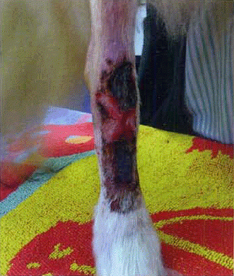
In cases of primary ischaemic injury, a pressure sore (Figure 1) or skin necrosis (Figure 2) may develop, the latter would require debridement.
Figure 1. The patient suffered a pressure sore on the lateral aspect of the carpus. It was very quickly resolved by the use of a ‘doughnut’ dressing to relieve the pressure
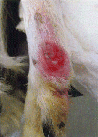
Figure 2. The patient's limb had been ‘casted’. Upon removal of the cast, substantial skin necrosis had occurred
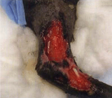
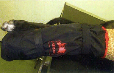
In more severe cases of secondary ischaemia, there is a risk of loss of digits or, in extreme cases, the loss of a limb (Figure 3).
Figure 3. A small avulsed wound was closed surgically and a light support dressing applied. The patient chewed the dressing the following day, was off her food and subdued. The client ignored these symptoms and the advice they had been given and returned for a routine appointment 3 days later: The nurse was presented with a dressing with an unpleasant odour, which was very painful to remove and had resulted in this severe injury to the limb
The early stages may manifest themselves in the patient as an excessive interest in the bandage, such as licking or chewing at it, and this should not be ignored – the dressing should be removed immediately.
One study (CitationAnderson and White, 2000) concluded that ‘ischaemic injury could be attributed to inadequate padding or uneven application of a bandage resulting in pressure points’. The authors strongly recommended that all owners receive instructions on home care and inspection of bandages. The majority of injuries are sustained within 24 to 48 hours of application, and removal of the bandage within that period would have prevented the injuries seen.
Where do we stand under our duty of care?
Adequate information must be given to clients regarding bandage aftercare (CitationMcKeating, 2013).
The Veterinary Defence Society (VDS) dealt with 74‘Duty of Care’ calls in relation to bandaging between 2010 and 2013. This highlights the need for good communication with clients when a patient is discharged with a bandage. Any defence can be weakened if insufficient warning of possible risks has not been given to the client. A print-out personalised with the practice details is the best way to ensure that clients have all the information they need to take home with them ().
Table 2. Example of a client infonnation sheet
They must be fully informed of the frequency of checks and how to protect the bandage. Traditionally, an empty ‘drip bag’ is the most commonly used protection (Figure 4); but there are now much more suitable breathable, waterproof alternatives, such as the Medipaw, available from JAK Marketing (Figure 5).
Figure 4. Drip bag in place
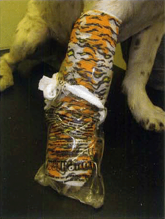
Figure 5. A Medipaw bandage cover in place over a large Robert-Jones dressing. It comes in various sizes and provides brilliant protection for the dressing
How can we prevent problems?
Training veterinary staff will decrease the overall risk. Understanding why problems can occur will lead to a better understanding of the steps to take to prevent them. Educating clients about bandage aftercare is also of utmost importance.
As most bandage injuries occur within 24 to 48 hours of their application, it could be deduced that if we changed the bandage within that period we would prevent any problems. In reality, that may not be practical for a number of reasons, which is why it is so important to ensure correct application of a bandage, to give correct advice and to ensure that the owner understands that they must not ignore any problems.
Determination of the frequency of bandage changes should be made on an individual basis, although three to four days’ wear without complication should be achievable if the correct technique is used.
The bandage should be checked frequently, and the practice should have regular contact with the owner following discharge. Owners should be educated on signs of bandage-related problems and implications and a return appointment should be scheduled before leaving the clinic ().
Limb dressings
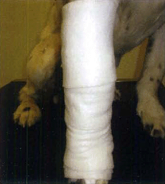
A consistent technique should be used as the basis for all limb dressings, including the Robert-Jones technique. An appropriate primary layer and topical agent should be used to promote the optimal healing environment. The combination of secondary and tertiary layer together determine the pressure on the wound (CitationNelissen, 2013)
Secondary layer
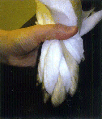
The toes (including dew claws) should be well padded. The preferred product is a synthetic padding (e.g. Orthoband, Millpledge) as it is less likely than cotton wool to change its shape in response to moisture and movement. Use long lengths and push to the base of the toe (Figure 6).
Figure 6. The toes should be adequately padded using a synthetic padding between each toe
The third and fourth distal phalanges should be left exposed to aid monitoring. The secondary layer should be started at the second phalange of the central toes working distally upwards with a 50 per cent overlap.
The pre-rolled synthetic padding is recommended, with a minimum size of 10cm. The wider the padding, the less pressure it can create (CitationNelissen, 2013).
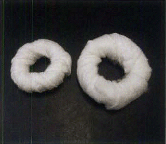
Incorporate ‘doughnut’ dressings as necessary to protect vulnerable areas such as stopper pads, hocks, elbow and bony prominences (Figure 7). They are quick and simple to make to any size and are excellent at relieving pressure (Figure 8).
Figure 7. ‘Doughnut’ in place on a hock
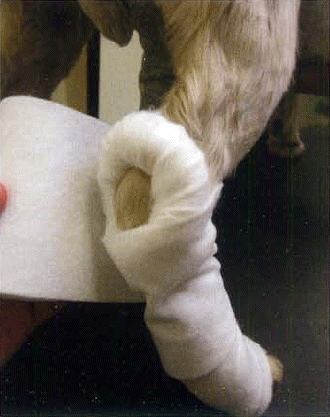
Figure 8. ‘Doughnuts’ can be used to protect vulnerable areas – such as hocks, elbow, stopper pads – from pressure sores. They are quickly and easily made by wrapping padding around the fingers, then folding the padding inside and round the circle made. They can be made to any size
Their usefulness for every dressing should not be underestimated. If a foot injury is present, a ‘doughnut’ should be used to relieve the pressure from the wound and the foot should be enclosed.
The padding layers should be repeated until a uniform radius is achieved (Figure 9). Extra padding should be added as necessary, rather than following the contours of the leg. This provides the narrower areas with extra padding to ensure an even distribution of tension throughout the leg.
Figure 9. Rather than following the exact shape of the leg, extra padding should be used to achieve a uniform radius
Tertiary layer
A tertiary layer of conforming bandage (e.g. Knit-fix; Millpledge) is then used to hold the secondary layer in place, working distally upwards from the toes as previously described. Two layers of cohesive bandage (e.g. Wrapz, Millpledge) can then be applied.
Although commonly used, adhesive tape should not be necessary. It can cause skin inflammation and be a focus of tension around the leg, as well as being painful and possibly damaging to the skin.
Each layer should be cut with scissors rather than torn (as this could put unnecessary tension on the dressing).
Why toes out?
Leaving the toes visible allows for monitoring of the foot. It increases the comfort for the patient as they can ‘feel’ their feet as they can spread their toes. It will also be less likely to result in skin damage to the foot if the dressing did become wet.
Discussion
Bandaging is a much more complex procedure than it is often given credit. It involves a lot of preparation, time and also cost to the owner. Gaining knowledge of the correct application of bandages is extremely worthwhile to prevent problems for the patient (and ourselves!).
We should aim to achieve best practice, defined as ‘a method or technique that has consistently shown results superior to those achieved by other means and that is used as a benchmark. Best practice can and does evolve as improvements are discovered’ (CitationNelissen, 2013).
The author feels this is a perfect attitude to have towards bandaging, and hopes that, by discovering advances in techniques, many patients will benefit by suffering from fewer complications.
Rules are not set in stone with approaches to bandaging. With practice and experience, we can see how improvements may be made. Bearing in mind the vast amount of inaccurate – often dangerous – online information that is available to clients on how to do their own bandages, we need to be seen to be providing a gold-standard service with a sound evidence base of knowledge on which we can base our techniques and choices.
Additional information
Notes on contributors
Caroline Calder
Caroline Calder RVN MBVNA
Caroline qualified in 2001 after attending Myerscough College, Preston, whilst working in Carlisle. For the last eight years, she has worked at a small animal practice in County Durham, recently taking on the role of head nurse. Since attending a CPD course in 2010, she has developed a keen interest in wound management and has more recently become involved in the creation of the ‘Bandaging Angels’ – a group set up by the Veterinary Wound Library to provide in-house CPD bandage training to practices.
References
- WILLIAMS, J. & MOORES, A. (2009) BSAVA Manual of Canine and Feline Wound Management and Reconstruction, p. 41. BSAVA, Cheltenham.
- NELISSEN, P. (2013) Bandaging Angels training session. Dick White Referrals, Newmarket August 2013.
- McKEATING, F. (2013) The Veterinary Defence Society Ltd Bandaging Angels training session. Dick White Referrals, Newmarket, August 2013.
- ANDERSON, D. M. & WHITE, R. A. S. (2000) Veterinary Surgery ‘Ischaemic Bandage Injuries: A case series and review of the literature.