
Abstract
Using the MOLAB® non-invasive analytical mobile laboratory, we studied a finely illuminated sixteenth-century Persian manuscript at the Fitzwilliam Museum, Cambridge, UK, in collaboration with its Department of Manuscripts and Printed Books. Three miniatures belonging to the manuscript, but ascribable to different periods, were analyzed in order to identify similarities and differences in the painting materials and techniques used by Safavid artists over a period of 150 years. The use of multiple analytical techniques indicated a common palette characterizing the three decorative schemes, along with some differences mainly regarding the pigment mixture used to obtain brown hues in the first scheme, as well as the presence of different mixtures in green and dark purple areas in the third scheme.
Introduction
In contrast to the study of Western European manuscripts and despite long-standing interest (Laurie, Citation1935), information on the materials and techniques used in the decoration of Persian illuminations is still for the most part lacking. The limited analytical evidence available regarding Persian and Islamic painting materials also reduces the possibility of establishing the extent of exchange and mutual influence between Eastern and Western painters, which is of indubitable interest and has been firmly established for other art forms such as glassmaking and textile dyeing (Berrie, Citation2007).
As a first step towards contributing to a corpus of analytical information about Persian painting, and with the goal of gaining a deeper understanding of a particularly interesting class of objects, a sixteenth-century Persian manuscript (MS 18-1948) from the Fitzwilliam Museum, Cambridge, UK, was investigated by the Museum's Department of Manuscripts and Printed Books through transnational access to the mobile analytical facilities provided by MOLAB®.Footnote1
Since manuscripts require particular care due to their intrinsic fragility and susceptibility to damage due to changes in environmental conditions, the MOLAB® non-invasive multi-technique approach (Miliani et al., Citation2010) offers a reliable and sensitive way to study the materials the manuscripts are made of, in situ. Furthermore, MOLAB® has extensive experience with studies of manuscripts of different provenance and periods, acquired during its investigations in many European museums and libraries (Miliani et al., Citation2012; Doherty et al., Citation2013; Buti et al., Citation2014; Sgamellotti et al., Citation2014). Three miniatures (fols. 1v, 67v and 202v) from Persian manuscript (MS 18-1948) were analyzed with the objective of identifying similarities and differences in materials and techniques used by Safavid artists over a period of 150 years by means of portable Raman, UV-Vis reflection and emission, X-ray fluorescence (XRF), and mid-infrared reflection spectroscopy, which allow for in-depth analysis of elemental and molecular properties of the investigated materials.
Manuscript 18-1948
As part of its rich collection of illuminated manuscripts, the Fitzwilliam Museum has a small but exceptionally beautiful selection of Persian manuscripts and Qu'rans, exemplifying many of the finest schools and styles of manuscript production from across the Islamic world, including this particularly fine sixteenth-century example.Footnote2 Manuscript 18-1948 was bequeathed to the Fitzwilliam Museum in the late 1940s and has remained untouched and largely unstudied since this time. Thanks to a grant from the Sumitomo Foundation, it has recently undergone comprehensive conservation treatment and technical research to better understand its true significance. The text is a copy of the twelfth-century epic Haft Paykar by the Persian poet Nizami, and the manuscript is truly a visual encyclopaedia of Safavid painting, representing three distinct styles. The main body of the manuscript was illuminated in Qazwin style ca. 1562 (); two miniatures taken from a manuscript executed ca. 1525 have been added as a double opening at the beginning of the volume (fols. 1v-2r, ), and two more miniatures added at the end of the volume come from a later manuscript of ca. 1675 (fols. 202v-203r, ). The complexity and decorative refinement of the first two miniatures showcase the technical achievements of Persian painting during the reign of Shah Tahmasp (r.1524-1576), whose return to Tabriz in 1522 transformed the city into a capital of the arts through his distinguished patronage. The illuminations in the main body of the manuscript demonstrate the elegance and detail of the 1560s, probably in response to Shah Tahmasp's withdrawal from luxury during this period. In contrast, the two final miniatures in the manuscript demonstrate the dissemination of styles, techniques, and compositions away from the main centres of Safavid painting and into the provinces. Technical analysis was carried out prior to undertaking the conservation treatment. Analyses were planned jointly by the Museum's researchers and the MOLAB® team, and were based on the preliminary results obtained by UV-Vis-NIR reflection spectrometry and XRF spectrometry carried out at the Fitzwilliam on eight miniatures representing all three decorative schemes.
Figure 1. MS 18-1948, fol. 67v. Persia, ca. 1562 © Fitzwilliam Museum, Cambridge.
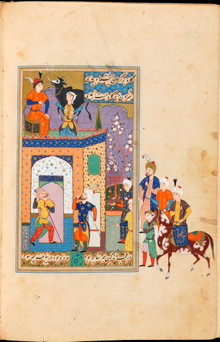
Figure 2. MS 18-1948, fol. 1v. Persia, ca. 1525 © Fitzwilliam Museum, Cambridge.
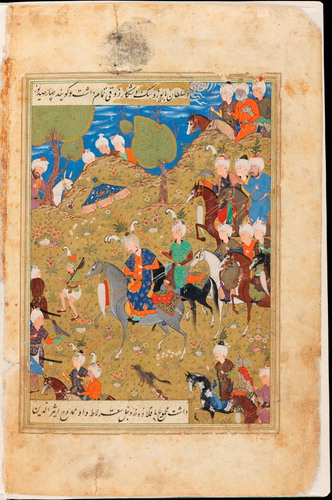
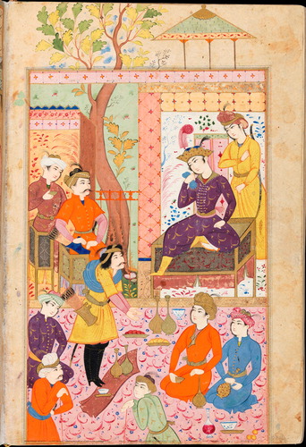
Figure 3. MS 18-1948, fol. 202v. Persia, ca. 1675 © Fitzwilliam Museum, Cambridge.
Experimental
Raman spectroscopy
The prototype system used by MOLAB® has been assembled from various components and has two excitation wavelengths: an Nd:YAG laser with excitation at 532 nm and a diode laser with excitation at 785 nm. The first uses a JASCO RMP-100 microprobe equipped with an Olympus objective (×50 or ×20), a CCD video camera for focusing and sample centring, and a 5 m quartz optic fibre with a core diameter of 100–200 μm. The second laser line uses an InPhotonics optic fibre system. The compact ORIEL MS125TM spectrograph is equipped with a Czerny-Turner type polychromator of about 100 mm focal length, with manual scanning, manual change of gratings, and an 1024 × 128 pixel CCD ANDOR detector Peltier-cooled to −50°C.
Mid-FTIR spectroscopy
Reflection mid-FTIR spectra were recorded using a compact portable Bruker Optics ALPHA-R spectrophotometer equipped with a Globar infrared radiation source, a Michelson interferometer (RockSolid(TM)) modified to work in any environmental condition and spatial orientation and a DLaTGS detector. An external reflection device permits working with an angle of incidence of approximately 20°. The instrument weighs 7 kg, and measures only 20 × 30 × 12 cm3. It is equipped with a USB high-resolution video camera for detailed visualization and monitoring of the area under analysis. Spectra were recorded using 200 interferograms across the range 7500–375 cm−1 with a spectral resolution of 4 cm−1. The correction of background absorption was carried out using a reflection spectrum of a reference gold surface. The areas examined measured approximately 28 mm2.
UV-Vis reflection and emission spectroscopy
UV-Vis reflection spectra as well as fluorescence emissions were recorded by the MOLAB® team with a self-assembled instrument which includes an excitation source consisting of a deuterium-halogen lamp (Avantes) and a highly sensitive AvaSpec-2048 CCD detector (200–1100 nm, Avantes) with a spectral resolution of 8 nm. The system has a dedicated fibre optic system, built up by Avantes following our specifications, whose probe allows measurements with an angle of 21° in order to minimize the specular component of the collected radiation. The probe area is less than 2 mm2. Measurements were carried out with an integration time of 600 ms and 10 averages for each acquisition. Ultra-compact diode lasers (Toptica Photonics AG, DE) integrated into the same apparatus were employed for fluorescence emission measurements: an ultraviolet source (375 nm, 18 mW max. nominal power) for uncoloured and yellow materials; a visible source (445 nm, 50 mW max. nominal power) for orange, red, and purple colorants; and a second visible source (640 nm, 100 mW max. nominal power) for green and blue dyes or pigments.
Reflection spectra in an extended spectral range, including the near- and short-wave infrared (UV-Vis-NIR-SWIR: 350–2500 nm) were recorded by the Fitzwilliam Museum researchers using a FieldSpec3 spectroradiometer, on loan from Analytik Ltd. The spectral resolution is 3 nm at 700 nm, and 10 nm at 1400 and 2100 nm. The instrument includes a modular Silicon array and two Peltier-cooled InGaAs detectors and is coupled to a fibre optic system with 25° field of view.
XRF spectrometry
The XRF equipment used by MOLAB® is composed of a miniaturized X-ray generator (EIS P/N 9910) equipped with a tungsten anode, and a silicon drift detector (resolution of about 150 eV at 5.9 keV) cooled with a Peltier element. The instrument permits the detection of elements with atomic number higher than silicon (Z > 14). The experimental parameters used were 38 kV and 10 µA; typical acquisition times were 120 s. The working distance between artwork and probe is approximately 2 cm, giving a beam diameter at the object's surface of about 4 mm.
Preliminary XRF of selected areas was carried out by Fitzwilliam Museum researchers using a hand-held Tracer IV-SD on loan from Bruker UK, equipped with a Rh tube and Pd collimator. Spectra were collected for 200 seconds, at either 40 kV and 16 μA, or 15 kV and 55 μA, without filters. Due to the difficulty in establishing the exact position and extent of the areas analyzed using this instrument, these XRF data were used only as additional information to support and confirm the identification of pigments made with other analytical methods.
Results and discussion
Out of the relatively small number of Islamic and Persian manuscripts whose decoration has been subjected to technical analysis, the majority contain only non-figurative decoration. While it is certainly possible that the same pigments were used both for figurative and non-figurative illuminations, the rich palette identified in the narrative compositions present in MS 18-1948 is best compared directly with similar types of miniatures contained in sixteenth- and seventeenth-century Persian manuscripts, such as those analyzed by Purinton & Watters (Citation1991), Jurado-López et al. (Citation2004), Clark & Mirabaud (Citation2006), and Muralha et al. (Citation2012). The following discussion is based on this premise.
The pigments identified in all three decorative schemes on MS 18-1948 are relatively homogeneous, as summarized in , and they largely correspond to those mentioned in contemporary technical treatises as reported by Purinton & Watters (Citation1991) and to those identified analytically by the previously mentioned authors. Any significant differences will be discussed below.
Table 1. Summary of pigments and mixtures identified on MS 18-1948. Tr. = traces
The second decorative scheme, used in the main body of the manuscript and exemplified by fol. 67v (), can be used as a reference for the prevalent palette, which is composed of:
- Natural ultramarine in blue areas, as shown by mid-FTIR band at 2340 cm−1 (Miliani et al., Citation2008) and UV-Vis reflection profiles (minimum absorption at 600 nm) followed by rise in reflectance with transition edge at 700 nm).
- Cinnabar (or its synthetic analogue vermillion) in red hues, identified by a sharp transition edge at 602 nm in the UV-Vis spectra and by a distinctive profile in Raman spectra (strong peaks at 254 and 342 cm−1, shoulder at 282 cm−1).
- Lead white (easily identified by Raman, mid-FTIR, and UV-Vis reflection spectroscopy), both used as a white pigment and found as a minor constituent in dark areas as well as in mixtures with other pigments to obtain lighter hues.
- An insect-derived anthraquinone dye based on UV-Vis reflectance and emission spectra (structured reflectance minimum between 530 and 590 nm, emission at 650 nm), used in pink and light purple areas.
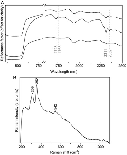
- Orpiment in yellow areas, as suggested by arsenic in the XRF spectra and confirmed by the Raman analysis (peaks at 309 and 352 cm−1 and broad shoulders at about 291 and 377 cm−1).
- Carbon-based pigments, often in mixture with lead white, in the black and grey areasFootnote3.
- Two metallic pigments, one containing mostly gold with traces of silver, and the second one containing mostly silver with traces of gold, both identified by XRF.
Figure 4. Identification of minium in orange areas. (A) UV-Vis-NIR spectra from orange areas on fols. 1v, 67v and 202v, showing evidence for the presence of minium (transition edge at about 565–572 nm) and for a lipid-containing material, characterized by intense absorption bands at about 1728, 1763, 2311 and 2352 nm. (B) Raman spectrum from an orange area on fol. 67v, showing the presence of orpiment in addition to minium to obtain a lighter hue.
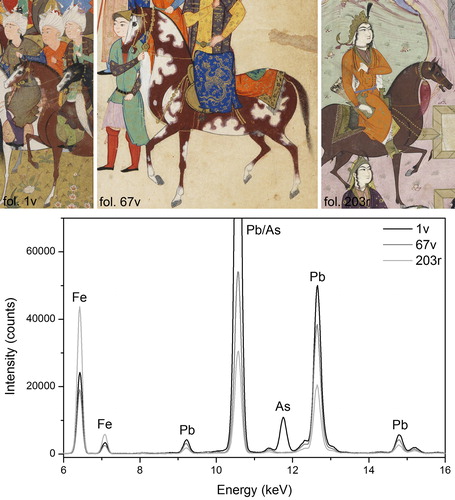
Earths/ochres were identified in brown areas, as shown by UV-Vis-NIR reflectance spectroscopy and by the high amounts of iron in the XRF spectra. The pigments were further characterized as haematite by Raman spectroscopy (peaks at 226, 290, and 407 cm−1). The brown horses present in the three schemes,Footnote4 in particular, provide an interesting comparison and one of the few significant differences identified (): the horses on fol. 1v were in fact painted using haematite in a mixture with orpiment, whose presence is suggested by arsenic in the XRF spectra and confirmed by Raman spectroscopy, and is not found in either of the two other schemes.
Figure 5. XRF spectra of brown horses from the three decorative schemes, all indicating the presence of an iron-bearing pigment (recognized as haematite from the Raman spectra, not shown). The presence of relevant amounts of arsenic in the spectrum from fol. 1v clearly differentiates the composition of the brown pigment used in the earliest decorative scheme, suggesting the presence of orpiment (confirmed by the Raman analysis).
Based on our limited literature survey, sixteenth- to seventeenth-century Persian painters seem to have obtained brown hues in a variety of ways, using for examples mixtures of minium (Muralha et al., Citation2012) or vermillion (Jurado-López et al., Citation2004). Only in a few cases is haematite mentioned as the main component of brown colourants (Clark & Mirabaud, Citation2006; Muralha et al., Citation2012), and never in addition to orpiment. A more systematic survey of the use of brown pigments by Safavid artists may lead to further insight into the painting techniques of individual painters and workshops, or point to geographical preferences based for example on the availability of local iron ores.
A copper-based pigment, most likely verdigris, was identified in green robes and in most architectural details in the first and second decorative schemes. The presence of this pigment was inferred on the basis of the high levels of copper in the XRF spectra and the characteristic shape of the reflection spectra in the extended UV-Vis-NIR-SWIR range, with a deep absorption between about 600 and 900 nm followed by a rise in reflectance up to a maximum around 1300 nm (Ricciardi et al., Citation2013). Interestingly, although it is the green pigment most commonly mentioned in the technical literature, and although it was identified by Purinton & Watters (Citation1991) and by Barkeshli & Ataie (Citation2002) in many of the miniatures included in their studies, verdigris has otherwise not been analytically identified recently in Persian and Islamic manuscripts, whether in the figurative (Jurado-López et al., Citation2004; Clark & Mirabaud, Citation2006; Muralha et al., Citation2012) or in the non-figurative decoration (Bruni et al., Citation2001; Hayez et al., Citation2004; Burgio et al., Citation2008; El Bakkali et al., Citation2012; Hamdan et al., Citation2012; Tanevska et al., Citation2014). One possible reason for this is the large preference given to Raman spectroscopy in these studies, by which the identification of verdigris, possible in theory, often proves difficult in practice.Footnote5 Several of these authors have speculated about the presence of verdigris in areas where green crystals could generally be observed but no Raman signal could be recorded, often due to the presence of a high fluorescence background. Such fluorescence might be due to the addition of saffron to verdigris, which appears to have been a common practice among medieval Persian artists (Barkeshli & Ataie, Citation2002). In the case of our study of MS 18-1948, the availability of UV-Vis-NIR-SWIR reflection spectroscopy in an extended spectral range was instrumental in allowing the tentative identification of verdigris, although further analyses would be needed to confirm this identification beyond any possible doubt.
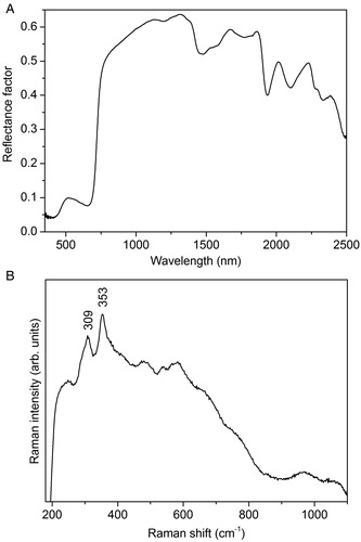
In all three decorative schemes, most green areas representing natural elements such as trees and grass were painted with vergaut, a commonly used mixture of indigo and orpiment (). The blue component was identified by the characteristic absorption at about 655 nm in UV-Vis reflection spectra, followed by a rise in reflectance (A), while the presence of arsenic in the XRF spectra and the characteristic Raman bands at 309 and 353 cm−1 and shoulders at about 293 and 377 cm−1 contributed to the identification of the yellow component (B). The use of vergaut in Persian manuscripts is well documented in technical treatises and has often been confirmed analytically. Other authors have also identified the selective use of an orpiment–indigo mixture in green areas depicting vegetation as opposed to a copper-based pigment to depict items of clothing (Muralha et al., Citation2012).Footnote6
Figure 6. Identification of vergaut in green areas. (A) UV-Vis-NIR spectrum showing the presence of indigo (B) Raman spectrum showing the presence of orpiment.
Finally, light green background areas on fol. 202v contain trace amounts of copper and no arsenic, and yield UV-Vis reflection spectra which resemble that of indigo.Footnote7 A mixture of malachite and indigo was identified by Muralha et al. (Citation2012) in the non-figurative decoration of a sixteenth-century manuscript, and it is possible that the same mixture was used here. Further analyses are needed to clarify these results.
A further difference in the palette of the third scheme (fol. 202v) lies in the presence on this folio of a very dark purple colour () obtained by mixing ultramarine blue, identified by mid-FTIR band at 2340 cm−1 (A), with an insect-derived anthraquinone dye, as indicated by the structured reflectance minimum between 530 and 570 nm (B) along with an emission at 650 nm. We have found no specific indication of how purple hues were generally obtained by Persian manuscript painters, and the only published analytical data concern the use of a mixture of ultramarine and cinnabar in a purple area (Muralha et al., Citation2012). As the identification of organic red colourants on manuscripts becomes progressively easier due to the availability of an ever-increasing number of non-invasive analytical methods, it is hoped that more meaningful comparisons will soon be possible.
Figure 7. Identification of a mixture of ultramarine blue (A, mid-FTIR spectrum) and an insect-derived dye (B, UV-Vis-NIR reflectance spectrum) in a dark purple robe on fol. 202v.

Conclusions
Access to the MOLAB® facility facilitated the thorough technical investigation of MS 18-1948, a finely decorated sixteenth-century Persian manuscript. The cross-disciplinary approach used by the joint MOLAB®-Fitzwilliam team was based on a close collaboration between scientists, conservators and art historians and proved invaluable in successfully addressing queries related to the manuscript's decorative materials. It has also raised further questions regarding the selective use of a lipid-containing paint binder in orange areas, which we hope to address more fully at a later stage. The comprehensive conservation treatment which followed the technical analysis will also be the object of a future publication.
Using a multi-technique spectroscopic in situ approach and portable, non-invasive instrumentation – including laser-based devices – this collaborative effort has produced a rare insight into the field of Persian manuscript painting. Few extensive analytical examinations of the figurative decoration of Persian manuscripts have been carried out to date, and therefore this work provides reliable comparative material on which future research can build.
Acknowledgements
The authors wish to thank the Sumitomo Foundation for funding the conservation of MS 18-1948. Thanks are also due to Analytik Ltd, and to Bruker UK for kindly loaning analytical equipment used in this work. Transnational access to the MOLAB® facility was funded by EU FP7 programme CHARISMA (Grant Agreement 228330). The IPERION CH.it project is gratefully acknowledged by the authors.
Notes
1 Access provided by the CHARISMA European project (www.charismaproject.eu).
2 This manuscript was being studied within the context of the cross-disciplinary research project MINIARE (www.miniare.org), which combines scientific and art-historical analysis in view of a comprehensive study of illuminated manuscripts, their makers, patrons, history, and broader context. One of the main outcomes of the project will be a major exhibition on ‘COLOUR: the art and science of illuminated manuscripts’ at the Fitzwilliam Museum (30 July–30 December 2016).
3 The presence of carbon-based blacks was inferred on the basis of near-infrared images acquired with a modified Canon 30D camera equipped with an X-nite 780 long-pass filter, and on the absence of analytical data suggesting any possible alternatives.
4 There is no horse depicted on fol. 202v, but there is one on fol. 203r, which was analyzed and is representative of the third decorative scheme.
5 We owe many thanks to Dr Lucia Burgio, from the Victoria and Albert Museum, for her insightful comments on the difficulties related to the identification of verdigris by Raman spectrometry.
6 Muralha et al. (Citation2012) also identified the use of a different mixture of indigo with yellow ochre to paint vegetation in a sixteenth-century Persian manuscript on which green clothes were instead found to contain emerald green, which is a nineteenth-century pigment. They suggest that this was added to the manuscript in an early attempt at restoration, and it is possible that the treatment may have been undertaken to remedy discoloration or pigment loss in green areas due to the use of verdigris by the sixteenth-century artist.
7 No Raman or mid-FTIR spectra were acquired in these green background area.
Related Research Data
References
- Barkeshli, M. & Ataie, G. H. 2002. pH Stability of Saffron used in Verdigris as an Inhibitor in Persian Miniature Paintings. Restaurator, 23: 154–64.
- Berrie, B. H. 2007. Pigments in Venetian and Islamic Painting. In: S. Carboni, ed. Venice and the Islamic World, 828–1797. New Haven: Yale University Press, pp. 140–45.
- Bruni, S., Cariati, F., Casadio, F. & Guglielmi, V. 2001. Micro-Raman Identification of the Palette of a Precious XVI Century Illuminated Persian Codex. Journal of Cultural Heritage, 4: 291–96.
- Burgio, L., Clark, R.J.H., Muralha, V. S. F. & Stanley, T. 2008. Pigment Analysis by Raman Microscopy of the Non-figurative Illumination in 16th- to 18th-Century Islamic Manuscripts. Journal of Raman Spectroscopy, 39: 1482–93.
- Buti, D., Domenici, D., Miliani, C., García Sáiz, C., Gómez Espinoza, T., Jímenez Villalba, F., Verde Casanova, A., Sabía de la Mata, A., Romani, A., Presciutti, F., Doherty, B., Brunetti, B.G. & Sgamellotti, A. 2014. Non-Invasive Investigation of a Pre-Hispanic Maya Screenfold Book: The Madrid Codex. Journal of Archaeological Science, 42: 166–78.
- Clark, R.J.H., & Mirabaud, S. 2006. Identification of the Pigments on a Sixteenth Century Persian book of Poetry by Raman Microscopy. Journal of Raman Spectroscopy, 37: 235–39.
- Doherty, B., Daveri, A., Clementi, C., Romani, A., Bioletti, S., Brunetti, B., Sgamellotti, A. & Miliani, C. 2013. The Book of Kells: A Non-Invasive MOLAB Investigation by Complementary Spectroscopic Techniques. Spectrochimica Acta Part A, 115: 330–36.
- El Bakkali, A., Lamhasni, T., Haddad, M., Ait Lyazidi, S., Sanchez-Cortes, S. & del Puerto Nevado, E. 2012. Non-Invasive Micro Raman, SERS and Visible Reflectance Analyses of Coloring Materials in Ancient Moroccan Islamic Manuscripts. Journal of Raman Spectroscopy, 44: 114–20.
- Hamdan, N. M., Alawadhi, H. & Jisrawy, N. 2012. Integration of μ-XRF and μ-Raman Techniques to Study Ancient Islamic Manuscripts. IOP Conference Series: Materials Science and Engineering, 37: 012006.
- Hayez, V., Denoël, S., Genadry, Z. & Gilbert, B. 2004. Identification of Pigments on a 16th Century Persian Manuscript by Micro-Raman Spectroscopy. Journal of Raman Spectroscopy, 35: 781–85.
- Jurado-López, A., Demko, O., Clark, R.J.H. & Jacobs, D. 2004. Analysis of the Palette of a Precious 16th Century Illuminated Turkish Manuscript by Raman Microscopy. Journal of Raman Spectroscopy, 35: 119–24.
- Laurie, A.P. 1935. Materials in Persian Miniatures. Technical Studies in the Field of Fine Arts, 3: 146–56.
- Miliani, C., Daveri, A., Brunetti, B.G. & Sgamellotti, A. 2008. CO2 Entrapment in Natural Ultramarine Blue. Chemical Physics Letters, 466, 148–51.
- Miliani, C., Rosi, F., Brunetti, B. G. & Sgamellotti, A. 2010. In situ Non-Invasive Study of Artworks: the MOLAB Multitechnique Approach. Accounts of Chemical Research, 43(6): 728–38.
- Miliani, C., Domenici, D., Clementi, C., Presciutti, F., Rosi, F., Buti, D., Romani, A., Laurencich Minelli, L. & Sgamellotti, A. 2012. Colouring Materials of Pre-Columbian Codices: Non-Invasive in situ Spectroscopic Analysis of the Cospi Codex. Journal of Archaeological Science, 39(3): 672–79.
- Muralha, V.S.F., Burgio, L. & Clark, R.J.H. 2012. Raman Spectroscopy Analysis of Pigments on 16–17th c. Persian manuscripts. Spectrochimica Acta Part A, 92: 21–28.
- Purinton, N. & Watters, M. 1991. A Study of the Materials Used by Medieval Persian Painters. Journal of the American Institute for Conservation, 30(2): 125–44.
- Ricciardi, P., Pallipurath, A. & Rose, K. 2013. ‘It's Not Easy Being Green’: a Spectroscopic Study of Green Pigments Used in Illuminated Manuscripts. Analytical Methods, 5: 3819–24.
- Sgamellotti, A., Brunetti, B.G. & Miliani, C. 2014. ‘Science and Art: The Painted Surface’ edited by Royal Society of Chemistry, London, ISBN: 9781849738187.
- Tanevska, V., Nastova, I., Minčeva-Šukarova, B., Grupče, O., Ozcatal, M., Kavčic, M. & Jakovlevska-Spirovska, Z. 2014. Vibrational Spectroscopy, 73: 127–37.
- Vagnini, M., Miliani, C., Cartechini, L., Rocchi, P., Brunetti, B. G. & Sgamellotti, A. 2009. FT-NIR Spectroscopy for Non-Invasive Identification of Natural Polymers and Resins in Easel Paintings. Analytical and Bioanalytical Chemistry, 395: 2107–18.