
Abstract
Purpose: Percutaneous coronary angioplasty (PCA) has been demonstrated to reduce mortality and morbidity and thereby improve the prognosis of patients undergoing acute myocardial infarctions (AMIs). However, this procedure paradoxically increases the initial damage as the result of a condition known as ‘myocardial reperfusion injury’. Oxidative stress may contribute to the mechanism of this injury. The goal of the present study was to ascertain whether high plasma ascorbate levels could ameliorate the reperfusion injuries that occur after the successful restoration of blood flow.
Methods: Patients from three clinical centers of the public health system were included in the study. The groups were formed by either-sex patients with a diagnosis of ST-segment elevation myocardial infarction with an indication for primary PCA. Only the patients who presented with their first myocardial infarction were enrolled. Ascorbate was administered through an infusion given prior to the restoration of the coronary flow, which was then followed by oral treatment with vitamin C (500 mg/12 hours) plus vitamin E (400 IU/day) for 84 days. The left ventricular ejection fraction (LVEF) was determined by using cardiac magnetic resonance on days 6 and 84 following the onset of the reperfusion. In addition, the microvascular function was assessed by an angiographic evaluation using the Thrombolysis In Myocardial Infarction (TIMI) myocardial perfusion grade (TMPG). The results were grouped according to the plasma ascorbate concentration achieved immediately following the onset of reperfusion into either the HA group (high ascorbate, >1 mmol/l) or the LA group (low ascorbate, <1 mmol/l). The biochemical parameters were analyzed throughout the protocol.
Results: The LVEF of the HA group was significantly higher than that of the LA group, values on day 84 in the HA group were 33% higher than those of the LA group. The amelioration of the LVEF was accompanied by an improvement in the microvascular dysfunction, after PCA, 95% of the patients in the HA group achieved a TMPG of 2–3, in the LA group only 79% of patients showed a TMPG of 2–3.
Conclusions: These data are consistent with the protective effect of high plasma levels of ascorbate against the oxidative challenge caused by reperfusion injury in patients subjected to PCA following an AMI. Further studies are needed to elucidate the mechanism accounting for this beneficial antioxidant effect.
Introduction
Acute myocardial infarction (AMI) has long been recognized as the first cause of mortality and morbidity at the global level. According to the World Health Organization, AMI alone accounts for 12.8% of the mortality worldwide. Citation1 Blood flow restoration is an essential aim of AMI treatment; however, reperfusion of the ischemic tissue can paradoxically increase the initial damage, a condition known as ‘myocardial reperfusion injury’ (MRI). Citation2 During the last three decades, several controlled clinical trials have been performed attempting to reduce MRI based on multiple antioxidant strategies. However, the results were disappointing in almost all of these cases, thus contributing to a decreased clinical interest in considering the role of antioxidant strategies in cardiovascular tertiary prevention. Citation3 Experimental evidence reports that MRI in AMI occurs most frequently during the few minutes following the onset of reperfusion, in association with the acute oxidative stress generated by the massive release of reactive oxidative species (ROS), primarily superoxide anions. Citation4,Citation5 Nitrosative stress, in which nitric oxide plays an important role, is another effect of revascularization. This nitrogen-centered radical rapidly reacts with oxygen radical superoxide anions to form the potent oxidant peroxynitrite. Citation6 The latter has the capacity to oxidize multiple target molecules, either directly or via the secondary generation of highly reactive radicals. Consequently, a significant structural alteration is caused in biomolecules such as lipids, proteins, and nucleic acids. Moreover, the formation of peroxynitrite is a key pathophysiological mechanism contributing to myocardial and endothelial dysfunction. Citation7
Reperfusion functional injuries include the development of arrhythmias, microvascular damage, myocardial stunning and, perhaps most importantly, cell death, directly accounting for the infarct size. Citation8 It has been suggested that reperfusion damage may account for up to 50% of the final size of a myocardial infarct Citation2 and is therefore damage that may be preventable. The prevention of MRI is potentially translatable to meaningful clinical outcomes, such as a decreased left ventricular ejection fraction (LVEF). The usefulness of LVEF as a measure of the impact of an AMI has been well supported in medical practice. Post-AMI LVEF is a strong mortality predictor, as documented by previous studies. Citation9,Citation10 In addition, the LVEF is a strong predictor for non-fatal complications after AMI, such as angina, re-infarction, and heart failure. Citation11,Citation12
It is notable that patients surviving the acute stage of AMI unfortunately display considerable morbidity and mortality in the medium–to-long term, in addition to suffering a marked deterioration in their quality of life. This decline is due not only to the acute myocardial injury per se but also to the subsequent biochemical and morphological changes occurring in the heart. Citation13,Citation14 Ventricular remodeling encompasses both the infarcted and non-infarcted myocardium. Citation15 Early myocardial remodeling is primarily due to the expansion of the infarcted area, and it begins immediately following the acute phase of AMI. Citation16 Over a period of time, changes also occur in the remote non-infarcted myocardium, as evidenced by the loss of myocytes from apoptosis, myocyte hypertrophy, and myocyte elongation, as well as myocardial fibrosis. Progressively, pathological cardiac remodeling is associated with chamber dilation, decreased contractile function, and dysrhythmias. Citation17 Therefore, the process of post-infarction maladaptive myocardial remodeling leads unavoidably to the development of heart failure.
The increased ROS and nitrogen reactive species, as well as the decreased antioxidant potential against this challenge, are all responsible for the condition of oxidative stress, accompanied by an inflammatory reaction. These processes contribute to increase the mechanical stress on the ventricular wall Citation18 and may represent a pharmacological target in the prophylaxis of reperfusion damage in AMI patients. Thus, the aim of the present study was to test the hypothesis that ascorbate administration prior to primary percutaneous coronary angioplasty (PCA) following AMI results in the amelioration of persistent left ventricular function impairment.
Materials and methods
Patient selection
The protocol consisted of a randomized, double-blind, placebo-controlled trial conducted in 53 either-sex patients with a diagnosis of ST-segment elevation myocardial infarction (STEMI) with an indication for primary PCA: (a) angina or the equivalent of at least 120 minutes duration and (b) an electrocardiogram with an STEMI that concerned more than two contiguous leads (>2 mm). The presentation should have been within 12 hours of the onset of symptoms. In addition, only the patients who presented with their first myocardial infarction were enrolled. The patient enrollment took place from September 2011 to July 2014. The cardiovascular units involved in the study included the University of Chile Clinical Hospital, San Juan de Dios Hospital, and San Borja Arriarán Clinical Hospital. The exclusion criteria included a history of renal or hepatic insufficiency, personal history of heart failure (New York Heart Association III, IV), cardiogenic shock, post-primary PCA Thrombolysis in Myocardial Infarction (TIMI) grade flow of <3, any serious medical comorbidity that indicated a life expectancy <6 months, pregnancy, renal stone formation history, and glucose 6-phosphate dehydrogenase deficiency.
All procedures performed in the study were in accordance with the ethical standards of the Institutional and/or National Research Committee and with the 1964 Helsinki Declaration and its later amendments or comparable ethical standards. Ethical approval of the study was obtained from the local Institutional Ethics Committees. Informed written consent was obtained from all participants before initiating any participation in the trial.
Study design
The participants were randomized at the time of enrollment to either the placebo or the vitamin-treatment groups. The randomization sequence was centrally generated, stored, and assigned by using online randomization software in random permuted blocks, each consisting of a box containing six treatment kits allocated in a placebo-to-vitamin treatment, in a 1:1 ratio for each participating center. The treatment was initiated immediately after the randomization. For subjects allocated to the vitamin-treatment group, the protocol began by receiving the administration of a unique oral dose of vitamin E as alpha-tocopherol (800 IU) and an intravenous infusion of vitamin C as sodium ascorbate (320 mmol/l) infused at a 10 ml/min flow rate during the initial hour and at a 3 ml/min rate during the following 2 hours. After the initial evaluation, the subjects in both groups were transported to the catheterization laboratory, in accordance with the standard protocol for AMI, and an angiography was performed to identify the infarct-related artery, percentage of stenosis, and the initial TIMI flow. PCA was performed according to the current guidelines of each cardiovascular center. Percutaneous access was performed in the radial or femoral artery, according to clinician's decision. Then, the catheter was inserted into the artery and threaded to the coronary vessel, once identified the area of stenosis balloon dilatation was performed only in the culprit artery, the stent devise implantation was decided according to need and judgment of the physician in charge of the procedure. The final TIMI flow and the stenting devices used were also registered (Table 1). After the PCA, oral doses of vitamin E (400 IU/day) and vitamin C (500 mg/12 hours) were taken by the patients for 84 continuous days. For the subjects allocated to the control group, the oral dose of vitamin E was replaced by vegetable oil capsules. The intravenous infusion of vitamin C was replaced by an equal volume of a sodium chloride solution having the same osmolality as the vitamin C infusion that was administered to the supplemented patients. The subsequent oral doses of vitamin E and vitamin C were replaced by vegetable oil (400 mg) and inert starch microgranules (500 mg), respectively. The pharmaceutical forms of vitamins C and E were indistinguishable from their respective placebos. To ascertain the effect of ascorbate on the clinical outcomes of the patients, it was decided to group the obtained results in relation to the plasma ascorbate levels reached immediately after achieving the coronary recanalization. Thus, results were obtained for the two groups of patients, those with low plasma ascorbate levels (<1 mmol/l) (LA group) and those with high plasma ascorbate levels (>1 mmol/l) (HA group).
Cardiac function assessment
The LVEF, the primary outcome of the preliminary results, was measured by cardiac magnetic resonance (CMR) at 6 and 84 days after the PCA. Both the determination and data obtained by these procedures were evaluated by two independent individuals. Adherence to the oral intake of vitamins C and E or the placebo was evaluated by weekly telephone contact by the nurse responsible for the trial patients' care.
Microvascular dysfunction assessment
The TIMI myocardial perfusion grade (TMPG) was used to indirectly evaluate the microvascular function after PCA, showing the filling and clearance of the contrast in the myocardium. The TMPG was assessed by analyzing the video recordings performed during the angiography. The imaging was recorded from the time of the first injection of the coronary arteries until venous filling was observed and continued for at least three cardiac cycles in the washout phase. Two independent operators who were unaware of the patients' characteristics analyzed the recordings. TMPG0 was defined as no apparent tissue-level perfusion (no ground-glass appearance of blush or opacification of the myocardium) in the distribution of the culprit artery. TMPG1 indicated the presence of myocardial blush but no clearance from the microvasculature (blush or a stain present on the next injection). TMPG2 indicated that the blush cleared slowly (the blush was strongly persistent and diminished minimally or not at all during three cardiac cycles of the washout phase). TMPG3 indicated that the blush began to clear during the washout (the blush was minimally persistent after three cardiac cycles of washout). Citation19 As previously described, TMPG 2 and 3 were considered to have open microvasculature. Citation20
Laboratory testing
Immediately before the coronary arteriography that confirmed the total coronary artery occlusion, a basal blood sample was drawn from the antecubital vein. Three more blood samples were obtained at different times: immediately after successful reperfusion, 6–8 hours after finishing the revascularization process, and before hospital discharge. Following the blood centrifugation (3000g for 10 minutes), the plasma supernatants were separated. The red blood cell sediment was washed twice with saline and then subjected to hemolysis with distilled water. The plasma and red blood cell lysates were stored at −80°C.
Oxidative stress-related parameters and myocardial damage biomarkers
The plasma vitamin C concentration was determined by the fluorometric method. Citation21 The intracellular redox status in the erythrocytes was assessed by measuring the oxidized glutathione (GSSG) and reduced glutathione (GSH) by fluorometry. Citation22 The thiol index was calculated from the GSH/GSSG ratio. The antioxidant capacity of the plasma was assessed by the ferric reducing ability of the plasma (FRAP) assay. Citation23 Lipid peroxidation was measured in the plasma samples through the determination of the 8-isoprostanes by a commercial enzyme immunoassay kit (Cayman Chemical Company, Ann Arbor, MI, USA). The creatine kinase-MB (CK-MB) was measured in the plasma using the standard methods of each cardiovascular unit.
Statistical analyses
The Shapiro–Wilk's test and distribution plots were used to test the normality of the distribution. The results were expressed as the mean ± the standard deviation for the parametric variables and the median plus the interquartile range for the non-parametric variables. Significant differences for the normally distributed variables were compared by Student's t-test, whereas non-normally distributed variables were compared using the Mann–Whitney U test Comparisons of the categorical variables among the different groups of the patients were performed by means of the chi-squared test. The Spearman coefficient was estimated in the correlation analyses. The difference between the groups was considered significant with a P value <0.05. All statistical analyses were performed using Stata version 10.1 (Stata Corporation, College Station, Texas, USA.).
Results
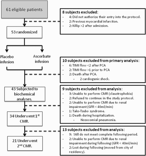
The study population consisted of 53 patients with comparable baseline characteristics (Table 2). A total of 43 patients underwent a biochemical analysis. Thirty-four patients underwent a CMR on the sixth day, and 21 patients completed the follow-up with a second CMR on the 84th day (Fig. ). Three patients died in the hospital, two from cardiogenic shock and one from nosocomial pneumonia. No patients showed any adverse event that was likely to be attributed to the antioxidant supplementation treatment.
Figure 1 Summary diagram of the enrolled patients to the randomized clinical trial. PCA, percutaneous coronary angioplasty; CMR, cardiac magnetic resonance; GFR, glomerular filtration rate.
Cardiac function
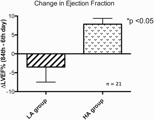
The LVEF assessed by the CMR showed no significant difference between the groups on day 6, but the values on day 84 in the HA group were 33% higher than those of the LA group (P < 0.05) (Fig. ). Additionally, to analyze the individual changes in the LVEF experienced by the patients in the two groups throughout the follow-up, the difference between the LVEF obtained at days 84 and 6 after the PCA was calculated (DeltaLVEF 84th − 6th). The changes in the LVEF obtained with this method showed significantly higher values for the HA group (P < 0.05) than for the LA group (Fig. ).
Figure 2 The LVEF accounting for the ventricle function assessed by CMR on day 6 and day 84 following PCA. Significant differences were found between the groups at day 84 and between the first and second CMR evaluation in the HA group. *P < 0.05.
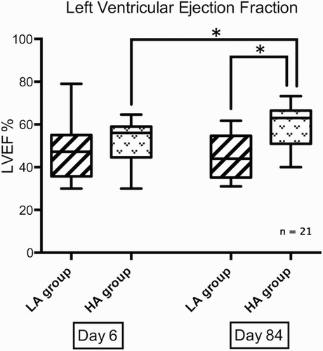
Figure 3 The LVEF differences between days 6 and 84 following PCA for each group. ΔLVEF% (84th − 6th day), LVEF on day 84 minus the LVEF on day 6. *P < 0.05.
Microvascular dysfunction
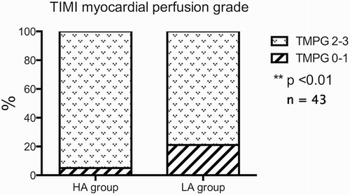
After PCA, 95% of the patients in the HA group achieved a TMPG of 2–3, and the other 5% remained at a TMPG of 0–1. Conversely, in the LA group, only 79% of patients showed a TMPG of 2–3, and 21% presented with a TMPG of 0–1 (P < 0.01) (Fig. ).
Figure 4 The microvascular dysfunction assessed by the TMPG following PCA. A properly functioning microvasculature was considered with a TMPG of 2 or 3. Significant differences were observed in the percentages of open microvasculatures after PCA between the groups. **P < 0.01.
Oxidative stress-related parameters
The mean plasma ascorbate levels (mmol/l) for the LA group were 0.03 ± 0.02 at baseline, 0.03 ± 0.04 immediately after the onset of reperfusion, 0.03 ± 0.03 at 6–8 hours after the onset of reperfusion, and 0.02 ± 0.01 at hospital discharge. In the HA group, these levels were 0.10 ± 0.20 at baseline, 9.79 ± 3.87 immediately after the onset of reperfusion, 1.79 ± 1.51 6–8 hours after reperfusion, and 0.06 ± 0.06 at hospital discharge. The group of HA patients showed significantly higher plasma ascorbate levels than the LA group both immediately after reperfusion and at 6–8 hours after the onset of reperfusion (P < 0.01).
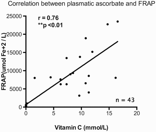
The antioxidant potential, as assessed by the plasma FRAP levels, did not show significant differences at the baseline or at hospital discharge. However, it is notable that immediately after the onset of reperfusion, patients in the HA group showed FRAP levels that were approximately 29 times higher than the respective LA values (P < 0.01). Moreover, at 6–8 hours after the onset of reperfusion, the FRAP values for the HA group were 4.8 times higher than the respective LA values (P < 0.01). Additionally, a positive association between the ascorbate and FRAP plasma levels was found immediately after the onset of reperfusion (Fig. ).
Figure 5 The correlation for the plasma antioxidant capacity assessed by FRAP and the vitamin C levels following the onset of reperfusion. A positive association showed a Spearman correlation rho index of 0.76. **P < 0.01.
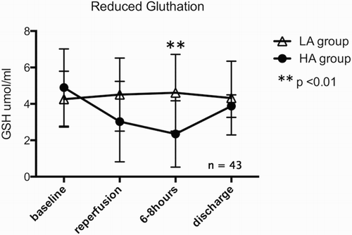
The erythrocyte GSH levels showed no significant differences between the groups at baseline, immediately after the onset of reperfusion or at hospital discharge. However, 6–8 hours after the onset of reperfusion, the GSH levels for the HA group were half of the respective LA values (P < 0.01) (Fig. ). A negative association was found consistently at these times between the plasma ascorbate and erythrocyte GSH levels, showing a correlation coefficient of −0.36 at the time of reperfusion and of −0.56 at 6–8 hours after the onset of reperfusion. In addition, there were no statistically significant differences between the groups in the GSH/GSSG ratio at baseline, at the time of reperfusion, or at discharge.
Figure 6 The erythrocyte GSH levels. Significant differences were observed at 6–8 hours following the onset of reperfusion. **P < 0.01.
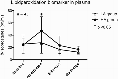
Concerning the lipid peroxidation, the 8-isoprostanes showed significant differences just after the onset of reperfusion (P < 0.05), which were greater in the HA group than in the LA group (P < 0.05) (Fig. ). No differences were observed for the other times of evaluation. In addition, the myocardial damage biomarker CK-MB showed no significant differences between the groups at baseline or at 6–8 hours after PCA.
Figure 7 The lipid peroxidation biomarker 8-isoprostane showed significant differences immediately after the onset of reperfusion. *P < 0.05.
Discussion
This study presents a novel strategy against reperfusion damage that is based on the administration of high doses of ascorbate to AMI patients who are subjected to PCA. The data reported here demonstrate for the first time that this therapy effectively ameliorates the persistent left ventricular impairment that usually occurs in these patients. In addition, the results of this trial provide evidence that an infusion of high doses of ascorbate followed by chronic oral supplementation with the antioxidant vitamins C and E is safe and positively influences the clinical outcome after AMI. The design of this study represents an original proposal for preventing MRI through supplementation with massive doses of ascorbate prior to the PCA procedure. At present, these results cannot be compared, but previous studies in other clinical settings have reported the safe administration of even higher ascorbate doses than we used in the present research. Citation24–Citation26 We attempted to abrogate the well-known contribution of oxidative stress in the mechanism of ischemia–reperfusion injury through the marked enhancement of the blood antioxidant potential at the onset of reperfusion. In fact, the FRAP values in the HA group were 29 times higher than those of the LA group, suggesting a lower vulnerability to an oxidative challenge of the former. Although to a lesser extent, this favorable condition was maintained for at least 6–8 hours following the onset of reperfusion, a phase likely characterized by the prevalence of a pro-oxidant state. It notable that despite this protective antioxidant condition, a paradoxical increase in the plasma 8-isoprostane levels was attained immediately after the reperfusion onset. Previous studies have documented the possible pro-oxidant effect caused by high doses of ascorbate; Citation27 thus, it is possible that the intervention performed in this study had two effects, a protective role against an ischemic–reperfusion injury and a minor pro-oxidant effect caused by the high concentrations of vitamin C that were achieved in the plasma. In another clinical setting of ischemia–reperfusion, it has been reported that intravenous ascorbate prior to vascular surgery increases the concentrations of ascorbate radicals and lipid hydroperoxides. This finding may suggest that the catalytic iron released into the circulation during the ischemic phase of the surgery in the presence of ascorbate may promote iron-induced lipid peroxidation. Citation28,Citation29 It is interesting that the higher lipid peroxidation levels in the patients in the HA group are not associated with myocardial damage, as assessed by the levels of CK-MB, the biomarker of this damage. However, high plasma ascorbate levels accompanied by decreased erythrocyte GSH levels were noted at 6–8 hours following the onset of reperfusion. A possible mechanistic explanation for this phenomenon is that the decrease in the GSH levels may be a consequence of the glutathione-dependent recycling of ascorbate that regenerates its antioxidant capacity in this manner. Citation30–Citation32 Nevertheless, further studies are required to determine the actual mechanism behind the antioxidant profile observed in the patients with high levels of plasma ascorbate.
A clear ventricle functional improvement induced by ascorbate might be suggested in the higher values of the LVEF three months after AMI in the patients showing high plasma ascorbate levels (Fig. ). It is noteworthy that in the CMRs performed at day 6, the ejection fraction was similar in both groups and that differences were not noted until day 84. The mechanism whereby the present intervention ameliorated the persistent ejection fraction impairment remains to be elucidated. An analysis of these results should take into consideration that previous studies performed with the use of oral antioxidants alone following AMI showed no improvement in terms of clinical outcomes. Citation3 Therefore, it may be plausible to assume that the high plasma ascorbate concentration that was achieved early during the reperfusion had a key role in triggering this beneficial effect. It is likely that the FRAP elevation due to high plasma ascorbate levels at the onset of reperfusion is able to exert a prophylactic effect against the lethal reperfusion caused by an angioplasty. Citation33 To our knowledge, there are no previous studies using an ascorbate dose capable of achieving the plasma concentrations that were reached in this study. In addition, it is noteworthy that the ascorbate administration resulted in the amelioration of the microvascular dysfunction (Fig. ). This derangement could be due to a combination of endothelial dysfunction, microvascular obstruction mediated by the downstream microembolism of platelets, de novo thrombosis, and neutrophil capillary plugging. When a severe microvascular dysfunction limits adequate perfusion despite the fact that a patient has undergone a successful angioplasty, the phenomenon is termed ‘no-reflow’. This is a specific type of vascular damage, first described by Kloner et al. in 1974, Citation34,Citation35 occurring when the complete removal of a coronary occlusion does not lead to the restoration of a normal coronary flow. Microvascular dysfunction has been associated with poor clinical outcomes after AMI, Citation36,Citation37 pathological cardiac remodeling, Citation38–Citation40 and ventricle functional impairment. Citation41–Citation44 It is known that oxidative stress is primarily responsible for the pathogenesis of microvascular dysfunction after ischemia–reperfusion. Citation45,Citation46 In this context, there is evidence suggesting that the plasma antioxidant depletion after AMI is strongly associated with the occurrence of the no-reflow phenomenon. Citation47 Thus, it is conceivable to put forward the hypothesis that antioxidant treatment has a beneficial role in the prevention of microvascular dysfunction. Our results are consistent with those of some studies demonstrating the benefits of antioxidant supplementation in decreasing the occurrence and extent of microvascular dysfunction. Citation48–Citation50
Limitations of the study
In the current study, we emphasized the assessment of cardiac function using the LVEF. However, other effects associated with the treatment were not evaluated. It is possible that when considering variables other than LVEF, such as the amelioration of the infarct size and myocardial remodeling, additional positive effects could be achieved by the antioxidant treatment. In addition, although the follow-up time of 84 days was sufficient to assess the evolution of the ventricular function, this monitoring did not allow the evaluation of the differences in the morbidity and mortality in the medium-to-long term. Another clinical limitation is that only those patients with a confirmed initial TIMI flow of 0 and reaching a TIMI flow of 3 after PCA were included in this study. Because of these selection criteria, an important section of the larger group of patients with myocardial infarctions was excluded from the analysis, causing the results to lack some external validity.
Table 1 Angiographic and procedural data.
Table 2 Baseline characteristics and drug use of the study groups.
Conclusions
In summary, the present study supports the view that high blood ascorbate levels reached prior to the reperfusion onset of AMI patients who were subjected to PCA followed by oral doses of vitamins C and E result in functional ventricle amelioration. This beneficial effect reported here for the first time is accompanied by an improvement in the microvascular blood flow, suggesting that the abrogation of oxidative stress is involved in the mechanisms whereby these effects occur. Enhancing the LVEF with a simple, innocuous, and low-cost pharmacological intervention may supply a cost-effective strategy for significantly improving the long-term prognosis of patients with acute myocardial infarctions and reducing the mortality and morbidity associated with AMI.
Disclaimer statements
Contributors All authors contributed equally.
Funding None.
Conflicts of interest The authors declare that they have no conflict of interest.
Ethics approval Paper has received ethical approval from University of Chile Ethical Committee, also ethical approval was obtained from all clinical Institutional Ethics Committees.
Acknowledgments
The authors thank the National Fund for Scientific and Technological Development (FONDECYT) for grant 1120594. The technical assistance of Diego Soto and the statistical analyses of PhD candidates Claudia Chavez and Daniela Albers are also acknowledged.
References
- World Health Organization (WHO) [Internet]. Methods and data sources for global causes of death 2000-2012. Global Health Estimates Technical Paper WHO/HIS/HSI/GHE/2014.7. 2014 Jun [cited 2015 Jun 12] Available from: http://www.who.int/healthinfo/global_burden_disease/GlobalCOD_method_2000_2012.pdf .
- Yellon DM , Hausenloy DJ . Myocardial reperfusion injury. N Engl J Med 2007;13(357):1121–35. doi: 10.1056/NEJMra071667
- Yizhou Y , Jing L , Zhongxiang Y . Effect of antioxidant vitamin supplementation on cardiovascular outcomes: A Meta-Analysis of Randomized Controlled Trials. PLos ONE 2013;8:e56803. doi: 10.1371/journal.pone.0056803
- Dhalla NS , Golfman L , Takeda S , Takeda N , Nagano M . Evidence for the role of oxidative stress in acute ischemic heart disease: a brief review. Can J Cardiol 1999;15:587–93.
- Xu Y , Liu B , Zweier JL , He G . Formation of hydrogen peroxide and reduction of peroxynitrite via dismutation of superoxide at reperfusion enhances myocardial blood flow and oxygen consumption in postischemic mouse heart. J Pharmacol Exp Ther 2008;327:402–10. doi: 10.1124/jpet.108.142372
- Hrabarova E , Juranek I , Soltes L . Pro-oxidative effect of peroxynitrite regarding biological systems: a special focus on high-molar-mass hyaluronan degradation. Gen Physiol Biophys 2011;30:223–38. doi: 10.4149/gpb_2011_03_223
- Liaudet L , Rosenblatt-Velin N , Pacher P . Role of peroxynitrite in the cardiovascular dysfunction of septic shock. Curr Vasc Pharmacol 2013;11:196–207.
- Rodrigo R , Libuy M , Feliú F , Hasson D . Molecular basis of cardioprotective effect of antioxidant vitamins in myocardial infarction. Biomed Res Int 2013;2013:437613. doi: 10.1155/2013/437613
- The Multicenter Postinfarction Research Group . Risk stratification and survival after myocardial infarction. N Engl J Med 1983;309:331–6. doi: 10.1056/NEJM198308113090602
- Rouleau JL , Talajic M , Sussex B , Potvin L , Warnica W , Davies RF ., et al. Myocardial infarction patients in the 1990s-their risk factors, stratification and survival in Canada: the Canadian Assessment of Myocardial Infarction (CAMI) study. J Am Coll Cardiol 1996;27:1119–27. doi: 10.1016/0735-1097(95)00599-4
- Odemuyiwa O , Malik M , Farrell T , Bashir Y , Poloniecki J , Camm J . Comparison of the predictive characteristics of heart rate variability index and left ventricular ejection fraction for all-cause mortality, arrhythmic events and sudden death after acute myocardial infarction. Am J Cardiol 1991;68:434–9. doi: 10.1016/0002-9149(91)90774-F
- Copie X , Hnatkova K , Staunton A , Fei L , Camm AJ , Malik M . Predictive power of increased heart rate versus depressed left ventricular ejection fraction and heart rate variability for risk stratification after myocardial infarction. Results of a two-year follow-up study. J Am Coll Cardiol 1996;27:270–6. doi: 10.1016/0735-1097(95)00454-8
- Van der Vleuten PA , Rasoul S , Huurnink W , van der Horst IC , Slart RH , Reiffers S , et al. The importance of left ventricular function for longterm outcome after primary percutaneous coronary intervention. BMC Cardiovasc Disord 2008;8:4. doi: 10.1186/1471-2261-8-4
- St John SM , Pfeffer MA , Moye L , Plappert T , Rouleau JL , Lamas G , et al. Cardiovascular death and left ventricular remodeling two years after myocardial infarction: baseline predictors and impact of long term use of captopril: information from the Survival and Ventricular Enlargement (SAVE) trial. Circulation 1997;96:3294–9. doi: 10.1161/01.CIR.96.10.3294
- Pfeffer MA , Braunwald E . Ventricular remodeling after myocardial infarction: experimental observations and clinical implications. Circulation 1990;81:1161–72. doi: 10.1161/01.CIR.81.4.1161
- Li WG , Zaheer A , Coppey L , Oskarsson HJ . Activation of JNK in the remote myocardium after large myocardial infarction in rats. Biochem Biophys Res Commun 1998;246:816–20. doi: 10.1006/bbrc.1998.8662
- Bolognese L , Cerisano G . Early predictors of left ventricular remodeling after acute myocardial infarction. Am Heart J 1999;138:S79–83. doi: 10.1016/S0002-8703(99)70325-X
- Chen W , Frangogiannis NG . The role of inflammatory and fibrogenic pathways in heart failure associated with aging. Heart Fail Rev 2010;15:415–22. doi: 10.1007/s10741-010-9161-y
- Gibson CM , Cannon CP , Murphy SA , Ryan KA , Mesley R , Marble SJ , et al. Relationship of TIMI myocardial perfusion grade to mortality after administration of thrombolytic drugs. Circulation 2000;101:125–30. doi: 10.1161/01.CIR.101.2.125
- Gibson CM , Cannon CP , Murphy SA , Marble SJ , Barron HV , Braunwald E . TIMI Study Group . Relationship of the TIMI myocardial perfusion grades, flow grades, frame count, and percutaneous coronary intervention to long-term outcomes after thrombolytic administration in acute myocardial infarction. Circulation 2002;105:1909–13. doi: 10.1161/01.CIR.0000014683.52177.B5
- Chung WY , Chung JK , Szeto YT , Tomlinson B , Benzie IF . Plasma ascorbic acid: measurement, stability and clinical utility revisited. Clin Biochem 2001;34:623–7. doi: 10.1016/S0009-9120(01)00270-3
- Hissin PJ , Hilf R . A fluorometric method for determination of oxidized and reduced glutathione in tissues. Anal Biochem 1976;74:214–26. doi: 10.1016/0003-2697(76)90326-2
- Benzie IF , Strain JJ . The ferric reducing ability of plasma (FRAP) as a measure of ‘antioxidant power’: the FRAP assay. Anal Biochem 1996;239:70–6. doi: 10.1006/abio.1996.0292
- Padayatty SJ , Sun AY , Chen Q , Espey MG , Drisko J , Levine M . Vitamin C: intravenous use by complementary and alternative medicine practitioners and adverse effects. Plos ONE 2010;5:e11414. doi: 10.1371/journal.pone.0011414
- Duconge J , Miranda-Massari JR , Gonzalez MJ , Jackson JA , Warnock W , Riordan NH . Pharmacokinetics of vitamin C: insights into the oral and intravenous administration of ascorbate. P R Health Sci J 2008;27:7–19.
- Stephenson CM , Levin RD , Spector T , Lis CG . Phase I clinical trial to evaluate the safety, tolerability, and pharmacokinetics of high-dose intravenous ascorbic acid in patients with advanced cancer. Cancer Chemother Pharmacol 2013;72:139–46. doi: 10.1007/s00280-013-2179-9
- Park SW , Lee SM . Antioxidant and prooxidant properties of ascorbic acid on hepatic dysfunction induced by cold ischemia/reperfusion. Eur J Pharmacol 2008;580:401–6. doi: 10.1016/j.ejphar.2007.11.023
- Buettner GR . The reaction of superoxide, formate radical, and hydrated electron with transferrin and its model compound, Fe(III)-ethylenediamine-N,N′-bis [2-(2-hydroxyphenyl) acetic acid] as studied by pulse radiolysis. J Biol Chem 1987;262:11995–8.
- Dabbagh AJ , Trenam CW , Morris CJ , Blake DR . Iron in joint inflammation. Ann Rheum Dis 1993;52:67–73. doi: 10.1136/ard.52.1.67
- Meister A . Glutathione-ascorbic acid antioxidant system in animals. J Biol Chem 1994;2690:9397–400.
- Winkler BS . Unequivocal evidence in support of the nonenzymatic redox coupling between glutathione/glutathione disulfide and ascorbic acid/dehydroascorbic acid. Biochim Biophys Acta 1992;1117:287–90. doi: 10.1016/0304-4165(92)90026-Q
- Winkler BS , Orselli SM , Rex TS . The redox couple between glutathione and ascorbic acid: a chemical and physiological perspective. Free Radic Biol Med 1994;17:333–49. doi: 10.1016/0891-5849(94)90019-1
- Rodrigo R , Hasson D , Prieto JC , Dussaillant G , Ramos C , León L , et al. The effectiveness of antioxidant vitamins C and E in reducing myocardial infarct size in patients subjected to percutaneous coronary angioplasty (PREVEC Trial): study protocol for a pilot randomized double-blind controlled trial. Trials 2014;15:192. doi: 10.1186/1745-6215-15-192
- Kloner RA , Ganote CE , Jennings RB . The 'no reflow' phenomenon after temporary coronary occlusion in the dog. J Clin Invest 1974;54:1496–508. doi: 10.1172/JCI107898
- Kloner RA , Rude RE , Carlson N , Maroko PR , DeBoer LW , Braunwald E . Ultrastructural evidence of microvasular damage and myocardial cell injury after coronary artery occlusion: which comes first? Circulation 1980;62:945–52. doi: 10.1161/01.CIR.62.5.945
- Ito H , Maruyama A , Iwakura K , Takiuchi S , Masuyama T , Hori M , et al. Clinical implications of the ‘no-reflow’ phenomenon: a predictor of complications and left ventricular remodeling in reperfused anterior wall myocardial infarction. Circulation 1996;93:223–8. doi: 10.1161/01.CIR.93.2.223
- Wu KC , Zerhouni EA , Judd RM , Lugo-Olivieri CH , Barouch LA , Schulman SP , et al. Prognostic significance of microvascular obstruction by magnetic resonance imaging in patients with acute myocardial infarction. Circulation 1998;97:765–72. doi: 10.1161/01.CIR.97.8.765
- Erlebacher JA , Weiss JL , Weisfeldt ML , Bulkley BH . Early dilation of the infarcted segment in acute transmural myocardial infarction: role of infarct expansion in acute left ventricular enlargement. J Am Coll Cardiol 1984;4:201–8. doi: 10.1016/S0735-1097(84)80203-X
- Sutton MG , Sharpe N . Left ventricular remodeling after myocardial infarction: pathophysiology and therapy. Circulation 2000;101:2981–8. doi: 10.1161/01.CIR.101.25.2981
- Lombardo A , Niccoli G , Natale L , Bernardini A , Cosentino N , Bonomo L , et al. Impact of microvascular obstruction and infarct size on left ventricular remodeling in reperfused myocardial infarction: a contrast-enhanced cardiac magnetic resonance imaging study. Int J Cardiovasc Imaging 2012;28:835–42. doi: 10.1007/s10554-011-9901-7
- Hombach V , Grebe O , Merkle N , Waldenmaier S , Höher M , Kochs M , et al. Sequelae of acute myocardial infarction regarding cardiac structure and function and their prognostic significance as assessed by magnetic resonance imaging. Eur Heart J 2005;26:549–57. doi: 10.1093/eurheartj/ehi147
- Choi CJ , Haji-Momenian S , Dimaria JM , Epstein FH , Bove CM , Rogers WJ , et al. Infarct involution and improved function during healing of acute myocardial infarction: the role of microvascular obstruction. J Cardiovasc Magn Reson 2004;6:917–25. doi: 10.1081/JCMR-200036206
- Nijveldt R , Beek AM , Hirsch A , Stoel MG , Hofman MB , Umans VA , et al. Functional recovery after acute myocardial infarction. J Am Coll Cardiol 2008;52:181–9. doi: 10.1016/j.jacc.2008.04.006
- Limalanathan S , Eritsland J , Andersen GØ , Kløw NE , Abdelnoor M , Hoffmann P . Myocardial salvage is reduced in primary PCI-treated STEMI patients with microvascular obstruction, demonstrated by early and late CMR. Plos ONE 2013;8:e71780. doi: 10.1371/journal.pone.0071780
- Garlick PB , Davies MJ , Hearse DJ , Slater TF . Direct detection of free radicals in the reperfused rat heart using electron spin resonance spectroscopy. Circ Res 1987;61:757–60. doi: 10.1161/01.RES.61.5.757
- Moens AL , Claeys MJ , Timmermans JP , Vrints CJ . Myocardial ischemia/reperfusion-injury, a clinical view on a complex pathophysiological process. Int J Cardiol 2005;100:179–90. doi: 10.1016/j.ijcard.2004.04.013
- Matsumoto H , Inoue N , Takaoka H , Hata K , Shinke T , Yoshikawa R , et al. Depletion of antioxidants is associated with no-reflow phenomenon in acute myocardial infarction. Clin Cardiol 2004;27:466–70. doi: 10.1002/clc.4960270809
- McNulty PH , Robertson BJ , Tulli MA , Hess J , Harach LA , Scott S , et al. Effect of hyperoxia and vitamin C on coronary blood flow in patients with ischemic heart disease. J Appl Physiol 2007;102:2040–5. doi: 10.1152/japplphysiol.00595.2006
- Molyneux CA , Glyn MC , Ward BJ . Oxidative stress and cardiac microvascular structure in ischemia and reperfusion: the protective effect of antioxidant vitamins. Microvasc Res 2002;64:265–77. doi: 10.1006/mvre.2002.2419
- Basili S , Tanzilli G , Mangieri E , Raparelli V , Di Santo S , Pignatelli P , et al. Intravenous ascorbic acid infusion improves myocardial perfusion grade during elective percutaneous coronary intervention: relationship with oxidative stress markers. JACC Cardiovasc Interv 2010;3:221–9. doi: 10.1016/j.jcin.2009.10.025