
Abstract
Background
The purpose of this study was to examine if L-leucine (Leu), β-hydroxy-β-methylbutyrate (HMB), or creatine monohydrate (Crea) prevented potential atrophic effects of myostatin (MSTN) on differentiated C2C12 myotubes.
Methods
After four days of differentiation, myotubes were treated with MSTN (10 ng/ml) for two additional days and four treatment groups were studied: 1) 3x per day 10 mM Leu, 2) 3x per day 10 mM HMB, 3) 3x per day 10 mM Crea, 4) DM only. Myotubes treated with DM without MSTN were analyzed as the control condition (DM/CTL). Following treatment, cells were analyzed for total protein, DNA content, RNA content, muscle protein synthesis (MPS, SUnSET method), and fiber diameter. Separate batch treatments were analyzed for mRNA expression patterns of myostatin-related genes (Akirin-1/Mighty, Notch-1, Ski, MyoD) as well as atrogenes (MuRF-1, and MAFbx/Atrogin-1).
Results
MSTN decreased fiber diameter approximately 30% compared to DM/CTL myotubes (p < 0.001). Leu, HMB and Crea prevented MSTN-induced atrophy. MSTN did not decrease MPS levels compared to DM/CTL myotubes, but MSTN treatment decreased the mRNA expression of Akirin-1/Mighty by 27% (p < 0.001) and MyoD by 26% (p < 0.01) compared to DM/CTL myotubes. shRNA experiments confirmed that Mighty mRNA knockdown reduced myotube size, linking MSTN treatment to atrophy independent of MPS. Remarkably, MSTN + Leu and MSTN + HMB myotubes had similar Akirin-1/Mighty and MyoD mRNA levels compared to DM/CTL myotubes. Furthermore, MSTN + Crea myotubes exhibited a 36% (p < 0.05) and 86% (p < 0.001) increase in Akirin-1/Mighty mRNA compared to DM/CTL and MSTN-only treated myotubes, respectively.
Conclusions
Leu, HMB and Crea may reduce MSTN-induced muscle fiber atrophy by influencing Akirin-1/Mighty mRNA expression patterns. Future studies are needed to examine if Leu, HMB and Crea independently or synergistically affect Akirin-1/Mighty expression, and how Akirin-1/Mighty expression mechanistically relates to skeletal muscle hypertrophy in vivo.
Electronic supplementary material
The online version of this article (doi:10.1186/1550-2783-11-38) contains supplementary material, which is available to authorized users.
Copyright comment
This article is published under license to BioMed Central Ltd. This is an Open Access article distributed under the terms of the Creative Commons Attribution License (http://creativecommons.org/licenses/by/4.0), which permits unrestricted use, distribution, and reproduction in any medium, provided the original work is properly credited. The Creative Commons Public Domain Dedication waiver (http://creativecommons.org/publicdomain/zero/1.0/) applies to the data made available in this article, unless otherwise stated.
Background
Myostatin (MSTN) is a key negative regulator of mature skeletal muscle myofiber growth [Citation1–Citation3]. In this regard, MSTN has been shown to reduce muscle protein synthesis by abrogating mTORC1 signaling [Citation4, Citation5] and increase muscle proteolytic mechanisms [Citation6, Citation7]. Furthermore, several studies have implicated physical inactivity-induced up-regulation in MSTN as a potential regulator in age-related skeletal muscle loss [Citation8–Citation13], and there is supporting evidence suggesting that serum and skeletal muscle MSTN are elevated with aging [Citation14, Citation15].
The effects of exercise have also been implicated in MSTN pathway signaling. Specifically, Louis et al. [Citation16] reported that MSTN mRNA expression is depressed within hours following either endurance or resistance exercise. Dalbo et al. [Citation17] similarly reported that resistance exercise decreased skeletal muscle myostatin mRNA levels up to 6 hours post-exercise. Likewise, recent evidence suggests that skeletal muscle MSTN increases three days following detraining from 90 days of resistance exercise; an event which preceded subtle but rapid type II fiber atrophy [Citation18].
While exercise reduces skeletal muscle myostatin expression, nutritional strategies to reduce myostatin signaling are also warranted. In this regard, select nutrients have been shown to increase skeletal muscle anabolic signaling mechanisms. Leucine is a well-known activator of mTOR signaling [Citation19–Citation21], and leucine has additionally been shown to reduce muscle proteolysis [Citation22]. The leucine metabolite beta-hydroxy- beta-methylbutyric acid (HMB) has also been well-described with regard to its effects on whole-body muscle mass accretion [Citation23–Citation25], as well as its ability to independently activate skeletal muscle mTOR signaling and reduce proteolytic signaling [Citation26–Citation28]. Creatine monohydrate has been less studied with regards to mTOR pathway modulation, although some evidence exists suggesting that creatine monohydrate is able to increase myotube differentiation through poorly understood mechanisms [Citation29]. Notwithstanding, ample literature has demonstrated that creatine monohydrate supplementation is able to increase muscle mass and strength [Citation30–Citation35].
While a plethora of literature reports the effects of these nutritional supplements on skeletal muscle anabolic and/or anti-catabolic mechanisms, no information to our knowledge is known regarding how or if these supplements can abrogate facets of MSTN signaling. Therefore, the purpose of this study was to determine whether differentiated/mature myotubes treated with leucine, HMB or creatine monohydrate in the presence of MSTN affected: a) myotube diameter, b) select anabolic indices (Protein: DNA, RNA: DNA), and c) the mRNA expression patterns of genes associated with myostatin signaling.
Methods
Cell culture methods
C2C12 myoblasts (graciously donated by RHA), passage no. 4–10, were maintained in growth medium (GM; DMEM, 10% FBS, 1% penicillin/streptomycin, 0.1% gentamycin) under standard culture conditions at 37°C in a 5% CO2 atmosphere. Myoblasts were grown on 145 mm plates (Griener Bio-One GmbH, Maybachstr, Frickenhausen, GER) at a density of 7.5 × 10⁵ in 10 ml of growth medium for protein analyses, or on 12-well plates (Griener Bio-One GmbH) at a density of 5 × 10⁵ for mRNA analyses. Differentiation was induced 48 h after myoblast growth reached 80%–90% confluency by removing the growth medium and replacing it with differentiation medium (DM; DMEM, 2% (vol/vol) horse serum, 1% penicillin/streptomycin). DM was then replaced every 24 h for 4 d.
Treatment methods (DM only, MSTN only, MSTN + Leucine, MSTN + HMB, MSTN + Creatine)
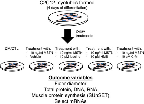
The study design is illustrated in Figure below. Briefly, after four days of differentiation, cells were treated three times per day with one of the following treatments for 48 h: 1) DM and vehicle (10 mM Tris-NaCl); denoted as ‘DM/CTL’ only, 2) 10 ng/ml rGDF-8 MSTN (R&D Systems, Minneapolis, MN, USA); denoted as ‘MSTN’, 3) 10 ng/ml MSTN and 10 μM (13 ug/ml) leucine (EMD Chemicals, Inc., San Diego, CA, USA); denoted as MSTN + Leu, 4) 10 ng/ml MSTN and 10 μM (13 ug/ml) of free acid HMB (Metabolic Technologies, Inc., Ames, IA, USA); denoted as ‘MSTN + HMB’, or 5) 10 ng/ml MSTN and 10 μM (12 ug/ml) creatine monohydrate (BodyBuilding.com, Boise, ID, USA); denoted as ‘MSTN + Crea’. The MSTN treatment dosage was based upon two prior studies showing that 10–30 ng/ml reduces myotube diameter in differentiated C2C12 myotubes [Citation36, Citation37]. The leucine, HMB and creatine monohydrate dosages were based upon prior C2C12 literature showing biological responses to similar dosages for each respective ingredient [Citation21, Citation38, Citation39]. On the last day of treatment, and 30–45 min prior to cell lysis, cells were pulse-labeled with 1 μM of puromycin hydrochloride (Millipore, Temecula, CA, USA) in phosphate-buffered saline for subsequent muscle protein synthesis (MPS) assessment.
Figure 1 Study design. After four days of differentiation, cells were treated for two days (three times per day) with differentiation media (DM) and vehicle (10 mM Tris-NaCl), 10 ng/ml recombinant GDF-8 MSTN, 10 ng/ml MSTN and 10 μM leucine, 10 ng/ml MSTN and 10 μM of HMB, or 10 ng/ml MSTN and 10 μM creatine monohydrate.
Cells grown on 145 mm plates were lysed using RIPA buffer (Tris base; pH 8.0, NaCl, NP-40, sodium deoxycholate, SDS) containing protease and phosphatase inhibitors (Ameresco, Solon, OH, USA). After cells were lysed, RIPA homogenates were analyzed for total protein, total DNA, and total RNA using a Qubit Fluorometer (Life Technologies, Grand Island, NY). RIPA homogenates were then spun down at 500xg for 5 min, and supernatants were stored for Western blotting analyses as described below.
Light Microscopy Imaging and ImageJ Analysis
Prior to cell lysis of the 145 mm plates, 10× digital images were obtained for n = 2 for each treatment using light microscopy (Nikon Eclipse Ci-L) and digital capture (Nikon DS-QilMc). Fiber diameters from 150–190 myotubes per condition were obtained and assessed using ImageJ (NIH, Bethesda, MD, USA).
Western blotting methods and analysis
In order to examine if MSTN treatment reduced muscle protein synthesis (MPS) rates compared to DM/CTL myotubes, the SUnSET method was employed [Citation40]. Briefly, RIPA homogenates from 145 mm plates were subjected to 4-20% SDS-polyacrylamide gel electrophoresis using pre-casted gels (C.B.S. Scientific Company, San Diego, CA, USA). Proteins were transferred to polyvinylidene difluoride membranes (Whatman™, Westran® Clear Signal), and membranes were blocked for 1 h at room temperature with 5% nonfat milk powder. Mouse anti-puromycin (1:5,000; Millipore) was then incubated with membranes overnight at 4°C in 5% bovine serum albumin, and the following day membranes were incubated with anti-mouse IgG secondary antibodies (Cell Signaling, Danvers, MA, USA) at room temperature for 1 h. Membranes were then developed using an enhanced chemiluminescent reagent (Amersham, Pittsburgh, PA, USA), and band densitometry was performed through the use of a UVP Imager and associated densitometry software (UVP, LLC, Upland, CA, USA).
RNA isolation and real-time PCR
RNA was isolated from myotubes grown on 12-well plates using Ribozol (Ameresco) per the manufacturer’s recommendations. 400 ng of cDNA was synthesized using a cDNA synthesis kit (Quanta, Gaithersburg, MD, USA) per the manufacturer’s recommendations. Real-time PCR was performed using mRNA specific primers {Akirin-1/Mighty: forward primer 5′- ATACAGTCACGGAGCTCCCT-3′, reverse primer 5′- ACTTGTTACACGCTCCGAGG-3′; Atrogin-1/MAFbx: forward primer 5′- CCATCCTCTTTCTTGCCCGT-3′, reverse primer 5′- ATCACTGTCCAACCTGGCTG-3′; MuRF-1: forward primer 5′- TGGGACAGATGAGGAGGAGG-3′, reverse primer 5′- TTTACCCTCTGTGGTCACGC-3′; beta-actin: forward primer 5′- GTGGATCAGCAAGCAGGAGT-3′, reverse primer 5′- ACGCAGCTCAGTAACAGTCC-3′; Notch-1: forward primer 5′- TGGACTGTTCTGTGCATCCC-3′, reverse primer 5′- TGGGGATCAGAGGCCACATA-3′; Ski: forward primer 5′- CCCACATGCCAGGATGACTT-3′, reverse primer 5′- GCTTTGCCAACTTCACCCAG-3′; MyoD: forward primer 5′- CCTGCCCTCCACATCCTTTT-3′, reverse primer 5′- GAAGGGGGAGAGTGGGGTAT-3′} and SYBR green chemistry (Quanta). Primer efficiency curves for all genes were generated and efficiencies ranged between 90% and 110%.
shRNA experiments to confirm that Akirin-1/Mighty affects myotube size
A separate batch of C2C12 myoblasts was seeded in 12-well plates at a density of 5 × 10⁵ cells per plate. Cells were grown to 80-90% confluency and then transfection growth media containing Lipofectamine 3000 (Life Technologies) was added to myoblasts per the manufacturer’s recommendations. Specifically, n = 4–5 wells were transfected with green fluorescent protein (GFP) reporter plasmids (GeneCopoeia, Rockville, MD, USA) containing either: a) scrambled shRNA, or b) Akirin-1/Mighty shRNA. Twenty-four hours after transfection, cells were differentiated as mentioned above. Cells were then allowed to differentiate for 4 days prior to imaging on an inverted fluorescent microscope (Olympus IX71). 10× digital fluoromteric FITC-filtered images were obtained for each transfection condition and myotube areas of GFP-positive myotubes were quantified using ImageJ. Cells were then lysed with Ribozol (Ameresco) per the manufacturer’s recommendations and Akirin-1/Mighty mRNA knock-down was confirmed using real-time PCR methods as mentioned above.
Statistics
All data are presented as means ± standard error. For all data, statistics were performed between treatments using an ANOVA with LSD post-hoc comparisons when applicable. All statistics were performed using IBM SPSS version 22.0 and significance was determined at p < 0.05.
Results
L-leucine, HMB and creatine monohydrate prevent myostatin-induced myotube atrophy
Two days of MSTN only treatment significantly reduced myotube diameter by approximately 30% compared to the DM/CTL condition (p < 0.001; Figure A). However, the MSTN + Leu, MSTN + HMB and MSTN + Crea treatments rescued this atrophy effect. MSTN treatment tended to decrease the protein: DNA compared to the DM/CTL condition (p = 0.084, Figure B; index of muscle hypertrophy [Citation41]). However, MSTN + Crea myotubes exhibited a significantly greater protein: DNA ratio compared to the MSTN only treatment (p < 0.01, Figure B). MSTN + Leu, MSTN + HMB, and MSTN + Crea myotubes all exhibited a greater RNA: DNA ratio compared to the MSTN only condition (Figure C; index of translational capacity and hypertrophic potential [Citation41]).
Figure 2 Efects of MSTN in the absence or presence of leucine, HMB, or creatine on markers of skeletal muscfle hypertrophy. Legend: Data are presented as means ± standard error. Data in A show that MSTN treatment decreases myotube diameter, while Leu, HMB, and Crea rescue MSTN-induced atrophy. Data in B & C are n = 5-6 plates per treatment. Muscle protein synthesis data in E are n = 4 treatments per plate; of note, MSTN+Leu, MSTN+HMB and MSTN+Crea samples were separately analyzed for MPS (data not shown). One-way ANOVA with LSD post-hoc comparisons performed where applicable; difference superscript letters = p < 0.05 and NS = no significant differences. In subfigure D of 10x light micrographs, white bar is 100 μm.
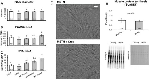
Interestingly, two days of MSTN treatment did not affect MPS rates compared to DM/CTL only-treated myotubes (Figure E). Thus, the MSTN-induced reduction in myotube diameter appears to be independent of MPS rates. Notably, MPS rates were also determined in a separate batch of DM/CTL (n = 2), MSTN + Leu (n = 6), MSTN + HMB (n = 6), and MSTN + Crea (n = 6) myotubes. Compared to DM/CTL myotubes, MSTN + Leu and MSTN + HMB caused non-significant increases in MPS rates (38% and 31%, respectively; data not shown), whereas MSTN + Crea did not increase MPS levels.
Myostatin-induced down-regulation of Akirin-1/Mighty mRNA is rescued by creatine treatments
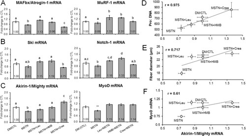
Given that MSTN-treated myotubes atrophied independent of MPS rates, we next examined if select atrogenes (MAFbx/atrogin-1, MuRF-1), MSTN signaling repressors (Notch-1, Ski), and/or MSTN transcriptional targets (Akirin-1/Mighty, MyoD) were affected at the mRNA level by MSTN treatment with or without leucine, HMB or creatine monohydrate treatments. Compared to DM/CTL myotubes, the MSTN-only treatment did not affect the mRNA expression patterns of MuRF-1, Ski, Notch-1. However, MAFbx/atrogin-1 mRNA levels were depressed compared to DM/CTL myotubes (Figure A/B).The MSTN-only treatment did depress Akirin-1/Mighty mRNA and MyoD mRNA levels by 27% (p < 0.001) and 26% (p < 0.01) compared to DM/CTL myotubes (Figure C). Moreover, MSTN-treated myotubes co-treated with leucine or HMB reversed MSTN-induced Akirin-1/Mighty mRNA down-regulation (p < 0.05); specifically, MSTN + Leu exhibited an 18% increase and MSTN + HMB exhibited a 27% increase in Akirin-1/Mighty mRNA compared to MSTN-treated myotubes. MSTN + Crea-treated myotubes exhibited a significant up-regulation in Akirin-1/Mighty mRNA levels by 36% compared to DM/CTL myotubes (p < 0.05) and 86% compared to MSTN-only treated myotubes (p < 0.001). The expression of Akirin-1/Mighty mRNA was modestly (0.50 < r < 0.80) to strongly (r > 0.80) correlated with select measured anabolic indices (Pro:DNA r = 0.98, p = 0.004; fiber diameter r = 0.72, p > 0.05; Figure D/E) as well as the early differentiation marker MyoD mRNA (r = 0.61, p > 0.05; Figure F).
Figure 3 Effects of MSTN in the absence or presence of leucine, HMB, or creatine on the expression of MSTN-related mRNAs. Legend: Data are presented as means ± standard error (n = 6–9 plates per treatment). Effects of MSTN with or without each ingredient on the mRNA expression of explicit atrogenes (A), MSTN signaling repressors (B), and MSTN transcriptional targets (C). Sub-figures D and E demonstrate that Akirin-1/Mighty mRNA expression was modestly-to-strongly correlated with measured hypertrophy variables. Sub-figure F shows that Akirin-1/Mighty mRNA expression was modestly correlated with the early differentiation marker MyoD mRNA. One-way ANOVA with LSD post-hoc comparisons performed in sub-figures A/B/C; difference superscript letters = p < 0.05. In sub-figure C, ** indicates that MSTN down-regulated MyoD mRNA compared to all other groups (p < 0.01).
Knockdown of Akirin-1/Mighty mRNA decreases myotube size
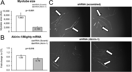
As mentioned above, Akirin-1/Mighty mRNA expression patterns between treatments exhibited modest to strong correlations to select hypertrophy indices. Thus, we next sought to determine if experimentally decreasing Akirin-1/Mighty mRNA using shRNA knockdown affected myotube size. Indeed, GFP-positive myotubes transfected with the Akirin-1/Mighty shRNA plasmid exhibited a drastic reduction in myotube area (−72%, p < 0.001; Figure A). Knockdown in transfected wells was also confirmed at the mRNA level (Figure B). Therefore, we posit that MSTN-induced atrophy observed in the current study is likely linked to the MSTN-induced down-regulation in Akirin-1/Mighty mRNA. Furthermore, leucine and HMB appear to prevent this down-regulation and creatine monohydrate increases myotube Akirin-1/Mighty mRNA levels in spite of MSTN treatment.
Figure 4 Experiment demonstrating Akirin-1/Mighty mRNA knockdown affects myotube size. Legend: Data are presented as means ± standard error (n = 4–5 plates per treatment). Effects of shRNA-mediated Akirin-1/Mighty mRNA knockdown on myotube size versus a scrambled shRNA control condition (A), and confirmation that Akirin-1 mRNA was reduced in shRNA-transfected myotubes (B). Photographs in sub-figure C are 10x representative images of GFP-positive cells (arrows) that were transfected with either a scrambled (CTL)-shRNA or Akirin-1/Mighty-shRNA.
Discussion
We used an in vitro approach to investigate whether leucine, HMB, or creatine monohydrate could rescue the atrophic effects of MSTN in C2C12 myotubes. MSTN has previously been shown to inhibit myoblast proliferation, myotube differentiation and protein synthesis in the C2C12 cell line [Citation42–Citation44]. Specifically, one of the aforementioned studies demonstrated that myotubes treated with high doses of recombinant MSTN (~1-6 μg/ml which is 100-600x the dose used in the current study) significantly depressed protein synthesis in myotubes [Citation42]. However, the relatively low concentration of MSTN treatments used in the current study reduced myotube size independent of muscle protein synthesis. This finding is in agreement with other studies which used MSTN treatment dosages similar to our study (10–30 ng/ml) and reported MSTN to reduce myotube diameter by inhibiting differentiation in C2C12 myotubes [Citation36, Citation37]. Thus, we hypothesize that MSTN-induced atrophy in the current study was likely due to diminished myotube differentiation rather than decreases in muscle protein synthesis and/or increases in muscle proteolysis; a hypothesis which is further supported by the MSTN-induced down-regulation in MyoD mRNA.
Interestingly, while myotubes treated with MSTN only showed a significant decrease in myotube diameter, this effect was reversed in all three treatment groups (MSTN + Leu, MSTN + HMB, and MSTN + Crea). There is ample evidence to suggest that leucine and HMB are able to increase muscle protein synthesis in vitro and in vivo[Citation26, Citation27, Citation45, Citation46]. Thus, it is difficult to reconcile why MSTN + Leu and MSTN + HMB treatments in the current study did not statistically increase muscle protein synthesis compared to DM/CTL myotubes. However, all treatments occurred in myotubes that were not amino acid deprived; this being a condition which may be obligatory for leucine and HMB to exert more profound muscle protein synthesis effects [Citation47]. Furthermore, the MSTN + Crea treated group demonstrated the greatest hypertrophic effect in spite of the fact that creatine monohydrate likely does not affect markers of muscle protein synthesis [Citation29] and/or muscle protein synthesis rates [Citation48]in vivo. Thus, we hypothesized that leucine, HMB and creatine monohydrate treatments all independently counteracted low-dose MSTN-induced atrophy through potential genetic mechanisms related to myotube differentiation.
Of the mRNAs measured in the current study select hypertrophic indices, including the protein: DNA and myofiber diameter, were strongly and modestly correlated with Akirin-1/Mighty gene expression, respectively. Furthermore, our main findings with Akirin-1/Mighty gene expression were as follows: 1) leucine and HMB can reverse MSTN-induced down-regulation in Akirin-1/Mighty mRNA; 2) in spite of MSTN treatment, creatine monohydrate up-regulated Akirin-1/Mighty mRNA while exhibiting the most potent anabolic effects; and 3) Akrin-1/Mighty mRNA knockdown via shRNA transfection reduced myofiber size. Hence, our findings support the hypothesis that the transcriptional modulation of Akirin-1/Mighty mRNA by leucine/HMB/creatine monohydrate may be a mechanism whereby these ingredients promote myotube hypertrophy in vitro in spite of MSTN treatments.
Our finding that each of these purported anabolic ingredients rescues MSTN-induced Akirin-1/Mighty mRNA down-regulation is indeed difficult to interpret from a mechanistic and practical viewpoint regarding the preservation of myofiber size. Furthermore, this is the first study to show that each of these ingredients affects (directly or indirectly) the mRNA expression of Akirin-1/Mighty mRNA. Resistance exercise has been shown to increase Akirin-1/Mighty mRNA expression patterns 6 h and 48 h following an acute exercise bout in rodents [Citation49]; of note these rodents were trained 6 weeks prior to the acute bout and the exercise-induced changes in Akrin-1/Mighty strongly predicted the hypertrophic response to the 6-week training bout. The authors concluded that, while Akrin-1/Mighty may play a role in the activation of satellite cells, how Akirin-1/Mighty promotes the hypertrophic response to exercise has yet to be determined. Notwithstanding, it appears that Akirin-1/Mighty plays a role in exercise-induced skeletal muscle hypertrophy. The expression of mRNA expression of akirin genes in skeletal muscle are sensitive to fasting and re-feeding in Artic charr [Citation50], and we have observed mixed gastrocnemius Akirin-1/Mighty mRNA to increase approximately 90% 3 h after rats fed 10 human equivalent grams of whey protein concentrate (which is ~12-14% leucine) when compared to 18-h fasted rats (p < 0.05; unpublished observations). Hence, our finding that purported anabolic ingredients directly or indirectly affect myotube Akirin-1/Mighty mRNA expression is not unfounded given that other hypertrophic stimuli (i.e., resistance exercise and protein feeding) have also been shown to increase the expression of this gene in skeletal muscle. In this regard, future research is needed to elucidate if: 1) decrements in Akirin-1/Mighty mRNA expression accompany and/or causal to muscle wasting conditions such as sarcopenia, cachexia, and disuse atrophy; and 2) if each of the anabolic ingredients studied herein mitigate these conditions through Akirin-1/Mighty gene expression changes.
While the exact mechanisms are unknown as to how Akirin-1/Mighty regulates muscle mass, it is a proven transcriptional target of myostatin [Citation49, Citation51, Citation52]. Recent evidence also suggests that the Akirin-1/Mighty gene modulates satellite cell proliferation and differentiation following muscle injury, and there is interplay between Akirin-1/Mighty down-regulation and the inhibition of differentiation-promoting genes such as MyoD [Citation53]. There is a paucity of literature examining how leucine and/or HMB affect Akirin-1/Mighty mRNA expression patterns, though recent in vivo evidence suggests that chronically consuming a leucine-rich pre-exercise beverage increases skeletal muscle MyoD and MRF4 mRNA [Citation54]. Furthermore, adding various concentrations (10–100 μg/ml) of HMB to serum-starved myoblasts has been shown to induce myoblast proliferation and MyoD expression, suggestive of enhanced myoblast differentiation [Citation55]. However, Akirin-1/Mighty mRNA was not assessed in the aforementioned studies making it difficult to reconcile whether these effects were mitigated through Akrin-1/Mighty mRNA gene expression.
Of particular interest was the ability of creatine monohydrate to promote the up-regulation of Akirin-1/Mighty mRNA in the presence of myostatin. Creatine monohydrate supplementation has been shown to reduce the catabolic response of hind limb immobilization in rats [Citation56]. Additionally, Johnston et al. [Citation57] have demonstrated that short-term creatine monohydrate supplementation (7 days) attenuates losses of muscle mass and strength during upper-arm immobilization in young men. Furthermore, prolonged creatine monohydrate supplementation has been reported to increase satellite cell proliferation and differentiation in resistance-trained subjects versus a placebo group [Citation58]. While the anabolic/anti-catabolic mechanisms of creatine monohydrate remain poorly understood, creatine monohydrate supplementation has been shown to increase cellular fluid retention and modulate the expression of myogenic transcription factors related to skeletal muscle hypertrophy [Citation59, Citation60]. With regards to the former mechanism, Häussinger [Citation61] reported that an increase in cellular fluid/swelling acts as an anabolic proliferative signal, whereas cell shrinkage is catabolic and anti-proliferative. However, creatine monohydrate has been shown to promote myotube hypertrophy in vitro by enhancing myotube differentiation compared to DM/CTL-treated myotubes [Citation38]; an effect the authors suggested may be independent of the intracellular osmolarity effects of creatine monohydrate. Therefore, the ability of creatine monohydrate to up-regulate Akirin-1/Mighty mRNA may be a primary mechanism involved in the ability of creatine monohydrate to stimulate skeletal muscle hypertrophy independent of muscle protein synthesis and/or its effects on osmolarity. In this regard, future research should continue to examine the ability of creatine monohydrate supplementation to modulate Akirin-1/Mighty mRNA expression in vivo.
Conclusions
We demonstrate that leucine, HMB, and creatine monohydrate reverse myostatin-induced atrophy in myotubes; this potentially results from the independent action of each ingredient modulating Akirin-1/Mighty mRNA expression. Furthermore, our findings suggest that, in spite of MSTN treatments, creatine monohydrate treatment up-regulates Akirin-1/Mighty mRNA which leads to a hypertrophic effect clearly independent of muscle protein synthesis. Future in vivo studies should continue to examine how leucine, HMB, and/or creatine monohydrate independently or synergistically affect Akirin-1/Mighty gene expression. More importantly, while Akirin-1/Mighty gene expression is needed for the maintenance of myofiber size as reported herein, further research is needed in order to examine how Akirin-1/Mighty gene expression mechanistically relates to skeletal muscle hypertrophy in vivo.
Abbreviations
| HMB | = | Beta-hydroxy-beta-methylbutyric acid |
| Leu | = | L-leucine |
| Crea | = | Creatine monohydrate |
| MSTN | = | Myostatin |
| DM | = | Differentiation media |
| CTL | = | Control |
| SUnSET | = | Surface sensing of translation. |
Authors’ original file for figure 1
Download PDF (108.4 KB)Authors’ original file for figure 2
Download PDF (369 KB)Authors’ original file for figure 3
Download TIFF Image (552.3 KB)Authors’ original file for figure 4
Download TIFF Image (618.8 KB)Acknowledgements
Internal funding from Auburn University’s School of Kinesiology was used to fund this study. The authors thank Dr. David Pascoe and Dr. Andreas Kavazis for intellectual input, as well as Dr. Heidi Kluess for allowing us to use her laboratory’s fluorescent microscope.
Competing interests
Besides SB, and JAR, none of the authors have non-finacial and/or financial competing interests. SB, and JAR are employed by Metabolic Technologies, Inc., but both authors intellectually contributed to study design and data write-up. Therefore, all co-authors agreed that their intellectual input into this project warranted co-authorship.
Authors’ contributions
CDF, BSF, RHA, VJD, SB, JAR, JMW, MDR. This person has made substantial contributions to conception and design, or acquisition of data, or analysis and interpretation of data. CBM, MDR. This person primarily was involved in drafting the manuscript or revising it critically for important intellectual content. CBM, CDF, BSF, RHA, VJD, SB, JAR, JMW, MDR. This person gave final approval of the version to be published. CBM, CDF, BSF, RHA, VJD, SB, JAR, JMW, MDR This person agrees to be accountable for all aspects of the work in ensuring that questions related to the accuracy or integrity of any part of the work are appropriately investigated and resolved.
References
- JackmanRWKandarianSCThe molecular basis of skeletal muscle atrophyAm J Physiol Cell Physiol2004287 4 C834 C843 1:CAS:528:DC%2BD2cXptVyisLc%3D https://doi.org/10.1152/ajpcell.00579.2003 15355854
- ChenYWGregoryCMScarboroughMTShiRWalterGAVandenborneKTranscriptional pathways associated with skeletal muscle disuse atrophy in humansPhysiol Genomics200731 3 510 520 1:CAS:528:DC%2BD2sXhtl2rt7zK https://doi.org/10.1152/physiolgenomics.00115.2006 17804603
- ElkinaYvon HaehlingSAnkerSDSpringerJThe role of myostatin in muscle wasting: an overviewJ Cachexia Sarcopenia Muscle20112 3 143 151 3177043 https://doi.org/10.1007/s13539-011-0035-5 21966641
- HulmiJJTannerstedtJSelanneHKainulainenHKovanenVMeroAAResistance exercise with whey protein ingestion affects mTOR signaling pathway and myostatin in menJ Appl Physiol (1985)2009106 5 1720 1729 1:CAS:528:DC%2BD1MXlvFOgsb8%3D https://doi.org/10.1152/japplphysiol.00087.2009
- GoodmanCAMcNallyRMHoffmannFMHornbergerTASmad3 induces atrogin-1, inhibits mTOR and protein synthesis, and promotes muscle atrophy in vivoMol Endocrinol201327 11 1946 1957 3805852 1:CAS:528:DC%2BC3sXhslKksLvP https://doi.org/10.1210/me.2013-1194 24002653
- McFarlaneCPlummerEThomasMHennebryAAshbyMLingNSmithHSharmaMKambadurRMyostatin induces cachexia by activating the ubiquitin proteolytic system through an NF-kappaB-independent, FoxO1-dependent mechanismJ Cell Physiol2006209 2 501 514 1:CAS:528:DC%2BD28XhtVClu7fL https://doi.org/10.1002/jcp.20757 16883577
- Joulia-EkazaDCabelloGMyostatin regulation of muscle development: molecular basis, natural mutations, physiopathological aspectsExp Cell Res2006312 13 2401 2414 https://doi.org/10.1016/j.yexcr.2006.04.012 16793037
- WuMFanninJRiceKMWangBBloughEREffect of aging on cellular mechanotransductionAgeing Res Rev201110 1 1 15 2888727 https://doi.org/10.1016/j.arr.2009.11.002 19932197
- DalboVJRobertsMDSunderlandKLPooleCNStoutJRBeckTWBembenMKerksickCMAcute loading and aging effects on myostatin pathway biomarkers in human skeletal muscle after three sequential bouts of resistance exerciseJ Gerontol A Biol Sci Med Sci201166 8 855 865 https://doi.org/10.1093/gerona/glr091 21665986
- SakumaKYamaguchiAMolecular mechanisms in aging and current strategies to counteract sarcopeniaCurr Aging Sci20103 2 90 101 1:CAS:528:DC%2BC3cXms1Clt7w%3D https://doi.org/10.2174/1874609811003020090 20158492
- LegerBDeraveWDe BockKHespelPRussellAPHuman sarcopenia reveals an increase in SOCS-3 and myostatin and a reduced efficiency of Akt phosphorylationRejuvenation Res200811 1 163 175B https://doi.org/10.1089/rej.2007.0588 18240972
- SiriettVSalernoMSBerryCNicholasGBowerRKambadurRSharmaMAntagonism of myostatin enhances muscle regeneration during sarcopeniaMol Ther200715 8 1463 1470 1:CAS:528:DC%2BD2sXhtVejt7vI https://doi.org/10.1038/sj.mt.6300182 17551508
- TobinJFCelesteAJMyostatin, a negative regulator of muscle mass: implications for muscle degenerative diseasesCurr Opin Pharmacol20055 3 328 332 1:CAS:528:DC%2BD2MXktlejur4%3D https://doi.org/10.1016/j.coph.2005.01.011 15907921
- KawadaSTachiCIshiiNContent and localization of myostatin in mouse skeletal muscles during aging, mechanical unloading and reloadingJ Muscle Res Cell Motil200122 8 627 633 1:CAS:528:DC%2BD38XltlSru74%3D https://doi.org/10.1023/A:1016366409691 12222823
- YarasheskiKEBhasinSSinha-HikimIPak-LoducaJGonzalez-CadavidNFSerum myostatin-immunoreactive protein is increased in 60–92 year old women and men with muscle wastingJ Nutr Health Aging20026 5 343 348 1:STN:280:DC%2BD38jhtFGlsQ%3D%3D 12474026
- LouisERaueUYangYJemioloBTrappeSTime course of proteolytic, cytokine, and myostatin gene expression after acute exercise in human skeletal muscleJ Appl Physiol (1985)2007103 5 1744 1751 1:CAS:528:DC%2BD2sXhtlyrtrzE https://doi.org/10.1152/japplphysiol.00679.2007
- DalboVJRobertsMDHassellSKerksickCMEffects of pre-exercise feeding on serum hormone concentrations and biomarkers of myostatin and ubiquitin proteasome pathway activityEur J Nutr201352 2 477 487 1:CAS:528:DC%2BC3sXis12qtLg%3D https://doi.org/10.1007/s00394-012-0349-x 22476926
- JespersenJGNedergaardAAndersenLLSchjerlingPAndersenJLMyostatin expression during human muscle hypertrophy and subsequent atrophy: increased myostatin with detrainingScand J Med Sci Sports201121 2 215 223 1:STN:280:DC%2BC3M3lsFemsw%3D%3D https://doi.org/10.1111/j.1600-0838.2009.01044.x 19903317
- AnthonyJCAnthonyTGKimballSRJeffersonLSSignaling pathways involved in translational control of protein synthesis in skeletal muscle by leucineJ Nutr2001131 3 856S 860S 1:STN:280:DC%2BD3M3hs1agug%3D%3D 11238774
- StipanukMHLeucine and protein synthesis: mTOR and beyondNutr Rev200765 3 122 129 https://doi.org/10.1111/j.1753-4887.2007.tb00289.x 17425063
- HaegensAScholsAMvan EssenALvan LoonLJLangenRCLeucine induces myofibrillar protein accretion in cultured skeletal muscle through mTOR dependent and -independent control of myosin heavy chain mRNA levelsMol Nutr Food Res201256 5 741 752 1:CAS:528:DC%2BC38Xnslaqt74%3D https://doi.org/10.1002/mnfr.201100695 22648621
- CombaretLDardevetDRieuIPouchMNBechetDTaillandierDGrizardJAttaixDA leucine-supplemented diet restores the defective postprandial inhibition of proteasome-dependent proteolysis in aged rat skeletal muscleJ Physiol2005569 Pt 2 489 499 1464228 1:CAS:528:DC%2BD28XhsVagtQ%3D%3D https://doi.org/10.1113/jphysiol.2005.098004 16195315
- NissenSSharpRRayMRathmacherJARiceDFullerJCJrConnellyASAbumradNEffect of leucine metabolite beta-hydroxy-beta-methylbutyrate on muscle metabolism during resistance-exercise trainingJ Appl Physiol (1985)199681 5 2095 2104 1:CAS:528:DyaK2sXjs1KqsA%3D%3D
- SlaterGJJenkinsDBeta-hydroxy-beta-methylbutyrate (HMB) supplementation and the promotion of muscle growth and strengthSports Med200030 2 105 116 1:STN:280:DC%2BD3M%2Fmt1Kgsg%3D%3D https://doi.org/10.2165/00007256-200030020-00004 10966150
- JowkoEOstaszewskiPJankMSacharukJZieniewiczAWilczakJNissenSCreatine and beta-hydroxy-beta-methylbutyrate (HMB) additively increase lean body mass and muscle strength during a weight-training programNutrition200117 7–8 558 566 1:CAS:528:DC%2BD3MXltVCqurk%3D https://doi.org/10.1016/S0899-9007(01)00540-8 11448573
- EleyHLRussellSTBaxterJHMukerjiPTisdaleMJSignaling pathways initiated by beta-hydroxy-beta-methylbutyrate to attenuate the depression of protein synthesis in skeletal muscle in response to cachectic stimuliAm J Physiol Endocrinol Metab2007293 4 E923 E931 1:CAS:528:DC%2BD2sXht1WltbjK https://doi.org/10.1152/ajpendo.00314.2007 17609254
- WilsonGJWilsonJMManninenAHEffects of beta-hydroxy-beta-methylbutyrate (HMB) on exercise performance and body composition across varying levels of age, sex, and training experience: A reviewNutr Metab (Lond)20085 1 https://doi.org/10.1186/1743-7075-5-1
- WilsonJMLoweryRPJoyJMWaltersJABaierSMFullerJCJrStoutJRNortonLESikorskiEMWilsonSMDuncanNMZanchiNERathmacherJbeta-Hydroxy-beta-methylbutyrate free acid reduces markers of exercise-induced muscle damage and improves recovery in resistance-trained menBr J Nutr2013110 3 538 544 1:CAS:528:DC%2BC3sXhtFChsLjO https://doi.org/10.1017/S0007114512005387 23286834
- DeldicqueLAthertonPPatelRTheisenDNielensHRennieMJFrancauxMEffects of resistance exercise with and without creatine supplementation on gene expression and cell signaling in human skeletal muscleJ Appl Physiol (1985)2008104 2 371 378 1:CAS:528:DC%2BD1cXjtlelt7Y%3D https://doi.org/10.1152/japplphysiol.00873.2007
- EarnestCPSnellPGRodriguezRAlmadaALMitchellTLThe effect of creatine monohydrate ingestion on anaerobic power indices, muscular strength and body compositionActa Physiol Scand1995153 2 207 209 https://doi.org/10.1111/j.1748-1716.1995.tb09854.x 7778463
- HultmanESoderlundKTimmonsJACederbladGGreenhaffPLMuscle creatine loading in menJ Appl Physiol (1985)199681 1 232 237 1:CAS:528:DyaK28XkvFemtL0%3D
- ClarksonPMRawsonESNutritional supplements to increase muscle massCrit Rev Food Sci Nutr199939 4 317 328 1:CAS:528:DyaK1MXlsFynsLo%3D https://doi.org/10.1080/10408699991279196 10442270
- FrancauxMPoortmansJREffects of training and creatine supplement on muscle strength and body massEur J Appl Physiol Occup Physiol199980 2 165 168 1:CAS:528:DyaK1MXjtlKnsbo%3D https://doi.org/10.1007/s004210050575 10408330
- BecqueMDLochmannJDMelroseDREffects of oral creatine supplementation on muscular strength and body compositionMed Sci Sports Exerc200032 3 654 658 1:CAS:528:DC%2BD3cXit1yrt7c%3D https://doi.org/10.1097/00005768-200003000-00016 10731009
- KreiderRBEffects of creatine supplementation on performance and training adaptationsMol Cell Biochem2003244 1–2 89 94 1:CAS:528:DC%2BD3sXht12jsb4%3D https://doi.org/10.1023/A:1022465203458 12701815
- Lach-TrifilieffEMinettiGCSheppardKIbebunjoCFeigeJNHartmannSBrachatSRivetHKoelbingCMorvanFHatakeyamaSGlassDJAn antibody blocking activin type II receptors induces strong skeletal muscle hypertrophy and protects from atrophyMol Cell Biol201434 4 606 618 3911487 https://doi.org/10.1128/MCB.01307-13 24298022
- TrendelenburgAUMeyerARohnerDBoyleJHatakeyamaSGlassDJMyostatin reduces Akt/TORC1/p70S6K signaling, inhibiting myoblast differentiation and myotube sizeAm J Physiol Cell Physiol2009296 6 C1258 C1270 1:CAS:528:DC%2BD1MXnsVClsLY%3D https://doi.org/10.1152/ajpcell.00105.2009 19357233
- DeldicqueLTheisenDBertrandLHespelPHueLFrancauxMCreatine enhances differentiation of myogenic C2C12 cells by activating both p38 and Akt/PKB pathwaysAm J Physiol Cell Physiol2007293 4 C1263 C1271 1:CAS:528:DC%2BD2sXht1WltbrO https://doi.org/10.1152/ajpcell.00162.2007 17652429
- SmithHJWykeSMTisdaleMJMechanism of the attenuation of proteolysis-inducing factor stimulated protein degradation in muscle by beta-hydroxy-beta-methylbutyrateCancer Res200464 23 8731 8735 1:CAS:528:DC%2BD2cXhtVCjs7vM https://doi.org/10.1158/0008-5472.CAN-04-1760 15574784
- GoodmanCAHornbergerTAMeasuring protein synthesis with SUnSET: a valid alternative to traditional techniques?Exerc Sport Sci Rev201341 2 107 115 3951011 https://doi.org/10.1097/JES.0b013e3182798a95 23089927
- Nader GA, Hornberger TA, Esser KA: Translational control: implications for skeletal muscle hypertrophy. Clin Orthop Relat Res. 2002, S178-S187. 403 Suppl
- TaylorWEBhasinSArtazaJByhowerFAzamMWillardDHJrKullFCJrGonzalez-CadavidNMyostatin inhibits cell proliferation and protein synthesis in C2C12 muscle cellsAm J Physiol Endocrinol Metab2001280 2 E221 E228 1:CAS:528:DC%2BD3MXjtVWqu7Y%3D 11158924
- RiosRCarneiroIArceVMDevesaJMyostatin is an inhibitor of myogenic differentiationAm J Physiol Cell Physiol2002282 5 C993 C999 1:CAS:528:DC%2BD38XjslWltLk%3D https://doi.org/10.1152/ajpcell.00372.2001 11940514
- McCroskerySThomasMMaxwellLSharmaMKambadurRMyostatin negatively regulates satellite cell activation and self-renewalJ Cell Biol2003162 6 1135 1147 2172861 1:CAS:528:DC%2BD3sXnsVWjsb0%3D https://doi.org/10.1083/jcb.200207056 12963705
- DardevetDSornetCBayleGPrugnaudJPouyetCGrizardJPostprandial stimulation of muscle protein synthesis in old rats can be restored by a leucine-supplemented mealJ Nutr2002132 1 95 100 1:CAS:528:DC%2BD38Xjt1emsA%3D%3D 11773514
- PimentelGDRosaJCLiraFSZanchiNERopelleEROyamaLMNascimento CMO dde MelloMTTufikSSantosRVbeta-Hydroxy-beta-methylbutyrate (HMbeta) supplementation stimulates skeletal muscle hypertrophy in rats via the mTOR pathwayNutr Metab (Lond)20118 1 11 1:CAS:528:DC%2BC3MXjslKnu7Y%3D https://doi.org/10.1186/1743-7075-8-11
- TalvasJObledAFafournouxPMordierSRegulation of protein synthesis by leucine starvation involves distinct mechanisms in mouse C2C12 myoblasts and myotubesJ Nutr2006136 6 1466 1471 1:CAS:528:DC%2BD28Xlt1WntLw%3D 16702305
- PariseGMihicSMacLennanDYarasheskiKETarnopolskyMAEffects of acute creatine monohydrate supplementation on leucine kinetics and mixed-muscle protein synthesisJ Appl Physiol (1985)200191 3 1041 1047 1:CAS:528:DC%2BD3MXmvV2qsb4%3D
- MacKenzieMGHamiltonDLPepinMPattonABaarKInhibition of myostatin signaling through Notch activation following acute resistance exercisePLoS One20138 7 e68743 3699505 1:CAS:528:DC%2BC3sXhtFGqsr7K https://doi.org/10.1371/journal.pone.0068743 23844238
- MacqueenDJKristjanssonBKJohnstonIASalmonid genomes have a remarkably expanded akirin family, coexpressed with genes from conserved pathways governing skeletal muscle growth and catabolismPhysiol Genomics201042 1 134 148 2888561 1:CAS:528:DC%2BC3cXhtlGjur%2FL https://doi.org/10.1152/physiolgenomics.00045.2010 20388840
- MarshallASalernoMSThomasMDaviesTBerryCDyerKBracegirdleJWatsonTDziadekMKambadurRBowerRSharmaMMighty is a novel promyogenic factor in skeletal myogenesisExp Cell Res2008314 5 1013 1029 1:CAS:528:DC%2BD1cXisVWjtLw%3D https://doi.org/10.1016/j.yexcr.2008.01.004 18255059
- SalernoMSDyerKBracegirdleJPlattLThomasMSiriettVKambadurRSharmaMAkirin1 (Mighty), a novel promyogenic factor regulates muscle regeneration and cell chemotaxisExp Cell Res2009315 12 2012 2021 1:CAS:528:DC%2BD1MXntVKjtbw%3D https://doi.org/10.1016/j.yexcr.2009.04.014 19406121
- DongYPanJSZhangLMyostatin suppression of Akirin1 mediates glucocorticoid-induced satellite cell dysfunctionPLoS One20138 3 e58554 3596298 1:CAS:528:DC%2BC3sXks1WnsL4%3D https://doi.org/10.1371/journal.pone.0058554 23516508
- ShelmadineBCookeMBufordTHudsonGReddLLeutholtzBWilloughbyDSEffects of 28 days of resistance exercise and consuming a commercially available pre-workout supplement, NO-Shotgun(R), on body composition, muscle strength and mass, markers of satellite cell activation, and clinical safety markers in malesJ Int Soc Sports Nutr20096 16 2731073 https://doi.org/10.1186/1550-2783-6-16 19656392
- KornasioRRiedererIButler-BrowneGMoulyVUniZHalevyOBeta-hydroxy-beta-methylbutyrate (HMB) stimulates myogenic cell proliferation, differentiation and survival via the MAPK/ERK and PI3K/Akt pathwaysBiochim Biophys Acta20091793 5 755 763 1:CAS:528:DC%2BD1MXmtVCjsrc%3D https://doi.org/10.1016/j.bbamcr.2008.12.017 19211028
- AokiMSLimaWPMiyabaraEHGouveiaCHMoriscotASDeleteriuos effects of immobilization upon rat skeletal muscle: role of creatine supplementationClin Nutr200423 5 1176 1183 1:CAS:528:DC%2BD2cXnslKlu70%3D https://doi.org/10.1016/j.clnu.2004.03.004 15380911
- JohnstonAPBurkeDGMacNeilLGCandowDGEffect of creatine supplementation during cast-induced immobilization on the preservation of muscle mass, strength, and enduranceJ Strength Cond Res200923 1 116 120 https://doi.org/10.1519/JSC.0b013e31818efbcc 19130643
- OlsenSAagaardPKadiFTufekovicGVerneyJOlesenJLSuettaCKjaerMCreatine supplementation augments the increase in satellite cell and myonuclei number in human skeletal muscle induced by strength trainingJ Physiol2006573 Pt 2 525 534 1779717 1:CAS:528:DC%2BD28XmtVait74%3D https://doi.org/10.1113/jphysiol.2006.107359 16581862
- WilloughbyDSRoseneJMEffects of oral creatine and resistance training on myogenic regulatory factor expressionMed Sci Sports Exerc200335 6 923 929 1:CAS:528:DC%2BD3sXlsVaku7s%3D https://doi.org/10.1249/01.MSS.0000069746.05241.F0 12783039
- HespelPOp’t EijndeBVan LeemputteMUrsoBGreenhaffPLLabarqueVDymarkowskiSVan HeckePRichterEAOral creatine supplementation facilitates the rehabilitation of disuse atrophy and alters the expression of muscle myogenic factors in humansJ Physiol2001536 Pt 2 625 633 2278864 1:CAS:528:DC%2BD3MXovVygsrg%3D https://doi.org/10.1111/j.1469-7793.2001.0625c.xd 11600695
- HaussingerDRothELangFGerokWCellular hydration state: an important determinant of protein catabolism in health and diseaseLancet1993341 8856 1330 1332 1:STN:280:DyaK3s3mtFWmsQ%3D%3D https://doi.org/10.1016/0140-6736(93)90828-5 8098459
Authors’ original submitted files for images
Below are the links to the authors’ original submitted files for images.