Abstract
Advances in medical ultrasound have made the modality widely applicable and the new cart-based and pocket size devices have allowed for relevant point-of-care (POC) ultrasound examinations in many medical specialties. POC ultrasonography is performed as a real-time examination assisting the physician in diagnosis, procedure or screening of the patient without life threatening delays. The examination can be performed at the bedside or wherever the patient may be present. Structured and focused protocols for simple clinical questions have been developed and implemented in the following specialties: Anesthesiology, Cardiology, Critical Care Medicine, Dermatology, Emergency Medicine, Neonatology, Gynecology and Rheumatology and many others. POC ultrasound, as well as ultrasound in general, is very user dependent and the need for quality assurance, formal education and practical training is obvious. With this in mind, POC ultrasound now really has the potential for becoming the physician's new personal universal examination tool. Patients admitted to emergency departments will be able to receive organ or symptom-guided initial focused ultrasound triage as part of the physician's first encounter with the patient. This will allow for more accurate referral, correct diagnosis and relevant screening in turn leading to better overall treatment results.
Keywords::
1. Introduction
Recent advances in ultrasound technology have fostered a new range of portable devices allowing physicians to perform ultrasonography at the point of care (POC) Citation[1]. The modality is rapidly expanding its application throughout most of the medical specialties, the critical care setting, emergency departments and also in the operating room Citation[2] and the pre-hospital setting Citation[3].
Point-of-care ultrasonography is a real-time examination performed with limited time consumption and assisting the physician in diagnosis, procedure or screening of the patient without life threatening delays Citation[1]. Furthermore, POC ultrasonography aids the physician in referral of patients to the correct specialist Citation[1].
2. POC cardiac ultrasound
Point-of-care cardiac ultrasound with regard to predefined clinical questions have been show to be comparable with formal echocardiography on high-end systems in numerous clinical settings including critical care Citation[4], inpatient Citation[5], and the ambulatory setting Citation[6], but the modality is not with perfect mobility since most systems have been cart based until recently. Cart-based examinations or POC cardiac ultrasounds have given rise to many focused protocols, like FATE Citation[7] and FEEL Citation[8], where the physician is able to follow a standardized examination protocol in order to assess basic cardiac function, chamber dimension and obvious pathology. The FATE protocol was developed in the 1990s and made widely available by distribution of the laminated FATE card. The POC cardiac ultrasound protocols have been applied in different clinical settings with promising results; most recently the FATE protocol was performed on a pocket system () and compared with a high-end cart-based system Citation[9]. This study was followed by a very similar study performed on inpatients from the medical department Citation[10].
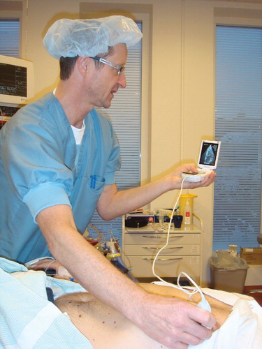
Figure 1. Pocket echocardiography at the bedside.
As a culmination of expert opinions and existing literature the European Association of Echocardiography has recently published a position statement on PE devices Citation[11]. Four recommendations are given on range of indication, documentation, education and patient information.
Point-of-care cardiac ultrasound has also been evaluated during life support and peri-resuscitation Citation[3], and recently the use of ultrasound has been mentioned in the European Resuscitation Council's guidelines Citation[12]. More ambition is needed in this area and great attention should be paid to education.
3. FAST examination
The Focused Assessment with Sonography for Trauma (FAST) Citation[13] examination is a POC protocol used for detection of intra-abdominal fluid in trauma patients. The protocol uses four predefined scanning positions for the detection of free intraperitoneal fluid, pericardial fluid and free fluid in the pelvis. A complete examination can be performed in a short span of time and the protocol has been shown to have a very high overall accuracy Citation[14]. The FAST examination is usually done on cart-based ultrasound systems, but the potential for handheld examination is obvious and has received some attention Citation[15].
4. The systematic ultrasound approach
The systematic ABCD approach to the critically ill patient has also been adopted by various ultrasound communities, now teaching different POC protocols for problem solving in emergency medicine.
The expansion of ultrasound into the critical care setting and the emergency departments is evolving at a rapid pace, but POC ultrasound is also expanding into many other specialties. A comprehensive review of all the POC ultrasound applications available is beyond the scope of this text, but a few will be mentioned: Dermatology – assessment of skin lesions and tumors, Neonatology – cranial and pulmonary assessments, Ophthalmology – corneal assessment and Rheumatology – monitoring of synovitis. In addition, procedural guidance is used in many specialties including some of the above mentioned and it seems as if the technology is expanding into every organ system of the human body.
5. Technological development
Pocket device development is still in progress, and until now only three manufactures have made ultra lightweight ‘pocket devices’; these devices have further increased the accessibility and mobility of POC ultrasound and take complete disregard of the setting.
The two first devices on the market were primarily intended for echocardiography Citation[9,16], as they were only equipped with a cardiac sector probe. The newest device on the market is the first smartphone-based device available. The smartphone device allows for a USB based connection of probes and is initially marketed with a low frequency probe for abdominal, Ob/Gyn and procedural guidance, and a high frequency probe for small organs and vascular scans.
The smartphone device became available very recently and no critical reviews or articles about it have been published. However, the literature on validation and feasibility of the pocket echocardiography (PE) devices is growing Citation[11]. It is unclear whether the PE devices are going to be an augmentation of the physical examination or a miniaturized version of the standard diagnostic echocardiography. The scientific literature and expert opinions will lead the way.
6. Education and documentation
POC ultrasound, as well as ultrasound in general, is very user dependent and the need for quality assurance, formal education and practical training is obvious. Many academic communities and professional societies have already undertaken this task, but there is still much work to be done. Some academic institutions have already foreseen the future and implemented the ultrasound as part of the curriculum for medical students, experiences from these institutions is pioneer work and should serve as an example for other institutions.
The daily routine documentation of findings is a key issue and needs to be structured in order to facilitate communication among physicians and optimize patient safety. This requires administrative work and organization.
The greatest challenge may be to provide scientific studies designed with meticulous methodology and focus on patient-centered outcomes. This is a key challenge in order to provide a safe and well-validated imaging modality for the patients.
7. Expert opinion
The array of ultrasound probes for pocket devices is now complete and allows full expansion of PE devices into all areas of POC ultrasound. Until now there is good agreement on image quality between PE examinations and examinations performed on high-end systems. However, the PE devices still lag behind in functionality. This leaves a great development potential for the manufactures to implement advanced functionality into PE devices, and allows the scientific community to review these entities. Although multiplane probes are already on the market, the invention of probes combining advantages from different probes into one is yet to be seen. Also, wireless functionality seems possible in the near future, since advances in probe technology have allowed for signal processing directly in the probe casing and, consequently, reduced need for wired data transmission. This could lead to many new opportunities of integrating ultrasound into existing equipment.
As many modern ultrasound devices have very advanced software the user interface is often very complex, simplification and online user guidance seem to be an obvious and feasible improvement. Even a software-assisted interpretation of images should be within reach.
The POC ultrasound examination now has the potential of becoming a part of the new standard for a physical examination. The new array of devices is small and inexpensive and has the potential of becoming a replacement for the stethoscope.
7.1 Ultrasound triage
In the future all physicians will be able to introduce a better triage of patients by integrating their physical examination with POC ultrasound. As most boundaries in the shape of accessibility and portability are gone, POC ultrasound now really has the potential for becoming the physician's new personal universal examination tool. Patients admitted to emergency departments will be able to receive organ or symptom-guided initial focused ultrasound triage as part of the physician's first encounter with the patient. This will allow for a more accurate referral, correct diagnosis and relevant screening in term leading to better overall treatment results.
Declaration of interest
The authors state no conflict of interest and have received no payment in preparation of this manuscript.
Bibliography
- Moore CL, Copel JA. Point-of-care ultrasonography. N Engl J Med 2011;364:749-57
- Johnson DW, Oren-Grinberg A. Perioperative point-of-care ultrasonography: the past and the future are in anesthesiologists' hands. Anesthesiology 2011;115:460-2
- Breitkreutz R, Price S, Steiger HV, Focused echocardiographic evaluation in life support and peri-resuscitation of emergency patients: a prospective trial. Resuscitation 2010;81:1527-33
- Vignon P, Frank MB, Lesage J, Hand-held echocardiography with Doppler capability for the assessment of critically-ill patients: is it reliable? Intensive Care Med 2004;30:718-23
- Martin LD, Howell EE, Ziegelstein RC, Hand-carried ultrasound performed by hospitalists: does it improve the cardiac physical examination? Am J Med 2009;122:35-41
- Vourvouri EC, Poldermans D, Deckers JW, Evaluation of a hand carried cardiac ultrasound device in an outpatient cardiology clinic. Heart 2005;91:171-6
- Jensen MB, Sloth E, Larsen KM, Schmidt MB. Transthoracic echocardiography for cardiopulmonary monitoring in intensive care. Eur J Anaesthesiol 2004;21:700-7
- Breitkreutz R, Uddin S, Steiger H, Focused echocardiography entry level: new concept of a 1-day training course. Minerva Anestesiol 2009;75:285-92
- Frederiksen CA, Juhl-Olsen P, Larsen UT, New pocket echocardiography device is interchangeable with high-end portable system when performed by experienced examiners. Acta Anaesthesiol Scand 2010;54:1217-23
- Andersen GN, Haugen BO, Graven T, Feasibility and reliability of point-of-care pocket-sized echocardiography. Eur J Echocardiogr 2011;12:665-70
- Sicari R, Galderisi M, Voigt JU, The use of pocket-size imaging devices: a position statement of the European Association of Echocardiography. Eur J Echocardiogr 2011;12:85-7
- Nolan JP, Soar J, Zideman DA, European resuscitation council guidelines for resuscitation 2010 Section 1. Executive summary. Resuscitation 2010;81:1219-76
- Scalea TM, Rodriguez A, Chiu WC, Focused Assessment with Sonography for Trauma (FAST): results from an international consensus conference. J Trauma 1999;46:466-72
- McKenney KL, Nunez DB Jr, McKenney MG, Sonography as the primary screening technique for blunt abdominal trauma: experience with 899 patients. AJR Am J Roentgenol 1998;170:979-85
- Kirkpatrick AW, Sirois M, Ball CG, The hand-held ultrasound examination for penetrating abdominal trauma. Am J Surg 2004;187:660-5
- Egan M, Ionescu A. The pocket echocardiograph: a useful new tool? Eur J Echocardiogr 2008;9:721-5