Abstract
Hypertriglyceridemia is a risk factor for atherosclerotic coronary heart disease. Very high triglyceride (TG) levels (≥500 mg/dl [5.65 mmol/l]) increase the risk of pancreatitis. One therapeutic option to lower TG levels is omega-3 fatty acids, which are derived from the oil of fish and other seafood. The American Heart Association has acknowledged that fish oils may decrease dysrhythmias, decrease sudden death, decrease the rate of atherosclerosis and slightly lower blood pressure, and has recommended fish consumption or fish oil supplementation as a therapeutic strategy to reduce cardiovascular disease. A prescription omega-3-acid ethyl esters (P-OM3) preparation has been available in many European nations for at least a decade, and was approved by the US FDA in 2004 to reduce very high TG levels (≥500 mg/dl [5.65 mmol/l]). Mechanistically, most evidence suggests that omega-3 fatty acids reduce the synthesis and secretion of very-low-density lipoprotein (VLDL) particles, and increase TG removal from VLDL and chylomicron particles through the upregulation of enzymes, such as lipoprotein lipase. Omega-3 fatty acids differ mechanistically from other lipid-altering drugs, which helps to explain why therapies such as P-OM3 have complementary mechanisms of action and, thus, complementary lipid benefits when administered with statins. Additional human studies are needed to define more clearly the cellular and molecular basis for the TG-lowering effects of omega-3 fatty acids and their favorable cardiovascular effects, particularly in patients with hypertriglyceridemia.
Increased circulating FFAs may be lipotoxic to muscle, liver and pancreas. When adipocytes become excessively enlarged, especially in the setting of visceral adiposity, adipocyte and adipose tissue dysfunction (i.e., ‘adiposopathy’) may result in adverse metabolic consequences. One of the manifestations of adiposopathy is a relative increase of intra-adipocyte lipolysis over that of intra-adipocyte lipogenesis, leading to a net release of FFAs, insulin resistance and diminished pancreatic insulin secretion, all leading to hyperglycemia and possible diabetes mellitus, as well as other metabolic diseases. Steatosis, or ‘fatty liver’, is another consequence of increased FFA delivery to the liver.
FFA: Free faty acid.
Adapted with permission from Future Medicine Ltd Citation[7].
![Figure 1. Relationship between adiposopathy (pathogenic adipose tissue) and metabolic disease.Increased circulating FFAs may be lipotoxic to muscle, liver and pancreas. When adipocytes become excessively enlarged, especially in the setting of visceral adiposity, adipocyte and adipose tissue dysfunction (i.e., ‘adiposopathy’) may result in adverse metabolic consequences. One of the manifestations of adiposopathy is a relative increase of intra-adipocyte lipolysis over that of intra-adipocyte lipogenesis, leading to a net release of FFAs, insulin resistance and diminished pancreatic insulin secretion, all leading to hyperglycemia and possible diabetes mellitus, as well as other metabolic diseases. Steatosis, or ‘fatty liver’, is another consequence of increased FFA delivery to the liver.FFA: Free faty acid.Adapted with permission from Future Medicine Ltd Citation[7].](/cms/asset/c0c4a04b-3a3b-4379-ae64-0628fd6f7f72/ierk_a_11210365_f0001_b.jpg)
Pathogenic adipose tissue, increased postprandial CHYL and increased VLDL particles may increase free FA delivery to the liver, and increase hepatic lipid content, which are substrates for TG synthesis and, thus, VLDL production. Most evidence supports that omega-3 fatty acids inhibit hepatic TG synthesis, decrease VLDL production/secretion and increase VLDL metabolism by: decreasing lipogenesis by decreasing the enzymatic conversion of acetyl CoA to FAs; increasing β-oxidation of FA; inhibiting both PAP (an enzyme that catalyzes that reaction of converting PA to DAG) and DGAT (an enzyme that catalyzes the final step in TG synthesis); potentially increasing the degradation of apolipoprotein B; and increasing LPL activity, which is an enzyme that increases the conversion of VLDL particles to LDL particles.
CHYL: Chylomicrons; DAG: diacylglycerol; DGAT: Diacylglycerol acyltransferase; FA: Fatty acid; LPL: Lipoprotein lipase; PA: Phosphatidic acid; PAP: phosphatidic acid phosphatase/phosphohydrolase; TG: Triglyceride; VLDL: Very low-density lipoprotein.
Adapted from Citation[98].
![Figure 2. Potential TG-lowering mechanisms of eicosapentaenoic acid and docosahexaenoic acid.Pathogenic adipose tissue, increased postprandial CHYL and increased VLDL particles may increase free FA delivery to the liver, and increase hepatic lipid content, which are substrates for TG synthesis and, thus, VLDL production. Most evidence supports that omega-3 fatty acids inhibit hepatic TG synthesis, decrease VLDL production/secretion and increase VLDL metabolism by: decreasing lipogenesis by decreasing the enzymatic conversion of acetyl CoA to FAs; increasing β-oxidation of FA; inhibiting both PAP (an enzyme that catalyzes that reaction of converting PA to DAG) and DGAT (an enzyme that catalyzes the final step in TG synthesis); potentially increasing the degradation of apolipoprotein B; and increasing LPL activity, which is an enzyme that increases the conversion of VLDL particles to LDL particles.CHYL: Chylomicrons; DAG: diacylglycerol; DGAT: Diacylglycerol acyltransferase; FA: Fatty acid; LPL: Lipoprotein lipase; PA: Phosphatidic acid; PAP: phosphatidic acid phosphatase/phosphohydrolase; TG: Triglyceride; VLDL: Very low-density lipoprotein.Adapted from Citation[98].](/cms/asset/4459a6e9-88fa-4c91-b673-ef960159ae21/ierk_a_11210365_f0002_b.jpg)
P-OM3 and atorvastatin lower triglyceride levels by different mechanisms. (A) Percentage change in the secretion rate of apoB-containing lipoproteins into the plasma. (B) Percentage change in the interconversion of apoB-containing lipoproteins. P-OM3, alone or in combination with atorvastatin, increased conversion of TG-rich lipoproteins to LDL.
*p < 0.01 compared with placebo group.
IDL: Intermediate-density lipoprotein; P-OM3: Prescription omega-3-acid ethyl esters; VLDL: Very-low-density lipoprotein.
Reproduced from Citation[118].
© 2002 American Diabetes Association.
![Figure 3. Effects of statins, fish oils and their combination on lipoprotein secretion rate (not lipid levels) and conversion.P-OM3 and atorvastatin lower triglyceride levels by different mechanisms. (A) Percentage change in the secretion rate of apoB-containing lipoproteins into the plasma. (B) Percentage change in the interconversion of apoB-containing lipoproteins. P-OM3, alone or in combination with atorvastatin, increased conversion of TG-rich lipoproteins to LDL.*p < 0.01 compared with placebo group.IDL: Intermediate-density lipoprotein; P-OM3: Prescription omega-3-acid ethyl esters; VLDL: Very-low-density lipoprotein.Reproduced from Citation[118].© 2002 American Diabetes Association.](/cms/asset/ec4846c8-b1d3-47f4-932a-9beb58bf4964/ierk_a_11210365_f0003_b.jpg)
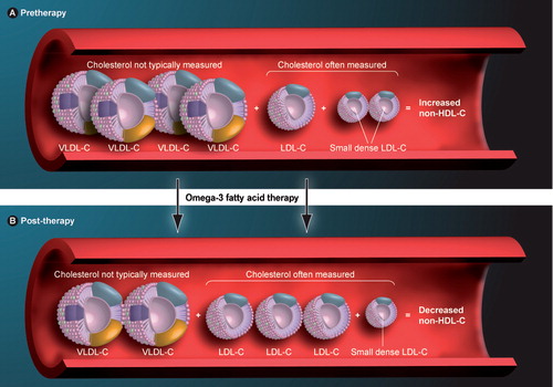
Non-HDL-C is reduced in many P-OM3 trials, concomitantly with an apparent paradoxical increase in LDL-C levels. This can be explained by P-OM3’s increased conversion of VLDL to LDL particles. Thus, in this case, P-OM3 resulted in a decrease in VLDL-C levels and decrease in VLDL particle size, and an increase in LDL-C levels and increase in LDL particle size, with a net decrease in the total cholesterol carried by atherogenic lipoproteins, as represented by non-HDL-C.
HDL-C: HDL cholesterol; LDL-C: LDL cholesterol; P-OM3: Prescription omega-3-acid ethyl esters; VLDL: Very-low-density lipoprotein.
Reproduced from Citation[92].
![Figure 4. Effect of P-OM3 on non-HDL-C in patients with triglycerides of 500 mg/dl.Non-HDL-C is reduced in many P-OM3 trials, concomitantly with an apparent paradoxical increase in LDL-C levels. This can be explained by P-OM3’s increased conversion of VLDL to LDL particles. Thus, in this case, P-OM3 resulted in a decrease in VLDL-C levels and decrease in VLDL particle size, and an increase in LDL-C levels and increase in LDL particle size, with a net decrease in the total cholesterol carried by atherogenic lipoproteins, as represented by non-HDL-C.HDL-C: HDL cholesterol; LDL-C: LDL cholesterol; P-OM3: Prescription omega-3-acid ethyl esters; VLDL: Very-low-density lipoprotein.Reproduced from Citation[92].](/cms/asset/f6abaa60-a385-4d8f-b346-76e2a6780133/ierk_a_11210365_f0004_b.jpg)
Many patients with hypertriglyceridemia have increased cholesterol carried by atherogenic particles, which is best assessed by measuring non-HDL-C levels. VLDL particles are considered to be atherogenic. Omega-3 fatty acid therapy decreases the cholesterol carried by VLDL particles, and is a cholesterol effect not typically measured in clinical practice. Omega-3 fatty acids may also decrease VLDL particle size. Conversely, omega-3 fatty acids may increase LDL-C levels, which is a lipid parameter that is often measured in clinical practice. This is thought to be due to the increased conversion of VLDL particles to LDL particles. Finally, omega-3 fatty acids may increase LDL particle size, which may render them less atherogenic. Overall, despite a potential increase in LDL-C levels, many studies have reported that P-OM3 reduces non-HDL-C, which may be a better predictor of atherosclerotic coronary heart disease risk than LDL-C alone.
HDL-C: High-density lipoprotein cholesterol; LDL-C: Low-density lipoprotein cholesterol; P-OM3: Prescription omega-3-acid ethyl esters; VLDL: Very-low-density lipoprotein.
Hypertriglyceridemia, which is defined as a triglyceride (TG) blood concentration of 150 mg/dl or higher Citation[1], is a common dyslipidemia encountered in clinical practice and occurs with or without elevated cholesterol levels. In the Framingham Offspring Study, 11.7% of women and 22.3% of men had TG levels that were higher than 200 mg/dl (2.26 mmol/l) Citation[2]. The Third National Health and Nutrition Examination Survey (NHANES III) of 8814 adult Americans found that 25% of women and 35% of men had a TG level of 150 mg/dl or higher (≥1.69 mmol/l) Citation[3]. The obesity epidemic Citation[201], along with its metabolic con-sequences, is an important contributor to the rising prevalence of hypertriglyceridemia Citation[4–7].
For patients with very high TG levels (≥500 mg/dl [5.65 mmol/l]), the initial therapeutic goal is to lower TG levels to prevent pancreatitis Citation[1], which is a potentially life-threatening complication of severe hyper-tri-glyceri-demia Citation[1,8,9]. The risk of pancreatitis is especially increased when TG levels are found to be above 1000 mg/dl (11.3 mmol/l) Citation[10]. When TG levels are above 1000 mg/dl (11.3 mmol/l), this is usually the result of a secondary cause of hypertriglyceridemia occurring in individuals with one of the more common genetic hyper-tri-glycerid-emic disorders (Box 1) such as familial hypertriglyceridemia and familial combined hyperlipidemia (FCH) Citation[202], both of which occur in 3% or fewer of the population. Familial hyper-tri-glycerid-emia (hyper-pre-beta-lipo-protein-emia) may possibly be due to the presence of a lipoprotein lipase (LPL) inhibitor that results in increased chylomicron and VLDL levels, and is clinically manifested by pancreatitis and eruptive xanthomas, especially when accompanied by secondary causes that exacerbate hypertriglyceridemia, such as hypothyroidism, uncontrolled diabetes mellitus or excessive alcohol intake with fatty liver Citation[11]. FCH is probably due to a variety of apolipoprotein defects, which results in elevations in TG (with the same potential symptoms, as mentioned previously), but also with elevated cholesterol and apo-lipo-protein B-100 (apoB-100) levels Citation[1,12]. HDL cholesterol (HDL-C) levels may be decreased, and LDL particles may be small and dense and, thus, potentially more atherogenic Citation[13]. FCH is the most common form of nonpolygenic, heritable dys-lipid-emia, and is found in 10–20% of survivors of myo-cardial infarction Citation[13] and approximately 20% of patients with coronary heart disease (CHD) under the age of 60 years Citation[14]. Both familial hypertriglyceridemia and FCH may increase CHD risk Citation[12].
Severe hypertriglyceridemia may also represent more rare, under-lying genetic dyslipidemias, such as LPL deficiency Citation[15] or homozygous apolipoprotein C-II (apoC-II) deficiency Citation[16], which occur in approximately 1:1,000,000 people (Box 1). In both cases, diagnosis usually occurs during childhood or young adulthood, with the presentation of recurrent pancreatitis, eruptive cutaneous xanthomata, hepatosplenomegaly and lipemia retinalis. Untreated TG levels are usually found to be greater than 2000 mg/dl Citation[1,201]. Both also result in a ‘chylo-micron-emia syndrome’, defined as elevated chylo-microns, marked increase in TG levels, and the clinical signs and symptoms described above.
While VLDL particles normally constitute approximately 90% of the TG-containing lipoproteins, and while levels of both VLDL and chylomicron-associated TG increase after meals Citation[17,18], it is the profound increase in TG levels associated with chylomicrons (most often in patients with an under-lying, inherent, genetic defect) that is most described to contribute to pancreatitis. Chylomicrons are TG-rich lipo-protein particles that predominantly carry post-prandial/post-absorptive TG. Marked increases in chylo-microns are hypo-thesized to impair circulatory flow in pancreatic capillary beds, leading to ischemia-induced disruption in acinar structure and exposing the TG-rich particles to pancreatic lipase, leading to necrosis, edema and inflammation Citation[19].
Therapeutic interventions to treat hypertriglyceridemia include increased physical exercise Citation[20,21] and a low-calorie diet with reduced consumption of high-glycemic index carbohydrates and alcohol Citation[22]. Other interventions, depending on the patient population, may include lipopheresis, heparin-ization and insulin Citation[23–25]. Statins and ezetimibe are approved lipid-altering drugs that may modestly reduce TG levels. However, they are mainly used to lower LDL cholesterol (LDL-C) levels. Other lipid-altering agents that are used more specifically to reduce TG levels include niacin, fibrates and omega-3 fatty acids.
Eicosapentaenoic acid (EPA) and docosahexaenoic acid (DHA) are long-chain, polyunsaturated, omega-3 fatty acids that effectively lower TG levels Citation[26–28]. EPA and DHA may be used as monotherapy, or as adjunctive therapy to fibrates and/or nicotinic acid to lower TGs to prevent pancreat-itis in patients with very high TG levels Citation[1]. Fish consumption and supplements are dietary sources of omega-3 fatty acids. A prescription combination of omega-3-acid ethyl esters (P-OM3; Lovaza™ Capsules, Reliant Pharmaceuticals, Inc.) is available that contains concentrated forms of EPA (∼465 mg), DHA (∼375 mg) and other omega-3 fatty acids (∼60 mg), for a total of at least 900 mg of omega-3 fatty acids per each 1-g gel capsule. P-OM3 is approved by the US FDA for the treatment of very high TGs (≥500 mg/dl [5.65 mmol/l]) in adult patients. This review examines the pathophysiology of hyper-tri-glycerid-emia and possible mechanisms for the TG-lowering effect of omega-3 fatty acids.
Pathophysiology of hypertriglyceridemia
Lipoproteins serve to transport varying types and varying amounts of lipids in the circulation, including TG, cholesterol and phospholipids () Citation[29]. The TGs found in lipoproteins are derived from dietary consumption, intestinal secretion and hepatic production Citation[29]. The term ‘triglyceride-rich lipoproteins’ (TRLs) most often refers to chylomicrons, VLDL and their remnants. Intermediate-density lipoproteins (IDLs) are often considered to represent VLDL remnants Citation[30,31].
Chylomicron particles deliver lipids derived from dietary fat consumption and intestinal absorption to peripheral and hepatic tissues. VLDL particles transport lipids from the liver to peripheral tissues Citation[29,31]. The enzyme LPL, located on the endothelial side of capillaries within fat and muscle tissue, hydrolyzes TG from both chylomicrons and VLDL into free fatty acids, resulting in the formation of chylomicron and VLDL remnants, respectively Citation[29,31]. These remnants may be atherogenic Citation[32,33]. Mutations in the LPL gene may impair lipolysis from these TRL and significantly increase TG levels; such mutations have been identified in patients with hyper-tri-glyceridemia-induced pancreatitis Citation[34,35].
Hyperchylomicronemia may occur due to rare genetic defects, resulting in postprandial hyper-tri-glycerid-emia, as has already been described. VLDL excess may also be due to genetic defects (Box 1). Beyond rare genetic defects, over-production of VLDL may have varying etiologies resulting in fasting hyper-tri-glycerid-emia. For example, adipose tissue is the major energy storage organ of the body, with calories predominantly stored in the form of TG. During times of positive caloric balance, adipo-cytes may become excessively enlarged and visceral adiposity may accumulate, resulting in pathologic adipocyte and adipose tissue dysfunction. Physio-logically, this adiposopathy results in adverse metabolic and immune consequences resulting in the onset or worsening of clinical metabolic diseases, such as Type 2 diabetes mellitus, hypertension and dyslipidemia Citation[36,37]. Thus, clinically, excessive and pathogenic adipocyte hyper-trophy and an increase in visceral adipose tissue (central obesity) are often associated with hyperglycemia, high blood pressure and hyper-tri-glycerid-emia (and low HDL-C levels), which represents a clustering of atherogenic risk factors often described as representing a ‘metabolic syndrome’ Citation[1].
One of the metabolic manifestations of adiposopathy is a relative increase of intra-adipocyte lipolysis over that of intra-adipo-cyte lipogenesis, leading to a net release of free fatty acids that may be ‘lipotoxic’ to body organs Citation[37]. In addition to contributing to the before-mentioned metabolic diseases, increased circulating free fatty acids may also contribute to hepatic steatosis Citation[38,39], which is a common clinical finding among patients with the components of the m-etabolic syndrome.
With specific regard to TGs, the increase in free fatty acid delivery to the liver increases TG synthesis Citation[40], which can lead to VLDL overproduction Citation[41]. Increased VLDL production is exacerbated if hepatic free fatty acid β-oxidation (metabolism) is impaired (e.g., through genetic limitations or with insulin resistance), thereby leaving more substrate for VLDL synthesis. Nonetheless, it is unknown if the increase in the hepatic cytoplasmic TG pool is truly rate-limiting for VLDL-TG or apoB-100 production Citation[41]. However, once hepatocyte TGs are packaged into VLDL particles, they are then secreted into the circulation Citation[42]. Fasting hyper-tri-glycerid-emia ensues, which may also be exacerbated if LPL-mediated lipolysis is impaired and/or the removal of remnant VLDL particles is delayed Citation[43].
In summary, severe hypertriglyceridemia occurs with increased chylomicrons, VLDL particles and/or their remnants, with causality and promotion being due to primary and secondary factors Citation[44,45]. Primary causes include genetic defects (Box 1)Citation[15,16,46–51], while common secondary contributors that may cause or exacerbate hypertriglyceridemia include pathogenic adipose tissue (visceral adiposity and adipocyte hyper-trophy), excessive and acute consumption of alcohol, consumption of high-glycemic index carbohydrates Citation[29,52], hyper-glycemia, hypothyroidism and nephrotic syndrome.
Lipid atherosclerotic risk factors & mechanisms of the potential atherogenicity of hypertriglyceridemia
Hypertriglyceridemia is a risk factor for CHD, particularly in women Citation[53], although it is unclear if hypertriglyceridemia is always an independent risk factor Citation[54,55]. What does seem clear is that when hypertriglyceridemia is combined with elevated total and LDL-C levels, then CHD risk is amplified Citation[56,57]. The increased CHD risk with combined hyperlipidemia may be due to several mechanisms, many of which may or may not be independent of one another.
Increased non-HDL-C levels
VLDL and its remnants carry cholesterol and, thus, constitute a component of non-HDL-C. Non-HDL-C is the sum of cholesterol carried by atherogenic lipoproteins (e.g., LDL, VLDL, IDL, lipoprotein (a) and lipoprotein remnants), and is thought to be a better predictor of CHD risk than LDL-C levels alone Citation[58–60]. Mechanistically, an increase in atherogenic lipoprotein levels enhances cholesterol delivery to endothelial plaques, promotes atherosclerotic progression, and increases the risk of plaque rupture resulting in an increased risk of a CHD event.
Increased apolipoprotein B-100 (apoB-100) levels
Another marker that is thought to be a better predictor of CHD risk than LDL-C levels alone is a measurement of apoB-100 Citation[58]. Each LDL, VLDL and IDL particle contains one apoB-100 molecule. Thus, apoB-100 reflects the number of circulating atherogenic lipoproteins, and this may account for why this measurement is a strong predictor of CHD risk Citation[61]. Increased TRL through increased VLDL particles and their remnants increases apoB-100 levels and, thus, may increase CHD risk.
Increased small, dense LDL particles
Elevated TG levels are often associated with, and may contribute to, small, dense LDL particles. The generation of small, dense LDL particles often results from an interplay of various enzymes, including LPL, hepatic lipase and cholesteryl ester transfer protein Citation[62]. Although all LDL particles are considered atherogenic, small, dense LDL particles may be more atherogenic than larger LDL particles. As with hypertriglyceridemia, not all analyses support that LDL particle size is an independent predictor of CHD Citation[63]. However, if small, dense LDL particles are more atherogenic, then this is likely because they may be more able to penetrate arterial walls and have less resistance to oxidative stress. Small, dense LDL particles may also be associated with increased thrombosis, which may increase CHD events.
Decreased HDL-C levels
High TG levels are often associated with, and may contribute to, low HDL-C levels Citation[1]. High HDL-C levels are generally associated with decreased CHD risk. Conversely, lower HDL-C levels may be associated with increased CHD risk Citation[1]. Mechanistically, if lower HDL-C levels are directly associated with increased CHD risk, it is likely due to decreased flux of cholesterol from athero-sclerotic plaques, or possibly due to other effects, such as a reduced anti-inflammatory response otherwise attributable to HDL particles.
Post- & preprandial hyperlipidemia & TRL remnant formation
Postprandial hypertriglyceridemia may be an independent risk factor for CHD, which suggests that chylomicrons (even though they contain apoB-48, not apoB-100) and their remnants may be atherogenic Citation[64–68]. If true, then an increase in atherogenicity through this mechanism may have practical consequences for clinicians and their patients. For example, patients may sometimes believe that as long as their fasting lipid levels are well-controlled with lipid-altering drug therapy (e.g., statins), then food choices and diet composition no longer affect their CHD risk. But if postprandial lipemia does contribute to atherosclerosis, then it is possible that even with lipid-altering drug administration, poor dietary habits may still increase CHD risk.
Similarly, preprandial increases in TRL remnants may also increase CHD risk, with some studies suggesting that VLDL remnants, or IDL, are strong and independent risk factors for atherosclerotic progression Citation[69]. Animal studies have suggested that the generation of very large TRLs may not necessarily be atherogenic because they are unable to penetrate arterial endothelia Citation[70]. However, when apoB-48-containing chylomicrons and apoB-100-containing VLDL particles undergo circulatory metabolism by lipoprotein lipase, then TRL remnants may be generated, resulting in smaller, more dense particles that are relatively depleted of TG, phospholipid and apoC, and enriched in cholesteryl esters and apoE Citation[32]. TRL remnants may promote atherogenesis through impairment of endothelium-dependent vasorelaxation, enhancement of platelet aggregation and subendothelial macrophage uptake resulting in foam cell formation.
Increased potential for thrombosis
Elevated (postprandial) TG levels may unfavorably affect the coagulation system, increasing plasminogen activator inhibitor-1, an inhibitor of fibrolysis. Factor VII may also be increased, which may also increase the risk of thrombosis Citation[71]. An increase in the risk of thrombosis increases the risk of CHD events.
Increased apolipoprotein C-III (apoC-III) levels
ApoC-III, an apolipoprotein found on chylomicron, VLDL, IDL and HDL particles, inhibits LPL activity. An elevated apoC-III level may be associated with increased CHD risk Citation[72]. This is most likely because it reflects the concentration of TRL. ApoC-III may also directly activate vascular endothelial cells, which promotes inflammatory cell adhesion and recruitment Citation[73] and, thus, may directly contribute to the inflammatory process of atherosclerosis.
Biochemistry & nutritional aspects of omega-3 fatty acids
Omega-3 fatty acids are polyunsaturated fats in which the first double bond counting from the terminal (omega) methyl group is at carbon 3 Citation[74]. Major omega-3 fatty acids include a-linolenic acid (ALA [18:3N-3]), EPA (20:5N-3) and DHA (22:6N-3) Citation[75,76]. In general, fatty acids are of varying sizes, which affects function. Short chain fatty acids usually have less than six carbons. An example would be butyric acid, which is a four-carbon fatty acid found in butterfat. Medium chain fatty acids usually have six to 12 carbons. An example would be lauric acid, a 12-carbon fatty acid that is the main component of coconut oil. ALA, EPA and DHA are all considered long-chain fatty acids because each have 12 carbons or more.
ALA is an ‘essential’ fatty acid, because it cannot be synthesized in humans and, thus, must be consumed in the diet. ALA is a plant-derived omega-3 fatty acid that can be converted to EPA and DHA in mammals Citation[76]. However, the conversion of ALA to EPA is modest (<1%) and the subsequent conversion of EPA to DHA is also very low Citation[76]. Thus, while not necessarily essential fatty acids, preformed EPA and DHA are best obtained through dietary sources.
The best dietary sources of EPA and DHA include fatty or oily marine seafood, such as salmon, herring, mackerel, halibut and tuna Citation[77]. Some fresh-water fish may contain significant amounts of omega-3 fatty acids, and include lake herring, lake trout, freshwater salmon and whitefish Citation[203]. The omega-3 fatty acid content of these fish may be increased with farming Citation[77]. Some concerns have been raised about the environmental impact and residual pesticide and antibiotic content of selected types of fish Citation[204]. However, the risks from contaminants potentially contained in oily fish consumption may be outweighed by the potential benefits Citation[78]. The American Heart Association (AHA) has acknowledged that EPA and DHA may decrease dysrhythmias, decrease sudden death, decrease the rate of atherosclerosis and slightly lower blood pressure, and has recommended fish consumption or fish oil supplementation as a therapeutic strategy to reduce cardiovascular disease Citation[77]. While reducing TG levels may have cardiovascular benefits, it is unclear as to how much (if any) of these before-mentioned benefits are related to omega-3 fatty acid’s TG-lowering effects and how much is due to TG-independent effects.
P-OM3 triglyceride-lowering effects & potential adverse experiences
Omega-3 fatty acids reduce TG levels in humans Citation[79–81]. The amount of EPA and DHA typically administered for the treatment of hypertriglyceridemia is 2–4 g/day Citation[77]. EPA and DHA have similar TG-lowering effects Citation[82], and lower both fasting Citation[83,84] and postprandial Citation[82,83] TG levels. P-OM3 (4 g/day for ≥6 weeks) significantly reduces TG levels in subjects with severe hypertriglyceridemia Citation[85]. While ALA is an omega-3 fatty acid, it does not significantly reduce TG levels at typically administered doses Citation[86,87].
P-OM3 may reduce TG levels more effectively than fish oil formulations containing less omega-3 fatty acids, and may have greater bioavailability Citation[88]. Owing to the requirements in achieving a prescription status, P-OM3 has undergone more rigorous safety and efficacy evaluation and verification than dietary supplement omega-3 fatty acids Citation[89–91]. Selected dietary supplement omega-3 fatty acids do not appear to contain contaminants in sufficient concentrations to pose a potential health risk Citation[89]. However, individual supplements vary considerably in the amount of their omega-3 fatty acid content. P-OM3 has undergone the processes necessary to achieve FDA approval, verifying its consistent omega-3 fatty acid content Citation[90].
The most common drug-related adverse experiences attributable to P-OM3 include a mild, numerical increase in eructations (belching) and dyspepsia Citation[92], which are substantially mitigated by the purification process used for P-OM3 Citation[89]. P-OM3 does not have any known, clinically significant drug interactions. Some reports have suggested that omega-3 fatty acids may impair platelet aggregation and increase bleeding times Citation[93,94]. Clinical trial data suggest that P-OM3 does not increase clinical bleeding, even in patients receiving warfarin anticoagulants, aspirin and other older antiplatelet agents Citation[89,95,96]. P-OM3 has also been described to sometimes increase levels of liver transaminases, such as alanine aminotransferase. Thus, alanine aminotransferase levels should be monitored periodically during P-OM3 therapy Citation[92]. Finally, studies of omega-3 fatty acids, including P-OM3, have often reported a transient increase in glucose levels, but not increases in measures of long-term glucose control, such as fructosamine or hemoglobin A1C Citation[27].
Omega-3 fatty acids: TG-lowering mechanisms
As discussed above, omega-3 fatty acids are well-known to reduce TG blood levels. However, the mechanisms by which EPA and DHA reduce serum TGs are not well-known or completely understood. Simply put, preclinical and clinical studies provide compelling evidence that EPA and DHA can reduce hepatic VLDL-TG synthesis/secretion and enhances TG clearance from circulating VLDL particles Citation[97]. Regarding hyperchylomicronemia, both EPA and DHA may equally accelerate chylomicron TG clearance by promoting increased lipoprotein lipase activity Citation[82].
Reduced VLDL-TG synthesis by omega-3 fatty acids
Several mechanisms have been proposed as to how omega-3 fatty acids may reduce TG synthesis, reduce the incorporation of TG into VLDL particles, and ultimately reduce TG secretion. Omega-3 fatty acids may decrease hepatic lipogenesis, increase β-oxidation of fatty acids, and increase degradation of apoB-100 Citation[97,98].
Decreased hepatic lipogenesis through a decreased enzymatic conversion of acetyl CoA to fatty acids
Peroxisome proliferator-activated receptors (PPARs) and sterol regulatory element-binding proteins (SREBPs) are transcription factors that play a major role in regulating lipid metabolism. The nuclear receptors liver X receptor (LXR)α and retinoid X receptor (RXR)α typically form a heterodimer that regulates expression of the SREBP-1c gene by binding to the SREBP-1c promoter Citation[99]. SREBPs regulate the expression of cholesterol-, fatty-acid-, and TG-synthesizing enzymes. Activation of the transcription factor SREBP-1c stimulates the synthesis of lipogenic enzymes such as acetyl-CoA carboxylase-1 (ACC1) and fatty-acid synthase (FAS) Citation[100].
Fish-oil feeding in mice is associated with significant decreases in hepatic SREBP-1c mRNA expression and decreases in TG levels Citation[101]. Fish oils may inhibit LXR/RXR heterodimer binding to the SREBP-1c gene promoter, thereby suppressing SREBP-1c mRNA expression Citation[102] and, thus, decreasing lipogenic enzyme activity. DNA microarray analysis from rat livers indicates that SREBP-1 gene expression is decreased with a DHA-enriched diet compared with low fat, high fat, or low fat plus fenofibrate diets Citation[103]. Data from HepG2 human hepatoma cells support the notion that EPA decreases TG synthesis by suppressing the expression of SREBP-1c mRNA and SREBP-1c protein Citation[104]. However, not all evidence entirely supports this proposed mechanism of TG-lowering by omega-3 fatty acids, in that rat studies suggest that EPA-induced suppression of SREBP-1c may be independent of LXRα Citation[105].
Increased β-oxidation of fatty acids
Fatty acids, which are substrates for TG synthesis, are degraded by the β-oxidation pathway. An increased rate of hepatic fatty acid oxidation can decrease the amount of fatty acids available for TG synthesis and decrease the amount of TG available for incorporation into VLDL particles. Rat studies show that EPA and/or DHA increase free fatty acid β-oxidation in peroxisomes and mitochondria Citation[98], leaving less substrate available for TG and VLDL synthesis. Evaluation of healthy human subjects taking 9 g of omega-3 fatty acids containing 5.4 g EPA and 3.6 g DHA per day Citation[106] also supports a faster rate of hepatic fatty acid oxidation. EPA binds to all PPAR subtypes (PPAR-α, -β and -γ) Citation[75], and PPAR-α may be involved in omega-3 fatty acid modulation of fatty acid β-oxidation. But, as before, not all evidence is supportive of this mechanism, in that other studies in rats Citation[98,103] and monkeys Citation[107] have shown that EPA and/or DHA had no significant effect on β-oxidation.
Inhibition of phosphatidic acid phosphatase/phosphohydrolase & diacylglycerol acyltransferase
Phosphatidic acid phosphatase/phosphohydrolase (PAP) is an enzyme that catalyzes the conversion of phosphatidic acid to diacylglycerol. Diacylglycerol acyltransferase (DGAT) is an enzyme that catalyzes the final step in TG synthesis. Both are key enzymes involved in TG synthesis in the liver. Results from preclinical studies are divided with regard to the effect of EPA and DHA on PAP and DGAT activity. Some studies show that EPA and DHA inhibit the activity of PAP and DGAT in rat liver microsomes; other studies show no such effect Citation[98]. Thus, the extent to which the TG-lowering effects of EPA and DHA depend on the inhibition of PAP and/or DGAT activity is unclear.
Enhanced TG clearance by omega-3 fatty acids
Omega-3 fatty acids may increase TG removal from circulating VLDL and chylomicron particles, through increased hydrolysis by LPL. Specifically, EPA and DHA may increase LPL activity, and, thus, increase LPL-mediated clearance of TRL Citation[82,108]. EPA increases PPAR-γ mRNA in cultured adipocytes Citation[109], and PPAR-γ mRNA levels in adipose tissue of obese subjects may be positively correlated with plasma EPA concentrations Citation[109]. Agonism of the transcription factor PPAR-γ may increase LPL activity in adipose tissue Citation[110]. Therefore, it is plausible that an increased LPL activity associated with EPA and DHA treatment may be due, in part, to increased activity of PPAR-γ.
Additionally, DHA may be a ligand for the farnesoid X receptor (FXR) Citation[111], which is a nuclear receptor found in the liver and intestine, and for which bile acids are a natural ligand. FXR may also play a role in lipid homeostasis. ApoC-III resides on the surface of VLDL and LDL particles and inhibits the activity of LPL, thereby slowing the clearance of TG-rich lipoproteins Citation[112]. Conversely, apoC-II activates LPL Citation[113]. FXR suppresses apoC-III gene expression Citation[114] and induces apoC-II Citation[115] and VLDL-receptor gene expression Citation[116], all of which may contribute to the TG-lowering action of FXR agonists. Although speculative, FXR-induced changes in the expression of apoC-II, apoC-III, and/or VLDL-receptor gene may also play a role in LPL activity and the TG-lowering effect of DHA. Irrespective of the mechanism, omega-3 fatty acids increase TRL clearance, and decrease their circulating half-life Citation[82].
Statins & P-OM3 reduce TG levels by different mechanisms
Coadministration of P-OM3 with statins improves the lipid profile in patients with hypertriglyceridemia to a greater extent than statin treatment alone Citation[117–120]. Statins inhibit hydroxymethylglutaryl coenzyme A reductase, the rate-limiting enzyme in cholesterol biosynthesis. Inhibition of cholesterol synthesis leads to reduced hepatic cholesterol content, which in turn increases LDL receptor expression and activity and, thus, clears more LDL-C from the circulation. LDL-C levels are reduced. Upregulated LDL receptors may also increase clearance of other TG-containing lipoproteins, at least partially accounting for the modest TG-lowering effects of statins. The degree of TG lowering with P-OM3 is generally similar in statin-treated patients compared with nonstatin-treated patients because the mechanisms of actions of P-OM3 differ from that of statins Citation[118]. Specifically, P-OM3 decreases the rate of VLDL secretion and increases the conversion of VLDL to IDL and LDL , while statins decrease apoB-containing lipo-proteins, such as VLDL, IDL and LDL Citation[118].
In patients with persistent hypertriglyceridemia after achieving LDL-C treatment goals, as might occur after statin administration in combined hyperlipidemic patients, it is then recommended that non-HDL-C (total cholesterol minus HDL-C) levels be reduced to values less than 30 mg/dl added to the LDL-C treatment goal. Thus, it is relevant that in a study of statin-treated patients with persistent hypertriglyceridemia, P-OM3 added to ongoing simvastatin therapy produced significant additional improvements in reducing non-HDL-C levels and other lipid and lipoprotein parameters to a greater extent than simvastatin alone & Citation[120]. Thus mechanistically, in patients treated with statins and P-OM3, LDL-C levels may be reduced as a result of the statin-induced increase in hepatic LDL receptor activity. IDL and VLDL remnants may be reduced by P-OM3 impairment of VLDL synthesis and secretion. VLDL may also have enhanced clearance through enhanced LPL activity (by P-OM3) and upregulation of LDL receptor (by statins). This is an illustrative example of complementary mechanisms of actions by these two lipid-altering drugs, which may be of benefit in patients with combined hyperlipidemia.
With regard to other lipid parameters, EPA and DHA administration is sometimes associated with a modest increase in HDL-C levels. LDL-C levels may be variably increased. As with fibrates, the degree of LDL-C elevations observed with P-OM3 treatment is generally related to the pretreatment TG levels. P-OM3 increases LDL-C levels the most in patients with the highest pretreatment TG levels . The reason for the increased LDL-C levels with omega-3 fatty acids is related to the increased conversion of VLDL particles to LDL particles . For example, weight loss in overweight subjects with hypertriglyceridemia has been shown to raise LDL-C, and this effect has been attributed to a reduction in the fractional catabolic rate of LDL Citation[121]. As reviewed earlier, owing to their complementary mechanisms of action, concurrent treatment with statins may mitigate the rise in LDL-C in patients with hypertriglyceridemia treated with P-OM3 Citation[120].
Expert commentary
Omega-3 fatty acids lower TG levels through decreased hepatic secretion of TG-containing lipoproteins and enhanced clearance of TG from circulating TG-containing lipoproteins. In combination with statins, omega-3 fatty acids are effective in improving many lipid parameters beyond that of statin alone, due to their complementary mechanisms of action.
Five-year view
Due to its unique benefits, interest continues to increase regarding new formulations of omega-3 fatty acids, such as potential combination agents with other lipid-altering drugs, such as niacin, fibrates and statins.
In addition to its therapeutic use for hypertriglyceridemia, omega-3 fatty acids, in general, have also been studied for potential efficacy in the treatment of numerous noncardiac conditions, such as inflammatory and arthritic disorders Citation[122–135], neurologic/neuropsychiatric disorders Citation[122,136–148], ophthalmic disorders Citation[149,150], women’s health issues Citation[135,151,152], cancer Citation[153–155] and other disorders Citation[156–158]. However, the benefits in treating many, if not most of these noncardiac disorders have yet to be definitively proven. Results of future clinical trials should better define the efficacy and safety of omega-3 fatty acid therapy in these conditions.
In contrast to noncardiovascular effects, the evidence supporting the cardiovascular benefits of omega-3 fatty acid therapy is more compelling Citation[27,77,159], and includes possible antidysrhythmic Citation[160–170], antiatherogenic Citation[82,171–179], antithrombotic Citation[172,180–184] and anti-inflammatory endothelial effects Citation[183,185–187]. However, yet again, more definitive evidence is needed in order to substantiate these potential benefits. As such, ongoing clinical trials are seeking to better define these potential beneficial effects of omega-3 fatty acids. Specific ongoing cardiac and noncardiac P-OM3 trials are registered at ClinicalTrials.gov Citation[205] and summarized in .
Table 1. Pharmacotherapy effect of lipid-altering drugs on triglycerides, LDL-C and HDL-C levels.
Table 2. Physical–chemical characteristics of lipoproteins.
Table 3. Clinical studies of 4 g/day prescription omega-3-acid ethyl esters for the treatment of patients with severe hypertriglyceridemia.
Table 4. Effects of prescription omega-3-acid ethyl esters plus simvastatin on lipid and lipoprotein parameters compared with simvastatin alone.
Table 5. Ongoing prescription omega-3-acid ethyl esters trials registered at The US NIH.
Box 1. Examples of factors contributing to hypertriglyceridemia.
Primary
| • | Familial hypertriglyceridemia (hyperprebetalipoproteinemia) | ||||
| • | Familial combined hyperlipidemia | ||||
| • | Lipoprotein lipase deficiency | ||||
| • | Apolipoprotein CII deficiency | ||||
| • | Familial dysbetalipoproteinemia | ||||
Secondary
| • | Adipocyte hypertrophy and/or visceral adipose tissue accumulation (adiposopathy) | ||||
| • | A positive energy-balance diet with a high fat or high glycemic index content | ||||
| • | Acute alcohol consumption with fatty liver | ||||
| • | Diabetes mellitus | ||||
| • | Hypothyroidism | ||||
| • | Nephrotic syndrome | ||||
Medications
| • | Antiretroviral regimens, especially for HIV disease | ||||
| • | Psychotropic medications, such as some phenothiazines and second-generation antipsychotics | ||||
| • | Bile acid sequestrants | ||||
| • | Non-selective β-blockers, thiazide diuretics | ||||
| • | Cyclophosphamide | ||||
| • | Oral estrogens | ||||
| • | Glucocorticosteroids | ||||
| • | Tamoxifen | ||||
| • | Isotretinoin | ||||
Key issues
| • | Severe hypertriglyceridemia (≥500 mg/dl [5.65 mmol/l]) should be treated to reduce the risk of pancreatitis. | ||||
| • | The omega-3 fatty acids, eicosapentaenoic acid (EPA) and docosahexaenoic acid (DHA) effectively lower triglyceride (TG) levels. | ||||
| • | In patients with persistent elevation of TG levels (>200 mg/dl [2.26 mmol/l]) while on statin therapy, the treatment goal is to reduce non-HDL-C levels in patients with persistent hypertriglyceridemia. | ||||
| • | In statin-treated patients, omega-3 fatty acids may effectively reduce non-HDL-C levels. | ||||
| • | The mechanisms of action of EPA and DHA are not completely known, but appear to include a combination of decreased hepatic secretion of TG-containing lipoproteins (very low-density lipoprotein) and enhanced clearance of TG from circulating TG-containing lipoproteins (VLDL and chylomicrons) from the bloodstream. | ||||
Financial & competing interests disclosure
Dr Harold Bays has served as a Clinical Investigator for, and has received research grants from, pharmaceutical companies including: Abbott, Amylin, Alteon, Arena, AstraZeneca, Aventis, Bayer, Boehringer Ingelheim, Boehringer Mannheim, Bristol Myers Squibb, Ciba Geigy, Daiichi Sankyo, Eli Lilly, Esperion, Fujisawa, GelTex, Genetech, GlaxoSmithKline, Hoechst Roussel, Hoffman LaRoche, InterMune, KOS, Kowa, Lederle, Marion Merrell Dow, Merck, Merck Schering Plough, Miles, Microbia, Novartis, Obecure, Orexigen, Parke Davis, Pfizer, Pliva, Purdue, Reliant, Roche, Rorer, Regeneron, Sandoz, Sanofi, Sciele, Searle, Shionogi, Schering Plough, SmithKline Beacham, Takeda, TAP, UpJohn, Upsher Smith, Warner Lambert and Wyeth-Ayerst. He has also served as a consultant, speaker, and/or advisor to, and for, pharmaceutical companies such as Abbott, Arena, AstraZeneca, Aventis, Bayer, Bristol Myers Squibb, Daiichi Sankyo, KOS, Merck, Merck Schering Plough, Metabasis Therapeutics, Microbia, Novartis, Nicox, Ortho-McNeil, Parke Davis, Pfizer, Reliant, Roche, Sandoz, Sanofi Aventis, Schering Plough, SmithKline Beacham, Takeda, UpJohn, and Warner Lambert. Funding from Reliant Pharmaceuticals, Inc., a subsidiary of SmithKline Beecham Corporation, helped support the preparation of this paper for publication. Dr Tighe is an employee of Scientiae, which provides editorial assistance to Reliant Pharmaceuticals. Dr Sadovsky has served as a consultant for Reliant. Dr Davidson has received grant/research support or honoraria, or served as a consultant or on the speakers’ bureau, for Abbott, AstraZeneca, Bristol-Myers Squibb, Kos and Reliant. The authors have no other relevant affiliations or financial involvement with any organization or entity with a financial interest in or financial conflict with the subject matter or materials discussed in the manuscript apart from those disclosed.
Writing assistance was utilized in the production of this manuscript. The authors thank Scientiae, LLC, and DesignWrite, LLC, for editorial assistance with the manuscript.
Related Research Data
References
- Expert Panel on Detection, Evaluation and Treatment of High Blood Cholesterol in Adults. Third Report of the National Cholesterol Education Program (NCEP) Expert Panel on Detection, Evaluation, and Treatment of High Blood Cholesterol in Adults (Adult Treatment Panel III) final report. Circulation106, 3143–3421 (2002).
- Meigs JB, D’Agostino RB Sr, Wilson PW, Cupples LA, Nathan DM, Singer DE. Risk variable clustering in the insulin resistance syndrome. The Framingham Offspring Study. Diabetes46, 1594–1600 (1997).
- Ford ES, Giles WH, Dietz WH. Prevalence of the metabolic syndrome among US adults: findings from the third national health and nutrition examination survey. JAMA287, 356–359 (2002).
- Chan DC, Barrett HP, Watts GF. Dyslipidemia in visceral obesity: mechanisms, implications, and therapy. Am. J. Cardiovasc. Drugs4, 227–246 (2004).
- Bays HE, Chapman RH, Grandy S. The relationship of body mass index to diabetes mellitus, hypertension and dyslipidaemia: comparison of data from two national surveys. Int. J. Clin. Pract.61, 737–747 (2007).
- Bays H, Blonde L, Rosenson R. Adiposopathy: how do diet, exercise and weight loss drug therapies improve metabolic disease in overweight patients? Exp. Rev. Cardiovasc. Ther.4, 871–895 (2006).
- Bays H, Ballantyne C. Adiposopathy: why do adiposity and obesity cause metabolic disease? Future Lipidol.1, 389–420 (2006).
- Athyros VG, Giouleme OI, Nikolaidis NL et al. Long-term follow-up of patients with acute hypertriglyceridemia-induced pancreatitis. J. Clin. Gastroenterol.34, 472–475 (2002).
- Yadav D, Pitchumoni CS. Issues in hyperlipidemic pancreatitis. J. Clin. Gastroenterol.36, 54–62 (2003).
- Kyriakidis AV, Raitsiou B, Sakagianni A et al. Management of acute severe hyperlipidemic pancreatitis. Digestion73, 259–264 (2006).
- Fortson MR, Freedman SN, Webster PD III. Clinical assessment of hyperlipidemic pancreatitis. Am. J. Gastroenterol.90, 2134–2139 (1995).
- Hopkins PN, Heiss G, Ellison RC et al. Coronary artery disease risk in familial combined hyperlipidemia and familial hypertriglyceridemia: a case–control comparison from the National Heart, Lung, and Blood Institute Family Heart Study. Circulation108, 519–523 (2003).
- Veerkamp MJ, de Graaf J, Bredie SJH, Hendriks JCM, Demacker PNM, Stalenhoef AFH. Diagnosis of familial combined hyperlipidemia based on lipid phenotype expression in 32 families: results of a 5-year follow-up study. Arterioscler. Thromb. Vasc. Biol.22, 274–282 (2002).
- Suviolahti E, Lilja HE, Pajukanta P. Unraveling the complex genetics of familial combined hyperlipidemia. Ann. Med.38, 337–351 (2006).
- Garcia-Otin AL, Civeira F, Peinado-Onsurbe J, Gonzalvo C, Llobera M, Pocovi M. Acquired lipoprotein lipase deficiency associated with chronic urticaria. A new etiology for type I hyperlipoproteinemia. Eur. J. Endocrinol.141, 502–505 (1999).
- Nauck MS, Nissen H, Hoffmann MM et al. Detection of mutations in the apolipoprotein CII gene by denaturing gradient gel electrophoresis. Identification of the splice site variant apolipoprotein CII-Hamburg in a patient with severe hypertriglyceridemia. Clin. Chem.44, 1388–1396 (1998).
- Heath RB, Karpe F, Milne RW, Burdge GC, Wootton SA, Frayn KN. Selective partitioning of dietary fatty acids into the VLDL TG pool in the early postprandial period. J. Lipid Res.44, 2065–2072 (2003).
- Schneeman BO, Kotite L, Todd KM, Havel RJ. Relationships between the responses of triglyceride-rich lipoproteins in blood plasma containing apolipoproteins B-48 and B-100 to a fat-containing meal in normolipidemic humans. Proc. Natl Acad. Sci. USA90, 2069–2073 (1993).
- Gan SI, Edwards AL, Symonds CJ, Beck PL. Hypertriglyceridemia-induced pancreatitis: A case-based review. World J. Gastroenterol.12, 7197–7202 (2006).
- Zhang JQ, Ji LL, Fogt DL, Fretwell VS. Effect of exercise duration on postprandial hypertriglyceridemia in men with metabolic syndrome. J. Appl. Physiol.103, 1339–1345 (2007).
- Nishijima H, Satake K, Igarashi K, Morita N, Kanazawa N, Okita K. Effects of exercise in overweight Japanese with multiple cardiovascular risk factors. Med. Sci. Sports Exerc.39, 926–933 (2007).
- Gardner CD, Kiazand A, Alhassan S et al. Comparison of the Atkins, Zone, Ornish, and LEARN diets for change in weight and related risk factors among overweight premenopausal women: the A TO Z Weight Loss Study: a randomized trial. JAMA297, 969–977 (2007).
- Monga A, Arora A, Makkar RP, Gupta AK. Hypertriglyceridemia-induced acute pancreatitis – treatment with heparin and insulin. Indian J. Gastroenterol.22, 102–103 (2003).
- Loo Chee-Chuen , Tan JYL. Decreasing the plasma triglyceride level in hypertriglycerideia-induced pancreatitis in pregnancy: a case report. Am. J. Obstet. Gynecol.187, 241–242 (2002).
- Gursoy A, Kulaksizoglu M, Sahin M et al. Severe hypertriglyceridemia-induced pancreatitis during pregnancy. J. Natl Med. Assoc.98, 655–657 (2006).
- Oh RC, Lanier JB. Management of hypertriglyceridemia. Am. Fam. Physician75, 1365–1371 (2007).
- Bays H, Stein EA. Pharmacotherapy for dyslipidaemia – current therapies and future agents. Expert Opin. Pharmacother.4, 1901–1938 (2003).
- Harper CR, Jacobson TA. An evidence-based approach to the use of combination drug therapy for mixed dyslipidemia. JCOM13, 57 (2006).
- Shils ME, Shike M, Ross AC, Caballero B, Cousins RJ. Lipids, sterols, and their metabolites. In: Modern Nutrition in Health and Disease (10th Edition). Shils ME, Shike M, Ross AC, Caballero B, Cousins RJ (Eds). Lippincott Williams & Wilkins (2007).
- Berg JM, Tymoczko JL, Stryer L. Biochemistry (6th Edition). WH Freeman and Company, NY, USA (2007).
- Ginsberg HN, Zhang YL, Hernandez-Ono A. Regulation of plasma triglycerides in insulin resistance and diabetes. Arch. Med. Res.36, 232–240 (2005).
- Cohn JS, Marcoux C, Davignon J. Detection, quantification, and characterization of potentially atherogenic triglyceride-rich remnant lipoproteins. Arterioscler. Thromb. Vasc. Biol.19, 2474–2486 (1999).
- Marcoux C, Hopkins PN, Wang T et al. Remnant-like particle cholesterol and triglyceride levels of hypertriglyceridemic patients in the fed and fasted state. J. Lipid Res.41, 1428–1436 (2000).
- Wilson DE, Hata A, Kwong LK et al. Mutations in exon 3 of the lipoprotein lipase gene segregating in a family with hypertriglyceridemia, pancreatitis, and non-insulin-dependent diabetes. J. Clin. Invest.92, 203–211 (1993).
- Jap TS, Jenq SF, Wu YC, Chiu CY, Cheng HM. Mutations in the lipoprotein lipase gene as a cause of hypertriglyceridemia and pancreatitis in Taiwan. Pancreas27, 122–126 (2003).
- Bays H, Abate N, Chandalia M. Adiposopathy: sick fat causes high blood sugar, high blood pressure and dyslipedmia. Future Cardiol.39–59 (2005).
- Bays H, Mandarino L, DeFronzo RA. Role of the adipocyte, free fatty acids, and ectopic fat in pathogenesis of Type 2 diabetes mellitus: peroxisomal proliferator-activated receptor agonists provide a rational therapeutic approach. J. Clin. Endocrinol. Metab.89, 463–478 (2004).
- Chitturi S, Farrell GC. Etiopathogenesis of nonalcoholic steatohepatitis. Sem. Liver Dis.21, 27–41 (2001).
- Svegliati-Baroni G, Candelaresi C, Saccomanno S et al. A model of insulin resistance and nonalcoholic steatohepatitis in rats: role of peroxisome proliferator-activated receptor-alpha and n-3 polyunsaturated fatty acid treatment on liver injury. Am. J. Pathol.169, 846–860 (2006).
- Julius U. Influence of plasma free fatty acids on lipoprotein synthesis and diabetic dyslipidemia. Exp. Clin. Endocrinol. Diabetes111, 246–250 (2003).
- Millar JS, Stone SJ, Tietge UJ et al. Short-term overexpression of DGAT1 or DGAT2 increases hepatic triglyceride but not VLDL triglyceride or apoB production. J. Lipid Res.47, 2297–2305 (2006).
- Ginsberg HN. New perspectives on atherogenesis: role of abnormal triglyceride-rich lipoprotein metabolism. Circulation106, 2137–2142 (2002).
- McKenney J. Dyslipidemias, atherosclerosis, and coronary heart disease. In: Applied Therapeutics: The Clinical Use of Drugs (8th Edition). Koda-Kimble MA, Young LY, Kradjan WA, Guglielmo BJ, Alldredge BK (Eds). Lipincott, Williams and Wilkins (2005).
- Yuan G, Al-Shali KZ, Hegele RA. Hypertriglyceridemia: its etiology, effects and treatment. CMAJ176, 1113–1120 (2007).
- Fung MA, Frohlich JJ. Common problems in the management of hypertriglyceridemia. CMAJ167, 1261–1266 (2002).
- Ayyobi AF, Brunzell JD. Lipoprotein distribution in the metabolic syndrome, Type 2 diabetes mellitus, and familial combined hyperlipidemia. Am. J. Cardiol.92, 27J–33J (2003).
- Dunbar RL, Rader DJ. Demystifying triglycerides: a practical approach for the clinician. Cleve. Clin. J. Med.72, 661–680 (2005).
- Vakkilainen J, Jauhiainen M, Ylitalo K et al. LDL particle size in familial combined hyperlipidemia: effects of serum lipids, lipoprotein-modifying enzymes, and lipid transfer proteins. J. Lipid Res.43, 598–603 (2002).
- Davignon J, Genest J Jr. Genetics of lipoprotein disorders. Endocrinol. Metab. Clin. North Am.27, 521–550 (1998).
- Nagasaka H, Kikuta H, Chiba H et al. Two cases with transient lipoprotein lipase (LPL) activity impairment: evidence for the possible involvement of an LPL inhibitor. Eur. J. Pediatr.162, 132–138 (2003).
- Smelt AH, de BF. Apolipoprotein E and familial dysbetalipoproteinemia: clinical, biochemical, and genetic aspects. Sem. Vasc. Med.4, 249–257 (2004).
- Siri PW, Krauss RM. Influence of dietary carbohydrate and fat on LDL and HDL particle distributions. Curr. Atheroscler. Rep.7, 455–459 (2005).
- Austin MA, Hokanson JE, Edwards KL. Hypertriglyceridemia as a cardiovascular risk factor. Am. J. Cardiol.81, 7B–12B (1998).
- Sarwar N, Danesh J, Eiriksdottir G et al. Triglycerides and the risk of coronary heart disease: 10,158 incident cases among 262,525 participants in 29 Western prospective studies. Circulation115, 450–458 (2007).
- Jones JD. Hypertriglyceridemia and coronary heart disease. Arch. Fam. Med.9, 189–190 (2000).
- Assmann G, Schulte H, Cullen P. New and classical risk factors the Munster heart study (PROCAM). Eur. J. Med. Res.2, 237–242 (1997).
- Stampfer MJ, Krauss RM, Ma J et al. A prospective study of triglyceride level, low-density lipoprotein particle diameter, and risk of myocardial infarction. JAMA276, 882–888 (1996).
- Sniderman AD. Apolipoprotein B versus non-high-density lipoprotein cholesterol: and the winner is.. Circulation112, 3366–3367 (2005).
- Ingelsson E, Sullivan LM, Murabito JM et al. Prevalence and prognostic impact of subclinical cardiovascular disease in individuals with the metabolic syndrome and diabetes. Diabetes56, 1718–1726 (2007).
- Miller M, Ginsberg HN, Schaefer EJ. Relative atherogenicity and predictive value of non-high-density lipoprotein cholesterol for coronary heart disease. Am. J. Cardiol. (2008) (In Press).
- Durrington PN. Can measurement of apolipoprotein B replace the lipid profile in the follow-up of patients with lipoprotein disorders? Clin. Chem.48, 401–402 (2002).
- Bays H, McKenney J, Davidson M. Torcetrapib/atorvastatin combination therapy. Exp. Rev. Cardiovasc. Ther.3, 789–820 (2005).
- Campos H, Moye LA, Glasser SP, Stampfer MJ, Sacks FM. Low-density lipoprotein size, pravastatin treatment, and coronary events. JAMA286, 1468–1474 (2001).
- Pastromas S, Terzi AB, Tousoulis D, Koulouris S. Postprandial lipemia: an under-recognized atherogenic factor in patients with diabetes mellitus. Int. J. Cardiol. (2007).
- Marschang P, Gotsch C, Kirchmair R, Kaser S, Kahler CM, Patsch JR. Postprandial, but not postabsorptive low-density lipoproteins increase the expression of intercellular adhesion molecule-1 in human aortic endothelial cells. Atherosclerosis186, 101–106 (2006).
- Vine DF, Takechi R, Russell JC, Proctor SD. Impaired postprandial apolipoprotein-B48 metabolism in the obese, insulin-resistant JCR:LA-cp rat: increased atherogenicity for the metabolic syndrome. Atherosclerosis190, 282–290 (2007).
- Lopez-Miranda J, Perez-Martinez P, Marin C, Moreno JA, Gomez P, Perez-Jimenez F. Postprandial lipoprotein metabolism, genes and risk of cardiovascular disease. Curr. Opin. Lipidol.17, 132–138 (2006).
- Zilversmit DB. Atherogenesis: a postprandial phenomenon. Circulation60, 473–485 (1979).
- Mack WJ, Krauss RM, Hodis HN. Lipoprotein subclasses in the Monitored Atherosclerosis Regression Study (MARS). Treatment effects and relation to coronary angiographic progression. Arterioscler. Thromb. Vasc. Biol.16, 697–704 (1996).
- Nordestgaard BG, Stender S, Kjeldsen K. Severe hypertriglyceridemia, large lipoproteins and protection against atherosclerosis. Scand. J. Clin. Lab. Invest. Suppl.186, 7–12 (1987).
- Silveira A. Postprandial triglycerides and blood coagulation. Exp. Clin. Endocrinol. Diabetes109, S527–S532 (2001).
- Sacks FM, Alaupovic P, Moye LA et al. VLDL, apolipoproteins B, CIII, and E, and risk of recurrent coronary events in the Cholesterol and Recurrent Events (CARE) trial. Circulation102, 1886–1892 (2000).
- Kawakami A, Aikawa M, Alcaide P, Luscinskas FW, Libby P, Sacks FM. Apolipoprotein CIII induces expression of vascular cell adhesion molecule-1 in vascular endothelial cells and increases adhesion of monocytic cells. Circulation114, 681–687 (2006).
- Rahm JJ, Holman RT. The relationship of single dietary polyunsaturated fatty acids to fatty acid composition of lipids from subcellular particles of liver. J. Lipid Res.169–176 (2007).
- Jump DB. The biochemistry of n-3 polyunsaturated fatty acids. J. Biol. Chem.277, 8755–8758 (2002).
- Arterburn LM, Hall EB, Oken H. Distribution, interconversion, and dose response of n-3 fatty acids in humans. Am. J. Clin. Nutr.83, 1467S–1476S (2006).
- Kris-Etherton PM, Harris WS, Appel LJ. Fish consumption, fish oil, omega-3 fatty acids, and cardiovascular disease. Circulation106, 2747–2757 (2002).
- Mozaffarian D, Rimm EB. Fish intake, contaminants, and human health: evaluating the risks and the benefits. JAMA296, 1885–1899 (2006).
- Harris WS, Ginsberg HN, Arunakul N et al. Safety and efficacy of Omacor in severe hypertriglyceridemia. J. Cardiovasc. Risk4, 385–391 (1997).
- Harris WS. n-3 fatty acids and lipoproteins: comparison of results from human and animal studies. Lipids31, 243–252 (1996).
- Lewis A, Lookinland S, Beckstrand RL, Tiedeman ME. Treatment of hypertriglyceridemia with omega-3 fatty acids: a systematic review. J. Am. Acad. Nurse Pract.16, 384–395 (2004).
- Park Y, Harris WS. Omega-3 fatty acid supplementation accelerates chylomicron triglyceride clearance. J. Lipid Res.44, 455–463 (2003).
- Williams CM, Moore F, Morgan L, Wright J. Effects of n-3 fatty acids on postprandial triacylglycerol and hormone concentrations in normal subjects. Br. J. Nutr.68, 655–666 (1992).
- Weintraub MS, Zechner R, Brown A, Eisenberg S, Breslow JL. Dietary polyunsaturated fats of the W-6 and W-3 series reduce postprandial lipoprotein levels. Chronic and acute effects of fat saturation on postprandial lipoprotein metabolism. J. Clin. Invest.82, 1884–1893 (1988).
- Bays H. Clinical overview of Omacor: a concentrated formulation of omega-3 polyunsaturated fatty acids. Am. J. Cardiol.98, 71i–76i (2006).
- Wendland E, Farmer A, Glasziou P, Neil A. Effect of alpha linolenic acid on cardiovascular risk markers: a systematic review. Heart92, 166–169 (2006).
- Wilkinson P, Leach C, Ah-Sing EE et al. Influence of alpha-linolenic acid and fish-oil on markers of cardiovascular risk in subjects with an atherogenic lipoprotein phenotype. Atherosclerosis181, 115–124 (2005).
- Bryhn M, Hansteen H, Schanche T, Aakre SE. The bioavailability and pharmacodynamics of different concentrations of omega-3 acid ethyl esters. Prostaglandins Leukot. Essent. Fatty Acids75, 19–24 (2006).
- Bays HE. Safety considerations with omega-3 fatty acid therapy. Am. J. Cardiol.99, 35C–43C (2007).
- Brunton S, Collins N. Differentiating prescription omega-3-acid ethyl esters (P-OM3) from dietarty-supplement omega-3-fatty acids. Curr. Med. Res. Opin.23, 1139–1145 (2007).
- Collins N, Tighe AP, Brunton SA, Kris-Etherton PM. Differences between dietary supplement and prescription drug omega-3 fatty acid formulations: a legislative and regulatory perspective. J. Am. Coll. Nutr. (2008) (In Press).
- Reliant Pharmaceuticals. Lovaza™ (omega-3-acid ethyl esters) capsules (2007).
- Vanschoonbeek K, Feijge MA, Paquay M et al. Variable hypocoagulant effect of fish oil intake in humans: modulation of fibrinogen level and thrombin generation. Arterioscler. Thrombos.: J. Vasc. Biol.24, 1734–1740 (2004).
- Mueller BA, Talbert RL. Biological mechanisms and cardiovascular effects of omega-3 fatty acids. Clin. Pharm.7, 795–807 (1988).
- Harris WS. Expert opinion: omega-3 fatty acids and bleeding-cause for concern? Am. J. Cardiol.99, 44C–46C (2007).
- Bender NK, Kraynak MA, Chiquette E, Linn WD, Clark GM, Bussey HI. Effects of Marine Fish Oils on the Anticoagulation Status of Patients Receiving Chronic Warfarin Therapy. J. Thromb. Thrombolysis5, 257–261 (1998).
- Harris WS, Miller M, Tighe AP, Davidson MH, Schaefer EJ. Omega-3 fatty acids and coronoary heart disease risk: clinical and mechanistic perspectives. Atherosclerosis (2007) (In Press).
- Harris WS, Bulchandani D. Why do omega-3 fatty acids lower serum triglycerides? Curr. Opin. Lipidol.17, 387–393 (2006).
- Yoshikawa T, Shimano H, Memiya-Kudo M et al. Identification of liver X receptor-retinoid X receptor as an activator of the sterol regulatory element-binding protein 1c gene promoter. Mol. Cell Biol.21, 2991–3000 (2001).
- Horton JD, Bashmakov Y, Shimomura I, Shimano H. Regulation of sterol regulatory element binding proteins in livers of fasted and refed mice. Proc. Natl Acad. Sci. USA95, 5987–5992 (1998).
- Le Jossic-Corcos C, Gonthier C, Zaghini I, Logette E, Shechter I, Bournot P. Hepatic farnesyl diphosphate synthase expression is suppressed by polyunsaturated fatty acids. Biochem. J.385, 787–794 (2005).
- Yoshikawa T, Shimano H, Yahagi N et al. Polyunsaturated fatty acids suppress sterol regulatory element-binding protein 1c promoter activity by inhibition of liver X receptor (LXR) binding to LXR response elements. J. Biol. Chem.277, 1705–1711 (2002).
- Kramer JA, LeDeaux J, Butteiger D et al. Transcription profiling in rat liver in response to dietary docosahexaenoic acid implicates stearoyl-coenzyme a desaturase as a nutritional target for lipid lowering. J. Nutr.133, 57–66 (2003).
- Zaima N, Sugawara T, Goto D, Hirata T. Trans geometric isomers of EPA decrease LXRalpha-induced cellular triacylglycerol via suppression of SREBP-1c and PGC-1β. J. Lipid Res.47, 2712–2717 (2006).
- Davidson MH. Mechanisms for the hypotriglyceridemic effect of marine omega-3 fatty acids. Am. J. Cardiol.98, 27i–33i (2006).
- Dagnelie PC, Rietveld T, Swart GR, Stijnen T, van den Berg JW. Effect of dietary fish oil on blood levels of free fatty acids, ketone bodies and triacylglycerol in humans. Lipids29, 41–45 (1994).
- Parks JS, Johnson FL, Wilson MD, Rudel LL. Effect of fish oil diet on hepatic lipid metabolism in nonhuman primates: lowering of secretion of hepatic triglyceride but not apoB. J. Lipid Res.31, 455–466 (1990).
- Khan S, Minihane AM, Talmud PJ et al. Dietary long-chain n-3 PUFAs increase LPL gene expression in adipose tissue of subjects with an atherogenic lipoprotein phenotype. J. Lipid Res.43, 979–985 (2002).
- Chambrier C, Bastard JP, Rieusset J et al. Eicosapentaenoic acid induces mRNA expression of peroxisome proliferator-activated receptor gamma. Obes. Res.10, 518–525 (2002).
- Leibowitz MD, Fievet C, Hennuyer N et al. Activation of PPARdelta alters lipid metabolism in db/db mice. FEBS Lett.473, 333–336 (2000).
- Zhao A, Yu J, Lew JL, Huang L, Wright SD, Cui J. Polyunsaturated fatty acids are FXR ligands and differentially regulate expression of FXR targets. DNA Cell Biol.23, 519–526 (2004).
- Shachter NS. Apolipoproteins C-I and C-III as important modulators of lipoprotein metabolism. Curr. Opin. Lipidol.12, 297–304 (2001).
- Jong MC, Hofker MH, Havekes LM. Role of ApoCs in lipoprotein metabolism: functional differences between ApoC1, ApoC2, and ApoC3. Arterioscler. Thromb. Vasc. Biol.19, 472–484 (1999).
- Claudel T, Inoue Y, Barbier O et al. Farnesoid X receptor agonists suppress hepatic apolipoprotein CIII expression. Gastroenterology125, 544–555 (2003).
- Kast HR, Nguyen CM, Sinal CJ et al. Farnesoid X-activated receptor induces apolipoprotein C-II transcription: a molecular mechanism linking plasma triglyceride levels to bile acids. Mol. Endocrinol.15, 1720–1728 (2001).
- Sirvent A, Claudel T, Martin G et al. The farnesoid X receptor induces very low density lipoprotein receptor gene expression. FEBS Lett.566, 173–177 (2004).
- Durrington PN, Bhatnagar D, Mackness MI et al. An omega-3 polyunsaturated fatty acid concentrate administered for one year decreased triglycerides in simvastatin treated patients with coronary heart disease and persisting hypertriglyceridaemia. Heart85, 544–548 (2001).
- Chan DC, Watts GF, Barrett PH, Beilin LJ, Redgrave TG, Mori TA. Regulatory effects of HMG CoA reductase inhibitor and fish oils on apolipoprotein B-100 kinetics in insulin-resistant obese male subjects with dyslipidemia. Diabetes51, 2377–2386 (2002).
- Nordoy A, Bonaa KH, Nilsen H, Berge RK, Hansen JB, Ingebretsen OC. Effects of simvastatin and omega-3 fatty acids on plasma lipoproteins and lipid peroxidation in patients with combined hyperlipidaemia. J. Intern. Med.243, 163–170 (1998).
- Davidson MH, Stein EA, Bays HE et al. Efficacy and tolerability of adding prescription omega-3 fatty acids 4 g/d to simvastatin 40 mg/d in hypertriglyceridemic patients: an 8-week, randomized, double-blind, placebo-controlled study. Clin. Ther.29, 1354–1367 (2007).
- Ginsberg HN, Le NA, Gibson JC. Regulation of the production and catabolism of plasma low density lipoproteins in hypertriglyceridemic subjects. Effect of weight loss. J. Clin. Invest.75, 614–623 (1985).
- Simopoulos AP. Omega-3 fatty acids in inflammation and autoimmune diseases. J. Am. Coll. Nutr.21, 495–505 (2002).
- Zamaria N. Alteration of polyunsaturated fatty acid status and metabolism in health and disease. Reprod. Nutrit. Develop.44, 273–282 (2004).
- Cardoso CR, Souza MA, Ferro EA, Favoreto S Jr, Pena JD. Influence of topical administration of n-3 and n-6 essential and n-9 nonessential fatty acids on the healing of cutaneous wounds. Wound Rep. Reg.12, 235–243 (2004).
- Logan AC. Omega-3 fatty acids and acne. Arch. Dermatol.139, 941–942 (2003).
- DiGiacomo RA, Kremer JM, Shah DM. Fish-oil dietary supplementation in patients with Raynaud’s phenomenon: a double-blind, controlled, prospective study. Am. J. Med.86, 158–164 (1989).
- Donadio JV Jr. Omega-3 polyunsaturated fatty acids: a potential new treatment of immune renal disease. Mayo Clin. Proc.66, 1018–1028 (1991).
- Devereux G, Seaton A. Diet as a risk factor for atopy and asthma. J. Allergy Clin. Immunol.115, 1109–1117 (2005).
- Donadio JV, Grande JP. The role of fish oil/omega-3 fatty acids in the treatment of IgA nephropathy. Semin. Nephrol.24, 225–243 (2004).
- Mickleborough TD. Dietary omega-3 polyunsaturated fatty acid supplementation and airway hyperresponsiveness in asthma. J. Asthma42, 305–314 (2005).
- Mickleborough TD, Rundell KW. Dietary polyunsaturated fatty acids in asthma- and exercise-induced bronchoconstriction. Eur. J. Clin. Nutr.59, 1335–1346 (2005).
- Wong KW. Clinical efficacy of n-3 fatty acid supplementation in patients with asthma. J. Am. Diet Assoc.105, 98–105 (2005).
- Simopoulos AP. Essential fatty acids in health and chronic diseases. Forum of Nutrition56, 67–70 (2003).
- Curtis CL, Rees SG, Cramp J et al. Effects of n-3 fatty acids on cartilage metabolism. Proc. Nutrit. Soc.61, 381–389 (2002).
- Saldeen P, Saldeen T. Women and omega-3 Fatty acids. Obstet. Gynecol. Surv.59, 722–730 (2004).
- Bourre JM. Dietary omega-3 Fatty acids and psychiatry: mood, behaviour, stress, depression, dementia and aging. J. Nutr. Health Aging9, 31–38 (2005).
- Lin PY, Su KP. A meta-analytic review of double-blind, placebo-controlled trials of antidepressant efficacy of omega-3 fatty acids. J. Clin. Psychiatry68, 1056–1061 (2007).
- Wozniak J, Biederman J, Mick E et al. Omega-3 fatty acid monotherapy for pediatric bipolar disorder: a prospective open-label trial. Eur. Neuropsychopharmacol.17, 440–447 (2007).
- Amminger GP, Berger GE, Schafer MR, Klier C, Friedrich MH, Feucht M. Omega-3 fatty acids supplementation in children with autism: a double-blind randomized, placebo-controlled pilot study. Biol. Psychiatr.61, 551–553 (2007).
- Peet M, Stokes C. Omega-3 fatty acids in the treatment of psychiatric disorders. Drugs65, 1051–1059 (2005).
- Young G, Conquer J. Omega-3 fatty acids and neuropsychiatric disorders. Reprod. Nutr. Develop.45, 1–28 (2005).
- Puri BK, Bydder GM, Counsell SJ et al. MRI and neuropsychological improvement in Huntington disease following ethyl-EPA treatment. Neuroreport13, 123–126 (2002).
- Schwarz S, Leweling H. Multiple sclerosis and nutrition. Multiple Sclerosis11, 24–32 (2005).
- Ayton AK, Azaz A, Horrobin DF. A pilot open case series of ethyl-EPA supplementation in the treatment of anorexia nervosa. Prostaglandins Leukot. Essent. Fatty Acids71, 205–209 (2004).
- Kitajka K, Sinclair AJ, Weisinger RS et al. Effects of dietary omega-3 polyunsaturated fatty acids on brain gene expression. Proc. Natl Acad. Sci. USA101, 10931–10936 (2004).
- Decsi T, Koletzko B. N-3 fatty acids and pregnancy outcomes. Curr. Opin. Clin. Nutr. Metab. Care8, 161–166 (2005).
- Alessandri JM, Guesnet P, Vancassel S et al. Polyunsaturated fatty acids in the central nervous system: evolution of concepts and nutritional implications throughout life. Reprod. Nutrit. Develop.44, 509–538 (2004).
- Bourre JM. Roles of unsaturated fatty acids (especially omega-3 fatty acids) in the brain at various ages and during ageing. J. Nutr. Health Aging8, 163–174 (2004).
- SanGiovanni JP, Chew EY. The role of omega-3 long-chain polyunsaturated fatty acids in health and disease of the retina. Prog. Ret. Eye Res.24, 87–138 (2005).
- Cho E, Hung S, Willett WC et al. Prospective study of dietary fat and the risk of age-related macular degeneration. Am. J. Clin. Nutr.73, 209–218 (2001).
- Facchinetti F, Fazzio M, Venturini P. Polyunsaturated fatty acids and risk of preterm delivery. Eur. Rev. Med. Pharmacol. Sci.9, 41–48 (2005).
- Gazvani MR, Smith L, Haggarty P, Fowler PA, Templeton A. High omega-3: omega-6 fatty acid ratios in culture medium reduce endometrial-cell survival in combined endometrial gland and stromal cell cultures from women with and without endometriosis. Fertil. Steril.76, 717–722 (2001).
- Terry PD, Rohan TE, Wolk A. Intakes of fish and marine fatty acids and the risks of cancers of the breast and prostate and of other hormone-related cancers: a review of the epidemiologic evidence. Am. J. Clin. Nutr.77, 532–543 (2003).
- Terry PD, Terry JB, Rohan TE. Long-chain (n-3) fatty acid intake and risk of cancers of the breast and the prostate: recent epidemiological studies, biological mechanisms, and directions for future research. J. Nutr.134, 3412S–3420S (2004).
- Albino AP, Juan G, Traganos F et al. Cell cycle arrest and apoptosis of melanoma cells by docosahexaenoic acid: association with decreased pRb phosphorylation. Cancer Res.60, 4139–4145 (2000).
- Cawood AL, Carroll MP, Wootton SA, Calder PC. Is there a case for n-3 fatty acid supplementation in cystic fibrosis? Curr. Opin. Clin. Nutr. Metab. Care8, 153–159 (2005).
- Tamizi fB, Tamizi B. Treatment of chronic fatigue syndrome by dietary supplementation with omega-3 fatty acids – a good idea? Med. Hypotheses58, 249–250 (2002).
- Nettleton JA, Katz R. n-3 long-chain polyunsaturated fatty acids in Type 2 diabetes: a review. J. Am. Diet Assoc.105, 428–440 (2005).
- Harris WS. Are omega-3 fatty acids the most important nutritional modulators of coronary heart disease risk? Curr. Atheroscler. Rep.6, 447–452 (2004).
- Mozaffarian D, Psaty BM, Rimm EB et al. Fish intake and risk of incident atrial fibrillation. Circulation110, 368–373 (2004).
- Ismail HM. The role of omega-3 fatty acids in cardiac protection: an overview. Front. Biosci.10, 1079–1088 (2005).
- Christensen JH. n-3 fatty acids and the risk of sudden cardiac death. Emphasis on heart rate variability. Dan. Med. Bull.50, 347–367 (2003).
- Schrepf R, Limmert T, Claus WP, Theisen K, Sellmayer A. Immediate effects of n-3 fatty acid infusion on the induction of sustained ventricular tachycardia. Lancet363, 1441–1442 (2004).
- Geelen A, Zock PL, Brouwer IA et al. Effect of n-3 fatty acids from fish on electrocardiographic characteristics in patients with frequent premature ventricular complexes. Br. J. Nutr.93, 787–790 (2005).
- Raitt MH, Connor WE, Morris C et al. Fish oil supplementation and risk of ventricular tachycardia and ventricular fibrillation in patients with implantable defibrillators: a randomized controlled trial. JAMA293, 2884–2891 (2005).
- Frost L, Vestergaard P. n-3 Fatty acids consumed from fish and risk of atrial fibrillation or flutter: the Danish Diet, Cancer, and Health Study. Am. J. Clin. Nutr.81, 50–54 (2005).
- Lee KW, Hamaad A, MacFadyen RJ, Lip GY. Effects of dietary fat intake in sudden death: reduction of death with omega-3 fatty acids. Curr. Cardiol. Rep.6, 371–378 (2004).
- GISSI-Prevenzione Investigators. Dietary supplementation with n-3 polyunsaturated fatty acids and vitamin E after myocardial infarction: results of the GISSI-Prevenzione trial. Gruppo Italiano per lo Studio della Sopravvivenza nell’Infarto miocardico. Lancet354, 447–455 (1999).
- Burr ML, Fehily AM, Gilbert JF et al. Effects of changes in fat, fish, and fibre intakes on death and myocardial reinfarction: diet and reinfarction trial (DART). Lancet2, 757–761 (1989).
- He K, Song Y, Daviglus ML et al. Accumulated evidence on fish consumption and coronary heart disease mortality: a meta-analysis of cohort studies. Circulation109, 2705–2711 (2004).
- Harris WS. n-3 fatty acids and serum lipoproteins: human studies. Am. J. Clin. Nutr.65, 1645S–1654S (1997).
- Abbey M, Clifton P, Kestin M, Belling B, Nestel P. Effect of fish oil on lipoproteins, lecithin:cholesterol acyltransferase, and lipid transfer protein activity in humans. Arteriosclerosis10, 85–94 (1990).
- Lu G, Windsor SL, Harris WS. Omega-3 fatty acids alter lipoprotein subfraction distributions and the in vitro conversion of very low density lipoproteins to low density lipoproteins. J. Nutr. Biochem.10, 151–158 (1999).
- Mori TA, Burke V, Puddey IB et al. Purified eicosapentaenoic and docosahexaenoic acids have differential effects on serum lipids and lipoproteins, LDL particle size, glucose, and insulin in mildly hyperlipidemic men. Am. J. Clin. Nutr.71, 1085–1094 (2000).
- Suzukawa M, Abbey M, Howe PR, Nestel PJ. Effects of fish oil fatty acids on low density lipoprotein size, oxidizability, and uptake by macrophages. J. Lipid Res.36, 473–484 (1995).
- Rivellese AA, Maffettone A, Vessby B et al. Effects of dietary saturated, monounsaturated and n-3 fatty acids on fasting lipoproteins, LDL size and post-prandial lipid metabolism in healthy subjects. Atherosclerosis167, 149–158 (2003).
- Calabresi L, Villa B, Canavesi M et al. An omega-3 polyunsaturated fatty acid concentrate increases plasma high-density lipoprotein 2 cholesterol and paraoxonase levels in patients with familial combined hyperlipidemia. Metabolism53, 153–158 (2004).
- Sanders TA. Dietary fat and postprandial lipids. Curr. Atheroscler. Rep.5, 445–451 (2003).
- von Schacky C. The role of omega-3 fatty acids in cardiovascular disease. Curr. Atheroscler. Rep.5, 139–145 (2003).
- Schmitz PG, McCloud LK, Reikes ST, Leonard CL, Gellens ME. Prophylaxis of hemodialysis graft thrombosis with fish oil: double-blind, randomized, prospective trial. J. Am. Soc. Nephrol.13, 184–190 (2002).
- Cartwright IJ, Pockley AG, Galloway JH, Greaves M, Preston FE. The effects of dietary omega-3 polyunsaturated fatty acids on erythrocyte membrane phospholipids, erythrocyte deformability and blood viscosity in healthy volunteers. Atherosclerosis55, 267–281 (1985).
- Kaminski WE, Jendraschak E, Kiefl R, von Schacky C. Dietary omega-3 fatty acids lower levels of platelet-derived growth factor mRNA in human mononuclear cells. Blood81, 1871–1879 (1993).
- Mayer K, Merfels M, Muhly-Reinholz M et al. Omega-3 fatty acids suppress monocyte adhesion to human endothelial cells: role of endothelial PAF generation. Am. J. Physiol. Heart Circ. Physiol.283, H811–H818 (2002).
- von Schacky C, Baumann K, Angerer P. The effect of n-3 fatty acids on coronary atherosclerosis: results from SCIMO, an angiographic study, background and implications. Lipids36(Suppl.), S99–S102 (2001).
- Mori TA, Beilin LJ. Omega-3 fatty acids and inflammation. Curr. Atheroscler. Rep.6, 461–467 (2004).
- Kim DN, Eastman A, Baker JE et al. Fish oil, atherogenesis, and thrombogenesis. Ann. NY Acad. Sci.748, 474–480 (1995).
- Sethi S. Inhibition of leukocyte-endothelial interactions by oxidized omega-3 fatty acids: a novel mechanism for the anti-inflammatory effects of omega-3 fatty acids in fish oil. Redox Rep.7, 369–378 (2002).
- Ginsberg HN. Lipoprotein metabolism and its relationship to atherosclerosis. Med. Clin. North Am.78, 1–20 (1994).
- McKeone BJ, Osmundsen K, Brauchi D et al. Alterations in serum phosphatidylcholine fatty acyl species by eicosapentaenoic and docosahexaenoic ethyl esters in patients with severe hypertriglyceridemia. J. Lipid Res.38, 429–436 (1997).
- Abe Y, El-Masri B, Kimball KT et al. Soluble cell adhesion molecules in hypertriglyceridemia and potential significance on monocyte adhesion. Arterioscler. Thromb. J. Vasc. Biol.18, 723–731 (1998).
- Pownall HJ, Brauchi D, Kilinc C et al. Correlation of serum triglyceride and its reduction by omega-3 fatty acids with lipid transfer activity and the neutral lipid compositions of high-density and low-density lipoproteins. Atherosclerosis143, 285–297 (1999).
- Westphal S, Orth M, Ambrosch A, Osmundsen K, Luley C. Postprandial chylomicrons and VLDLs in severe hypertriacylglycerolemia are lowered more effectively than are chylomicron remnants after treatment with n-3 fatty acids. Am. J. Clin. Nutr.71, 914–920 (2000).
- Stalenhoef AF, de GJ, Wittekoek ME, Bredie SJ, Demacker PN, Kastelein JJ. The effect of concentrated n-3 fatty acids versus gemfibrozil on plasma lipoproteins, low density lipoprotein heterogeneity and oxidizability in patients with hypertriglyceridemia. Atherosclerosis153, 129–138 (2000).
Websites
- World Health Organization. Global Strategy on Diet, Physical Activity and Health. www.who.int/dietphysicalactivity/publications/facts/obesity/en/. Accessed January 2, 2008.
- Familial lipoprotein lipase deficiency. Genetics Home Reference: US National Library of Medicine. Updated August 2005. http://ghr.nlm.nih.gov/condition=familiallipoproteinlipasedeficiency. Accessed November 26, 2007.
- USDA National Nutrient Database. USDA Agricultural Research Service/Nutrient Data Laboratory. www.nal.usda.gov/fnic/foodcomp/search. Accessed November 26, 2007.
- Seafood and your health: fact sheet. Monterey Bay Aquarium Seafood Watch. www.montereybayaquarium.org/cr/cr_seafoodwatch/sfw_health.asp. (Accessed November 26, 2007).
- ClinicalTrials.gov: US National Institutes of Health. http://clinicaltrials.gov. (Accessed November 26, 2007).