

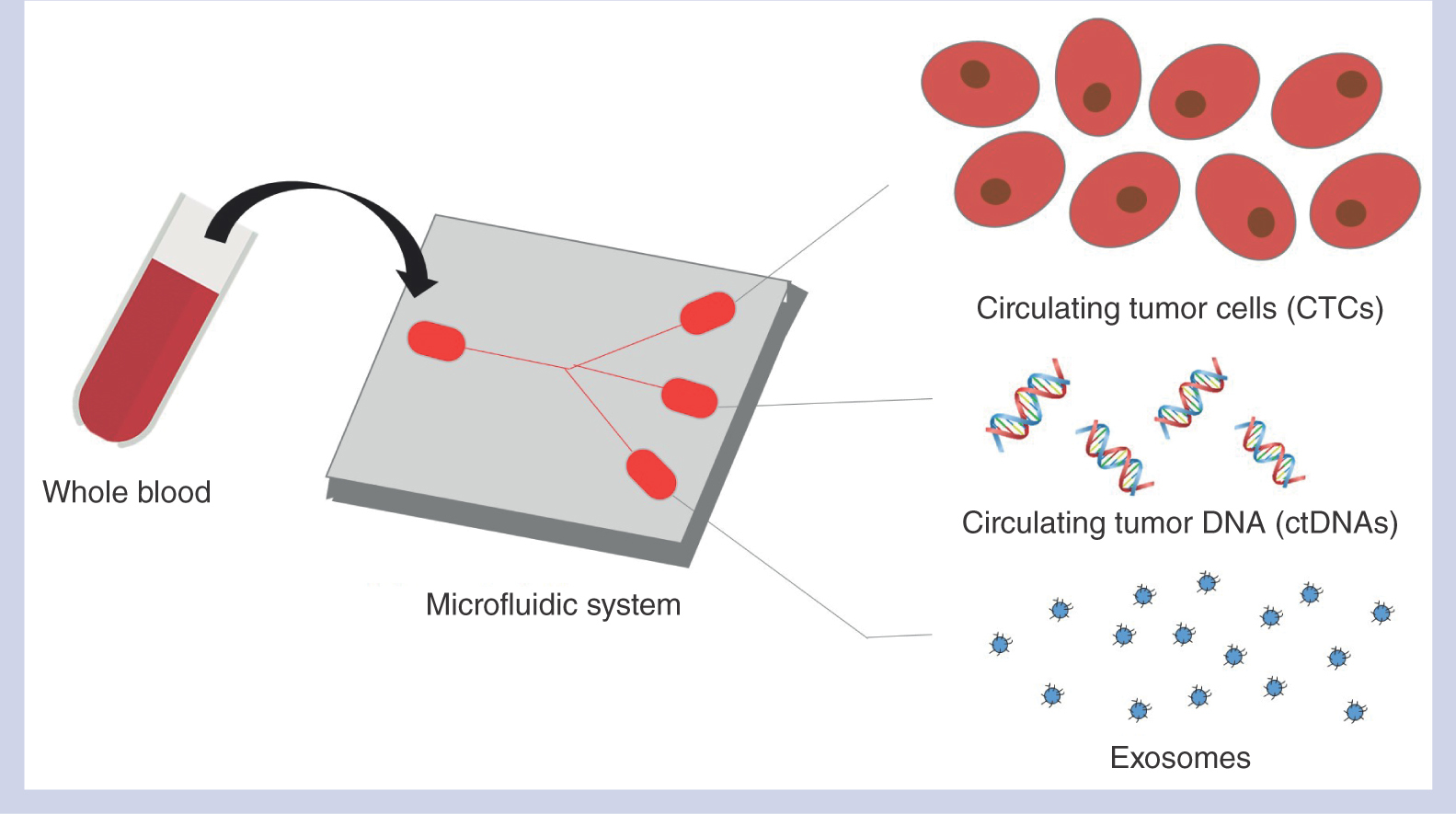
Cancer diagnosis has made paradigm-shift advancements in recent years with the development of sensitive, non-invasive liquid biopsy technologies. Dubbed as the less or noninvasive alternative to tumor biopsy, this approach has received significant attention and gained traction in both academia and medical institutions. Liquid biopsy employs various technologies to sieve out unique tumor-derived biomarkers found in the peripheral blood of patients. Mounting evidence has suggested close association between biomarker profiles and patient disease burden, thus enabling more accurate disease detection and monitoring. Importantly, the minimal invasivness of the technique and readily obtainable biofluids allow for more timely and targeted interventions. However, the scarcity of tumor-derived biomarkers residing within the bloodstream poses a major hurdle to targeted capture. The holy grail hence lies in an effective approach to isolate these biomarkers in their most native forms for further downstream processing and analysis.
Microscale technologies employing microfluidics is especially appealing as it enables laminar flow with amplified secondary forces, allowing significant migration of cells of different sizes within the microchannels. Notably, this makes it possible to capture and isolate tumor-derived cells from peripheral blood. For example, circulating tumor cells (CTCs) in blood were found to originate either from the primary tumor or secondary metastatic site [Citation1], and the enumeration of CTCs alone could be used to correlate prognosis. Attracted by its clinical significance, many researchers have invested enormous efforts in ensuring capture with high purity and high throughput. As most CTCs are distinctly larger than other blood cells, numerous physical size-based microfluidic technologies have been successfully developed and commercialized in the past decade [Citation1]. Recent efforts are now leaning towards the design of special geometries within the microchannels to isolate single CTCs into different compartments. This technology enables single-cell analysis that seeks to shed light on tumor composition and heterogeneity. More importantly, resolution at the single-cell level provides insights into drug interactions within heterogeneous cell populations.
Beyond CTCs, there exists a wealth of information that remains elusive to researchers and clinicians focusing on cancer diagnostics. It is posited that tumor cell lines release DNA fragments into the circulation for distant microenvironment remodeling [Citation2]. In fact, this circulating cell-free tumor DNA (ctDNA) is one of the most investigated analytes of liquid biopsy. Studies have suggested its potential for early cancer detection and cancer recurrence prediction. In a translational study by Cohen et al., the combination of several biomarkers including ctDNA analysis provided sensitivities ranging from 69% to 98% for five different cancer types with specificity greater than 99% [Citation3]. However, similar to CTCs, ctDNA exists in trace amounts, from less than 0.1% to more than 10% of the total circulating free DNA [Citation4]. Techniques interrogating ctDNA require extremely high sensitivity. Here, it is worthwhile to note that microfluidics has obvious advantages to isolate specific nucleic acids found in circulation with reduced reagent amounts and minimal dead volume. The advancements of next-generation sequencing methodologies seek to elicit further information on the tumor with the effective capture of ctDNA on a chip.
Apart from ctDNA being found freely in the bloodstream, researchers discovered that certain disease-specific nucleic acids are closely associated with extracellular vesicles. These nanosized vesicles, or exosomes, were initially thought to be waste disposal units but recent evidence suggests their pivotal role in cancer initiation, progression and metastasis. Importantly, the spherical bilipid membrane vesicles protect its content from degradation and are responsible for intercellular communication. By transferring its exosomic contents containing DNAs, RNAs and proteins to the host cell, it is able to perform distant microenvironment modelling. Unlike previous circulating biomarkers, exosomes are stably present in all bodily fluids, which makes them very appealing for point-of-care applications. Furthermore, circulating microvesicles could exist in abundance in cancer patients (∼1–3 trillion per ml of plasma). In fact, exosomes are found to increase by a few folds in cancer patients [Citation1], and could provide crucial information that complements the biodata derived from CTCs and ctDNAs. Exosomal contents have thus emerged as promising biomarkers comparable or even superior to the other analytes. For example, Melo et al. provided evidence for a cell surface proteoglycan as a reliable biomarker for detection of early pancreatic cancer specific to KRAS mutations [Citation5].
Similar to CTCs, the challenge therefore lies in an efficient isolation of exosomes for further analysis. However, owing to their size, circulating microvesicles are perhaps more difficult to capture than previous analytes. Conventional methods require many hours of repeated centrifugation using expensive ultracentrifuges before the microvesicles can be pelleted and retrieved. Furthermore, the high forces may disrupt the exosomic contents, leading to crucial information loss. This labor-intensive process impeded the true potential of microvesicles to complement liquid biopsy diagnostics until recently. Microfluidic approaches similar to CTCs isolation have been adapted for exosomes capture. For example, immunoaffinity approaches similar to CTC capture have been designed for exosome isolation. Microvesicles typically possess transmembrane proteins (e.g., CD63, CD9, CD81 and HSP70) that could be targeted for capture. More crucially, microfluidics increases the surface area-to-volume ratio, allowing more effective capture. However, owing to the wall-induced lift force, these nanosized vesicles are often found away from the walls of the microchannel, making immunoaffinity capture difficult. Therefore, these techniques mostly function at low throughputs.
At its miniscule size, separation by physical forcefields in microstructures is even more challenging. Researchers have utilized nanotechnologies to develop nanoporous structures, nanopillars or nanochannels to separate exosomes from the larger apoptotic bodies. However, this may not be viable due to the abundance of the exosomes and larger bodies, which may lead to passage clogging. To overcome this, Yeo et al. built a microfluidic centrifugal nanoparticles separation and extraction (μCENSE) platform, which uses centrifugal forces to separate microvesicles within minutes [Citation6]. The entire assembly is similar to having multiple microcentrifuges loaded in a platform. These microcentrifuges can then be processed at a significantly reduced speed and automatically sorted by size into different compartments. The entire process may be fully automated and is at least a hundredfold faster than the conventional ultracentrifuge. This strongly suggests the possibility of isolating exosomes in a timely manner using standard laboratory equipment.
Recent ctDNA and CTC tests have yielded promising results to accurately determine the nature and stage of disease, prognosis and treatment options for patients [Citation7,Citation8]. With the advances in exosomes isolation, liquid biopsy diagnostics integrating various technologies may soon be realized and may well achieve the vision of personalized oncology. Yet, despite its enormous potential and positive outlook, blood-based tests for cancer diagnostics may not replace the incumbent technology just yet. Cancer is often characterized by high intertumor and intratumor heterogeneity, and this is highly representative in the biomarkers as well. As such, this heterogeneity of cancer often makes diagnosis difficult. Also, this may be further confounded by the presence of circulating cells, DNA and extracellular vesicles that may not originate from the tumor, but may be associated with other diseases.
Despite significant interest from clinicians, few microfluidic technologies have successfully translated into clinical studies. Apart from regulatory barriers and manufacturing hurdles, commercial platforms require the development of user-friendly interfaces and the calibration of the system to cater for different patient’s profiles. Therefore, substantial efforts are still necessary in the translation of such research technologies from bench to bedside. These efforts could hardly be achieved by the sole researcher and require synergistic collaboration between clinicians, engineers, scientists, regulatory partners and commercial leads. With these issues carefully considered, the smooth transition of liquid biopsy technologies into clinical settings may be realized in the near future.
The clinical workflow for cancer patients in the future may involve a simple phlebotomy, where peripheral blood can be collected and inserted into an integrated liquid biopsy instrument for immediate processing and analysis. Within this system, multiple circulating biomarkers may be isolated, retrieved and analyzed across a large cohort of patients to determine cancer profile and disease burden. More importantly, with the biodata derived from multiple biomarkers, the clinician can recommend an effective treatment for the patient and monitor disease progression at different intervals. This will significantly improve the workflow of cancer therapy with the possibilities of personalized drug efficacy regimes based on the profile of individuals, paving the way to become the cornerstone of precision medicine and cancer research.
Author contributions
JCY and CTL prepared and wrote the manuscript. All authors have given approval to the final version of the manuscript.
Acknowledgments
We thank Helen Morrison and Zhao-Qi Wang for support; and Kyle J Roux for providing BiolD2 cDNA through Addgene.
Financial competing interests disclosure
The authors have no relevant affiliations or financial involvement with any organization or entity with a financial interest in or financial conflict with the materials discussed in the manuscript. This includes employment, consultancies, honoraria, stock ownership or options, expert testimony, grants or patents received or pending, or royalties.
No writing assistance was utilized in the production of this manuscript.
Additional information
Funding
References
- Sun Y , HaglundTA, RogersAJ, GhanimAF, SethuP. Review: Microfluidics technologies for blood-based cancer liquid biopsies. Analytica Chimica Acta1012, 10–29 (2018).
- Zhang W , XiaW, LvZ, NiC, XinY, YangL. Liquid biopsy for cancer: circulating tumor cells, circulating free DNA or exosomes?Cell. Physiol. Biochem.41(2), 755–768 (2017).
- Cohen JD , LiL, WangYet al. Detection and localization of surgically resectable cancers with a multi-analyte blood test. Science359(6378), 926–930 (2018).
- Pi C , ZhangM-f, PengX-x, ZhangY-c, XuC-r, ZhouQ. Liquid biopsy in non-small cell lung cancer: a key role in the future of personalized medicine?Exp. Rev. Mol. Diag.17(12), 1089–1096 (2017).
- Melo SA , LueckeLB, KahlertCet al. Glypican-1 identifies cancer exosomes and detects early pancreatic cancer. Nature523, 177 (2015).
- Yeo JC , Kenry, ZhaoZ, ZhangP, WangZ, LimCT. Label-free extraction of extracellular vesicles using centrifugal microfluidics. Biomicrofluidics12(2), 024103 (2018).
- Babayan A , PantelK. Advances in liquid biopsy approaches for early detection and monitoring of cancer. Genome Med.10(1), 21 (2018).
- Wan JCM , MassieC, Garcia-CorbachoJet al. Liquid biopsies come of age: towards implementation of circulating tumour DNA. Nat. Rev. Cancer17, 223 (2017).

