
Abstract
Because of the high mortality from myocardial infarction and stroke, there is a great demand for finding novel methods of diagnosis, prevention and treatment of these diseases. Most of the current tests measure important determinants of thrombosis such as platelet function, coagulation and fibrinolysis in isolation; therefore, a global test measuring the actual thrombotic status would be more useful in clinical conditions. We obtained considerable experience by using the global thrombosis test, which determines the actual thrombotic status by taking into account the measured platelet reactivity, coagulation and fibrinolytic activities. In animal experiments, we found significant correlation between the ex vivo global thrombosis test measurements and the in vivo thrombotic status. The published evidence for the benefit of an antithrombotic diet with regular physical exercise is also described.
Plain Language Summary
There is a great concern in the general population how to detect the risk of thrombotic events and prevent the high mortality from stroke, myocardial infarction, sudden death and cancer-associated thrombosis. Our experience on antithrombotic fruits and vegetables intake and regular exercise assessed by the global thrombosis test suggested a potentially unique way of preventing these life-threatening diseases. In addition, global thrombosis testing may offer some benefit in detecting risk of thrombotic of forthcoming thrombotic events in cancer and COVID-19 virus-infected patients.
The use of an overall in vivo thrombosis/thrombolysis test in experimental animals
Because of the severity of thromboembolic diseases such as myocardial infarction and stroke, there is a great demand for finding way of prevention and treatment of such diseases. For this purpose, finding a pathologically relevant tests suitable for the assessment of the actual thrombotic status is crucially important. At present, various tests exist claiming to assess determinants of thrombosis such as platelet reactivity, coagulation and fibrinolysis in isolation, but the common shortcoming of these test is the lack of relevance to the pathology of arterial thrombotic diseases. Most of these tests measure platelet function to various thrombotic stimuli and as such suitable for monitoring antiplatelet therapy while these tests do not detect endogenous fibrinolysis. In the latter field, d-dimer measurement is the predominant laboratory test [Citation1–8]. Finding a test which assesses reactivity of platelets, coagulation and fibrinolysis is justified by the ‘Virchow’s triad’ demonstrating the complexity of the interaction between platelets, other blood cells, vessel wall and blood flow in thrombus formation and lysis. This recognition requires the use of a global test, which takes into account of all contributors to the thrombotic process. For these reasons we employed the global thrombosis test (GTT) in animal experiments, where the conditions are reproducible and can be standardized.
Numerous animal models of thrombosis and thrombolysis (fibrinolysis) exist. Yamamoto and colleagues have employed helium–neon (He–Ne) laser-induced thrombosis/fibrinolysis model in rats and mice since the year 1989 [Citation9–20]. Ruby biolaser-induced thrombosis model was first published by Arfors and colleagues [Citation21], followed by He–Ne laser-induced thrombosis model in rodents by Kovacs, Gorog and colleagues [Citation22–26]. In our experience the He–Ne laser-induced animal thrombosis model is the most reliable technique providing reproducible and reliable results.
Earlier Furie and coworkers used the He–Ne laser-induced thrombosis model in mice to analyze thrombotic mechanism at molecular level [Citation27–29]. He–Ne laser-induced thrombosis/fibrinolysis animal model is shown in . In brief, the mesenteric or pial microvessel of anesthetized rat or the carotid artery of anesthetized mouse was exposed, and Evans blue dye was injected through the veins and then the exposed blood vessel was irradiated with laser beam. The injected dye specifically absorbs the laser energy, converts it to heat and as such burns the blood vessel from inside inducing thrombus formation. Thrombus formation in rat mesenteric vessels or in the carotid artery of mice having irradiated by laser was video monitored (https://drive.google.com/file/d/1EMaD-Rwt_lDlc_99rwy_q2WeSIWaX-wF/view?usp=sharing); (https://drive.google.com/file/d/1VNMjAjbQFRF18ZgdxEsRSzqpFX0gyh-W/view?usp=sharing). Severity of thrombotic reaction in the irradiated rat mesenteric microvessel was expressed by the number of irradiations required to cause complete occlusion of blood flow. In mouse carotid artery, the video recording in every 10 s in the first 10 min after irradiation was performed and the total size of emboli following irradiation was measured (). In thrombolytic activity measurement, the size of mural thrombus formed at the irradiation site was calculated with the formula shown in [Citation30–34].
Figure taken from Figure 1 from [Citation35] (original article is open access [CC-BY]).
CCD: Charged-coupled device.
![Figure 1. Helium–neon laser-induced thrombosis/thrombolysis (fibrinolysis) system.Figure taken from Figure 1 from [Citation35] (original article is open access [CC-BY]).CCD: Charged-coupled device.](/cms/asset/16c05042-e73e-4381-a303-f477b29f68ff/ifso_a_12364471_f0001.jpg)
(A) Thrombus size measurement of thrombus in mouse carotid artery. (B) Index of thrombogenicity measurement.
Figure taken from from [Citation35] (original article is open access [CC-BY]).
T: Thrombus; W: Vessel wall.
![Figure 2. Thrombosis in mouse carotid artery.(A) Thrombus size measurement of thrombus in mouse carotid artery. (B) Index of thrombogenicity measurement.Figure taken from Figure 1 from [Citation35] (original article is open access [CC-BY]).T: Thrombus; W: Vessel wall.](/cms/asset/de97a7c6-e2dc-417f-b0c2-542a3897c70d/ifso_a_12364471_f0002.jpg)
Thrombus area is calculated by delineating thrombus using computer (A). Subsequently, thrombus size (B) is obtained by multiplying gray scale and the area. Thrombolysis rate is compared with that at the start.
L: Lumen; T: Thrombus; V: Vessel wall.
Figure reproduced with permission from [Citation32] (Copyright © 2001, © 2001 S. Karger AG, Basel).
![Figure 3. Thrombolysis measurement in rat mesenteric microvessel.Thrombus area is calculated by delineating thrombus using computer (A). Subsequently, thrombus size (B) is obtained by multiplying gray scale and the area. Thrombolysis rate is compared with that at the start.L: Lumen; T: Thrombus; V: Vessel wall.Figure reproduced with permission from [Citation32] (Copyright © 2001, © 2001 S. Karger AG, Basel).](/cms/asset/33023e61-1ba1-418f-b7b1-0478431ddde9/ifso_a_12364471_f0003.jpg)
Overall ex vivo thrombosis/thrombolysis (fibrinolysis) test in experimental animals & in humans
Tests to assess thrombotic status using native (nonanticoagulated) blood and high shear forces were established by Baumgartner et al. and Sakariassen et al. [Citation35–41]. Their original shear rate dependent thrombosis chambers are most useful for investigating human thrombotic mechanisms under blood flow conditions and for monitoring human antithrombotic drugs. The haemostatometer were invented by Kovacs and Gorog to measure high shear-induced thrombosis and the subsequent thrombolysis [Citation42–46]. The haemostatometer and GTT tests, in which platelets were activated solely by high shear, proved to be useful assessing thrombus formation under high shear condition and proved to be clinically useful in assessing thrombotic status of patients. These tests were also suitable for selecting fruits and vegetables having antithrombotic activity for use as an antithrombotic diet in humans [Citation47–53].
Description of GTT test
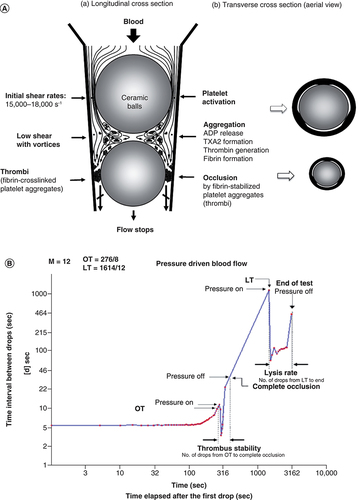
GTT test tube has a conical section in which two ceramic ball bearings are placed (A). Because of three flat segments formed on the inner surface of the tube, three narrow gaps exist adjacent to the ball bearings. When blood is added to the tube, it flows through the narrow gaps by the ball bearings and the droplets are collected in a reservoir downstream. While passing through the gaps by the upper (larger) ball bearing, platelets are exposed to high shear stress and become activated. In the space between the two ball bearings, platelet aggregates and thrombin is generated from the activated platelets. As fibrin-stabilized thrombi reach the lower ball bearing, blood flow gradually reduced and finally arrested. The instrument detects the time interval (d, s) between two consecutive blood drops falling into the reservoir. At the start of the test, flow is rapid and hence (d) is small. Subsequently, the flow rate decreases and hence (d) increases. When the actual default d ≥15 s is reached, this time is displayed as occlusion time (OT s). Subsequently the flow is completely arrested. After OT, a preset ‘thrombi stabilization period’ follows during which the sensors ignore counting the blood drops. This time allows stabilization of the fully occlusive thrombi. Eventually, due to endogenous thrombolysis (fibrinolysis), flow is partially restored as indicated by detection of the first blood drop after OT. This is recorded as lysis time (LT s). Typical pattern is shown in B.
(A) Principle of ex vivo GTT. Platelets are activated under high shear condition at the upper gaps created along the inner of a conical plastic tube. Activated platelets form fibrin-stabilized platelet aggregates under low shear condition between two balls. Fibrin-stabilized platelet aggregates occlude the lower gaps. (B) Pattern obtained by GTT (GTT-3).
GTT: Global thrombosis test; LT: Lysis time OT: Occlusion time.
GTT3 test starts by placing disposable test tube into the heated channel of the instrument and setting the instrument in standby mode, waiting for the blood sample to start the measurement (URL: https://drive.google.com/file/d/1Up-MgDCFPEBWYN3hKrmCobMCm47XpTp8/view). Blood is withdrawn from the antecubital vein, while the tourniquet constriction should be limited to a short time, only while the needle is inserted into the vein. To avoid activation of platelets and coagulation during withdrawal of blood, the ‘two-syringe blood taking technique’ is used. The first withdrawn 3–4 ml blood is used for laboratory tests and only the 4 ml blood in the second syringe is used for GTT measurement. The nonanticoagulated blood sample must be transferred from the syringe into the GTT test tube within <15 s from the withdrawal. With the transfer of blood into the test tube, test starts automatically and along the measurement reaching OT/thrombus stability then LT/rate of thrombolysis, these data are displayed and simultaneously recorded in an SD card for the possibility of displaying the measurement in a graph form later by a special algorithm ‘GTT-Draw’ [Citation54–62].
Thrombotic status is determined by the balance between thrombotic & fibrinolytic activities
The importance of balance between thrombotic and fibrinolytic activities determining the overall thrombotic status was confirmed in our experiments testing various fruit and vegetable varieties for antithrombotic activity. First, thrombotic (OT) and fibrinolytic activities (LT) of fruit and vegetables were measured by ex vivo GTT using rat blood. Next, the predicted antithrombotic activity was confirmed by in vivo He–Ne laser-induced thrombosis test in mice. These results are shown in . Grape varieties were classified as antithrombotic and prothrombotic varieties. Cabernet sauvignon (A) increased OT suggesting antithrombotic activity and decreased LT suggesting antithrombotic activity. The overall results showed being antithrombotic. On the contrary, Neo muscat (B) decreased OT suggesting prothrombotic activity and increased LT suggesting prothrombotic activity (A). These predictions were demonstrated by He–Ne laser-induced thrombosis test (B [Citation50]). The same ex vivo and in vivo correlation has been described in carrot varieties [Citation49]. Similar correlation was shown in congenital diabetic prothrombotic Goto-Kakizaki rat and antithrombotic Otsuka Long Evans Tokushima Fatty rat [Citation63,Citation64].
(A) Assessment of antithrombotic/prothrombotic activity by ex vivo test (GTT). (B) Assessment by in vivo test (He–Ne laser-induced thrombosis test). (A) Cabernet sauvignon. (B) Neo muscat.
(Figure taken from from [Citation50] (original article is open access [CC-BY]).
GTT: Global thrombosis test; LT: Lysis time OT: Occlusion time.
![Figure 5. Ex vivo and in vivo test.(A) Assessment of antithrombotic/prothrombotic activity by ex vivo test (GTT). (B) Assessment by in vivo test (He–Ne laser-induced thrombosis test). (A) Cabernet sauvignon. (B) Neo muscat.(Figure taken from Figure 1 from [Citation50] (original article is open access [CC-BY]).GTT: Global thrombosis test; LT: Lysis time OT: Occlusion time.](/cms/asset/a35a4658-6c11-4b33-ad29-ea34a6de47af/ifso_a_12364471_f0005.jpg)
Contribution from endothelium to thrombosis
Prothrombotic status of spontaneously hypertensive stroke prone rats was confirmed by the in vivo laser-induced thrombosis test. In contrast, platelet reactivity measured by ex vivo shear-induced thrombosis test using native blood was inhibited rather than enhanced [Citation65,Citation66]. To look into this contradiction, contribution from endothelium to thrombosis was examined in vivo in mice using the flow-mediated vasodilation test. Western-style high-fat diet and Japanese-style low fat diet were given to mice and thrombotic status was measured by the in vivo laser-induced thrombosis test. Results show that high-fat diet caused prothrombotic condition. High-fat diet did not affect the ex vivo shear-induced thrombosis test but induced endothelial dysfunction [Citation67–71]. Based on our finding, we suggested to couple the GTT test with the endothelial function evaluation using the flow-mediated vasodilation test.
Comparing thrombotic status of healthy people & cancer patients in different countries & under different conditions
OT as measured by GTT in British people is shorter than that of Japanese and LT of British is shorter than that of Japanese. This finding shows that in British subject thrombus forms rapidly but dissolved quickly, while in Japanese people thrombus forms and dissolved at a lower rate [Citation72]. People’s thrombotic status appears to be different in different countries, and for this reason epidemiological survey is necessary to have respective standard OT and LT (). Aging and habitual smoking enhance thrombotic status through inhibiting spontaneous fibrinolytic activity. This was obvious in the elderly [Citation73,Citation74]. Overwork of doctors at overnight causes prothrombotic status due to enhancing thrombosis and inhibiting fibrinolysis. This was not detected by APTT, PT-INR and PAI-1 measurements [Citation75].
Thrombotic and fibrinolytic activities in Westerners and Japanese, measured by GTT.
GTT: Global thrombosis test; J: Japanese; LT: Lysis time OT: Occlusion time; W: Westerners.
Reprinted from [Citation72] with permission from Elsevier.
![Figure 6. Comparison of thrombotic status between different races.Thrombotic and fibrinolytic activities in Westerners and Japanese, measured by GTT.GTT: Global thrombosis test; J: Japanese; LT: Lysis time OT: Occlusion time; W: Westerners.Reprinted from [Citation72] with permission from Elsevier.](/cms/asset/0cc8226d-d391-48b5-94c1-16b781b72469/ifso_a_12364471_f0006.jpg)
The cause and mechanism of cancer-associated thrombosis has been investigated over the years. Thrombotic status of patients with cancers and that without cancers are shown in . OT of patients with cancers (n = 48) was slightly shorter than that of patients without cancer (n = 17) but the difference did not reach significant level (p = 0.069). This might be due to the difference in number between the two groups. On the contrary, LT in patients with cancers were significantly prolonged (p < 0.0001). Overall thrombotic status of patients with cancers is higher, they are more prothrombotic than that of patients without cancers [Citation76]. GTT measurement may be helpful in finding the most effective treatment of thrombotic complications in cancer patients. As distribution patterns of OT and LT are different in people from different countries, GTT testing and monitoring therapy would offer personalized medication.
Enhanced thrombotic status in patients with cancers.
Figure taken from [Citation76]; was orally presented by Dr. Shioyama at The 25th Kinki Thrombosis Research Society Meeting. Osaka, Japan (2020).
![Figure 7. Thrombotic status of patients with and without cancers.Enhanced thrombotic status in patients with cancers.Figure taken from [Citation76]; was orally presented by Dr. Shioyama at The 25th Kinki Thrombosis Research Society Meeting. Osaka, Japan (2020).](/cms/asset/1e5c1fdb-cb7d-40c7-b372-d8728c2a725a/ifso_a_12364471_f0007.jpg)
Prevention of thrombotic diseases by daily intake of antithrombotic fruits, vegetable varieties & physical exercise
Antithrombotic effect of Mediterranean diet and various fruits and vegetables has been reported. Results of investigation aimed to find the specified ingredients responsible for the antithrombotic effect were also published [Citation77–86]. We were the first reporting that the antithrombotic effect of fruits and vegetables is dependent on the varieties, but not on species [Citation49–52]. GTT enables to classify fruit and vegetable varieties to antithrombotic and prothrombotic ones. Short- or long-term intake of antithrombotic fruits and vegetable varieties decreases thrombotic status in humans [Citation87,Citation88]. Further, we confirmed that the antithrombotic effect of fruits and vegetables are independent from antioxidants and polyphenols [Citation49–51].
Government authorities generally recommend physical exercise to prevent stroke and cardiovascular disease, but sometimes exercise causes sudden death (exercise paradox) [Citation89–99]. Yamamoto et al. suggested that regular measurement of thrombotic status of individuals and doing exercise in intensity and time length well-matched to individuals are beneficial. Acute strenuous exercise significantly shortens OT, but long term regular mild exercise increases OT. There was no significant difference in the rate of fibrinolysis between the two types of exercise [Citation100–102]. These results suggest that acute and strenuous exercise may be dangerous and may cause sudden death. In contrast, long term regular mild exercise may be beneficial by having antithrombotic effect.
Supplement
Coronavirus associated thrombosis
Thromboprophylaxis in coronavirus (COVID-19) pandemic seems to be justified [Citation103]. In addition, vaccine-induced immune thrombocytopenia was also reported [Citation104] which suggests a prothrombotic condition. The cause and mechanism of thromboembolic episodes associated with COVID-19 and vaccination are not clear, blood cells (erythrocytes, leucocytes, platelets) and endothelial cells may be involved. Currently the most recommended laboratory tests to detect prothrombotic condition are the d-dimer, PT, APTT, fibrinogen and platelet count measurements [Citation105–107]. Non-plasmin enzymes may also be involved in fibrinolysis in vivo [Citation108] Limitations of testing these biomarkers suggest the use of global thrombotic status measurement in people infected with COVID-19. The clinical benefit of current tests like thromboelastography and d-dimer measurements still needs to be established [Citation109,Citation110]. In testing nonanticoagulated whole blood with the GTT test in normal and cardiac patients, Yamamoto, Kovacs, Gorog and colleagues have demonstrated that leucocytes play a significant role in thrombosis/fibrinolysis [Citation111]. GTT using nonanticoagulated whole blood is suitable to assess thrombotic status and guide antithrombotic medication in COVID-19-associated pulmonary thromboembolic disorders.
Limitations of tests using native blood
Starting the GTT test within 15–20 s after withdrawal of blood samples requires the presence of highly trained, experienced phlebotomist, the GTT instrument and the patient at the site of test. Such conditions are difficult to fulfill in the present system of laboratory testing in hospitals. Video shown in this article may be helpful for those who want to use GTT in clinical practice.
Conclusion & future perspective
Adopting a long-term use of an experimentally proven antithrombotic diet and monitored regular physical exercise to put a stop to arterial thrombotic disorders, is certainly an interesting and challenging possibility. For this approach to be successful, it is important to find a test or tests that can monitor thrombotic status under pathologically relevant conditions. Compared with the common platelet function or coagulation tests currently being used, the shear-induced global thrombosis and fibrinolysis test (GTT) performed with native (nonanticoagulated) blood is suitable for the concept. Now that the pathologically relevant tools of testing and monitoring thrombotic status of individuals have been found, further investigations need to focus on the organization of large-scale and monitored trials that include both healthy individuals and those at risk of atherothrombotic diseases and cardiovascular events. The outcome of such trials may justify the everyday consumption of an antithrombotic diet with regular physical exercise, and this would be a simple and economical way of prevention of arterial thrombotic diseases.
To date, global thrombosis test (GTT) is the most suitable test for assessing thrombotic status of patients with thrombotic disorders.
Interest in prevention of thrombotic disorders by antithrombotic vegetable varieties and physical exercise is increasing. A pathologically relevant point-of-care global tests for the assessment of global thrombotic status is needed.
The GTT enables the simultaneous measurement of thrombosis and endogenous fibrinolysis under high shear stress. This technique proved to be suitable for screening fruits and vegetables for antithrombotic effect, making possible to establish an antithrombotic diet and also to individualize the need of physical exercise in people at risk of thrombotic events.
GTT-monitored large-scale trials are needed to verify the beneficial effect of an antithrombotic diet with regular physical exercise in the prevention of arterial thrombotic events.
The GTT may clarify the mechanism of association of thrombotic events and cancer and help in the treatment of COVID-19-associated thromboembolic disorders.
Author contributions
J Yamamoto designed the research project. M Murakami, K Otsui, Y Ijiri, M Shimizu, H Ikarugi, W Shioyama and J Yamamoto contributed to the implementation of research and creation of images. The manuscript was made with KS Sakariassen.
Edboard disclosure
KS Sakariassen is a member of the Future Science OA Editorial Board. They were not involved in any editorial decisions related to the publication of this article, and all author details were blinded to the article’s peer reviewers as per the journal’s double blind peer review policy.
Acknowledgments
The authors would like to thank colleagues who engaged in our previous research.
Financial & competing interests disclosure
The authors have no relevant affiliations or financial involvement with any organization or entity with a financial interest in or financial conflict with the subject matter or materials discussed in the manuscript. This includes employment, consultancies, honoraria, stock ownership or options, expert testimony, grants or patents received or pending, or royalties.
No writing assistance was utilized in the production of this manuscript.
References
- AnghelL , SascăuR , RaduR , StătescuC. From classical laboratory parameters to novel biomarkers for the diagnosis of venous thrombosis. Int. J. Mol. Sci.21(6), 1920–1937 (2020).
- GueYX , GorogDA. Importance of endogenous fibrinolysis in platelet thrombus formation. Int. J. Mol. Sci.18(9), 1850–1864 (2017).
- KoltaiK , KesmarkyG , FeherG , TiboldA , TothK. Platelet aggregometry testing: molecular mechanisms, techniques and clinical implications. Int. J. Mol. Sci.18(8), 1803–2003 (2017).
- PanicciaR , PrioraR , LiottaAA , AbbateR. Platelet function tests: a comparative review. Vasc. Health Risk Manag.11, 133–148 (2015).
- OkaforON , GorogDA. Endogenous fibrinolysis: an important mediator of thrombus formation and cardiovascular risk. J. Am. Coll. Cardiol.65(16), 1683–1699 (2015).
- PabingerI , ThalerJ , AyC. Biomarkers for prediction of venous thromboembolism in cancer. Blood122(12), 2011–2018 (2013).
- KhaleghiM , SaleemU , McBaneRD , JrMosley TH , KulloIJ. African American ethnicity is associated with higher plasma levels of D-dimer in adults with hypertension. J. Thromb. Haemost.7(1), 34–40 (2009).
- HarrisonP. Platelet function analysis. Blood Rev.19(2), 111–123 (2005).
- YamamotoJ , IizumiH , HirotaRet al.Effect of physical training on thrombotic tendency in rats: decrease in thrombotic tendency measured by the He-Ne laser-induced thrombus formation method. Haemostasis19(5), 260–265 (1989).
- YamamotoJ , IshiiI , SasakiYet al.Antithrombotic effect of ticlopidine on He-Ne laser-induced thrombus formation in rat mesenteric microvessels. Haemostasis22(3), 147–152 (1992).
- YamamotoJ , IshiiI , ChikamoriAet al.Effect of long-term aerobic exercise on helium-neon-laser-induced thrombogenesis in rat mesenteric arterioles and platelet aggregation. Haemostasis23(3), 129–134 (1993).
- SasakiY , MoriiS , YamashitaT , YamamotoJ. Antithrombotic effect of argatroban on the pial vessels of the rat: a study with He-Ne laser-induced thrombus formation. Haemostasis23(2), 104–111 (1993).
- SasakiY , OyamaT , OkitaN , GiddingsJC , SekiJ , YamamotoJ. Cerebral prothrombotic state in spontaneously hypertensive rats of different breeds. Haemostasis26(2), 79–84 (1996).
- SasakiY , SekiJ , GiddingsJC , YamamotoJ. Effects of NO-donors on thrombus formation and microcirculation in cerebral vessels of the rat. Thromb. Haemost.76(1), 111–117 (1996).
- YamamotoJ , IshiiI , OkitaNet al.The differential involvement of von Willebrand factor, fibrinogen and fibronectin in acute experimental thrombosis in rat cerebral and mesenteric microvessels. Jpn J. Physiol.47(5), 431–441 (1997).
- YamashitaT , TsujiT , MatsuokaA , GiddingsJC , YamamotoJ. The antithrombotic effect of synthetic low molecular weight human factor Xa inhibitor, DX-9065a, on He-Ne laser-induced thrombosis in rat mesenteric microvessels. Thromb. Res.85(1), 45–51 (1997).
- YamashitaT , TsudaY , KonishiYet al.The antithrombotic effect of potent bifunctional thrombin inhibitors based on hirudin sequence, P551 and P532, on He-Ne laser-induced thrombosis in rat mesenteric microvessels. Thromb. Res.90(5), 199–206 (1998).
- NagamatsuY , TsujiokaY , HashimotoM , GiddingsJC , YamamotoJ. The differential effects of aspirin on platelets, leucocytes and vascular endothelium in an in vivo model of thrombus formation. Clin. Lab. Haematol.21(1), 33–40 (1999).
- IjiriY , MiuraM , HashimotoMet al.A new model to evaluate the diet-induced prothrombotic state, using He-Ne laser-induced thrombogenesis in the carotid artery of apolipoprotein E-deficient and low-density lipoprotein receptor-deficient mice. Blood Coagul. Fibrinolysis13(6), 497–504 (2002).
- SasakiY , KobaraN , HigashinoS , GiddingsJC , YamamotoJ. Astaxanthin inhibits thrombosis in cerebral vessels of stroke-prone spontaneously hypertensive rats. Nutr. Res.31(10), 784–789 (2011).
- ArforsKE , HintHC , DhallDP , MathesonNA. Counteraction of platelet activity at sites of laser-induced endothelial trauma. Br. Med. J.16(4), 430–431 (1968).
- KovácsIB , CsalayL , GörögP. Laser-induced thrombosis in the microcirculation of the hamster cheek pouch and its inhibition by acetylsalicyclic acid. Microvasc. Res.6, 194–201 (1973).
- KovácsIB , Tigyi-SebesA , TrombitásKet al.Evans blue: an ideal energy-absorbing material to produce intravascular microinjury by HE-NE gas laser. Microvasc. Res.10, 107–124 (1975).
- KovácsIB , SebesA , TrombitásKet al.Proceedings: improved technique to produce endothelial injury by laser beam without direct damage of blood cells. Thromb. Diath. Haemorrh.34(1), 331 (1975).
- GörögP , KovácsIB. Antiarthritic and antithrombotic effects of topically applied dimethyl sulfoxide. Ann. NY Acad. Sci.243, 91–97 (1975).
- KovácsIB , GörögP. Laser-induced thrombosis test suitable for pharmacological screening studies. Microvasc. Res.18(3), 403–412 (1979).
- FalatiS , GrossP , Merrill-SkoloffGet al.Real-time in vivo imaging of platelets, tissue factor and fibrin during arterial thrombus formation in the mouse. Nat. Med.8(10), 1175–1181 (2002).
- FurieB , FurieBC. Mechanisms of thrombus formation. N. Engl. J. Med.359(9), 938–949 (2008).
- FurieB , FurieBC. The molecular basis of platelet and endothelial cell interaction with neutrophils and monocytes: role of P-selectin and the P-selectin ligand, PSGL-1. Thromb. Haemost.74(1), 224–227 (1995).
- KawanoM , WatanabeS , SasakiY , GiddingsJC , YamamotoJ. Adjuvant effect of argatroban on staphylokinase induced thrombolysis of platelet rich thrombi in rat mesenteric venules in vivo. Thromb. Res.86(2), 115–126 (1997).
- YamashitaT , KitamoriK , HashimotoM , WatanabeS , GiddingsJC , YamamotoJ. Conjunctive effects of the 5HT (2) receptor antagonist, sarpogrelate, on thrombolysis with modified tissue plasminogen activator in different laser-induced thrombosis models. Haemostasis30(6), 321–332 (2000).
- HashimotoM , WatanabeS , OiwaKet al.Enhanced thrombolysis induced by argatroban or activated protein C in the presence or absence of staphylokinase, measured in an in vivo animal model using mesenteric arterioles. Haemostasis31(2), 80–89 (2001).
- HashimotoM , YamashitaT , OiwaK , WatanabeS , GiddingsJC , YamamotoJ. Enhancement of endogenous plasminogen activator-induced thrombolysis by argatroban and APC and its control by TAFI, measured in an arterial thrombolysis model in vivo using rat mesenteric arterioles. Thromb. Haemost.87(1), 110–113 (2002).
- HashimotoM , OnobayashiY , OiwaK , GiddingsJC , YamamotoJ. Enhanced endogenous thrombolysis induced by a specific factor Xa inhibitor, DX-9065a, evaluated in a rat arterial thrombolysis model in vivo. Thromb. Res.106(2), 165–168 (2002).
- YamamotoJ , IjiriY , IkarugiH , OtsuiK , InoueN. Sakariassen KS. Prevention of thrombotic disorders by antithrombotic diet and exercise: evidence by using global thrombosis tests. Future Sci. OA4(4), FSO285 (2018).
- BaumgartnerHR. Effects of anticoagulation on the interaction of human platelets with subendothelium in flowing blood. Schweiz. Med. Wochenscher.106, 1367–1368 (1976).
- BarstadRM , RoaldHE , CuiW , TurittoVT , SakariassenKS. A perfusion chamber developed to investigate thrombus formation and shear profiles in flowing native blood at the apex of well-defined stenoses. Arterioscler. Thromb.14, 1984–1991 (1994).
- KirchhoferD , TschoppTB , HadváryP , BaumgartnerHR. Endothelial cells stimulated with tumor necrosis factor-alpha express varying amounts of tissue factor resulting in inhomogenous fibrin deposition in a native blood flow system. Effects of thrombin inhibitors. J. Clin. Invest.93(5), 2073–2083 (1994).
- SakariassenKS , AartsPAMM , de GrootPG , HoudijkWPM , SixmaJJ. A perfusion chamber developed to investigate platelet interaction in flowing blood with human vessel wall cells, their extracellular matrix and purified components. J. Lab. Clin. Med.102, 522–535 (1983).
- SakariassenKS , TurittoVT , BaumgartnerHR. Recollections of the development of flow devices for studying mechanisms of hemostasis and thrombosis in flowing whole blood. J. Thromb. Haemost.2(10), 1681–1690 (2004).
- SakariassenKS , OrningL , TurittoVT. The impact of blood shear rate on arterial thrombus formation. Future. Sci. OA1(4), FSO30 (2015).
- RatnatungaCP , EdmondsonSF , ReesGMet al.High-dose aspirin inhibits shear-induced platelet reaction involving thrombin generation. Circulation85(3), 1077–1082 (1992).
- GorogDA , KovacsIB. Thrombotic status analyser. Measurement of platelet-rich thrombus formation and lysis in native blood. Thromb. Haemost.73(3), 514–520 (1995).
- NakajimaS , NoguchiT , TakaTet al.A global platelet test of thrombosis and thrombolysis detects a prothrombotic state in some patients with non-insulin dependent diabetes and in some patients with stroke. Platelets11(8), 459–466 (2000).
- IkarugiH , TakaT , NakajimaSet al.Norepinephrine, but not epinephrine, enhances platelet reactivity and coagulation after exercise in humans. J. Appl. Physiol.86(1), 133–138 (1985).
- MurakiT , SasakiY , GiddingJC , IshiiH , KanekoT , YamamotoJ. Antithrombotic effect of FK506 versus prothrombotic effect of cyclosporine in vivo. Transplantation60(3), 308–310 (1995).
- YamamotoJ , NaemuraA , UraMet al.Testing various fruits for anti-thrombotic effect: i. Mulberries. Platelets17(8), 555–564 (2006).
- YamamotoJ , TakaT , YamadaKet al.Tomatoes have natural anti-thrombotic effects. Br. J. Nutr.90(6), 1031–1038 (2003).
- YamamotoJ , NaemuraA , IjiriYet al.The antithrombotic effects of carrot filtrates in rats and mice. Blood Coagul. Fibrinolysis19(8), 785–792 (2008).
- IwasakiM , MurakamiM , IjiriY , ShimizuM , YamamotoJ. Are all wines made from various grape varieties beneficial in the prevention of myocardial infarction and stroke?Future Sci. OA7(2), FSO649 (2020).
- MorishitaM , NaemuraA , TamuraYet al.Mechanism of the experimental antithrombotic effect of some apple varieties involves enhanced endogenous thrombolytic activity. Interv. Med. Appl. Sci.4(3), 115–124 (2012).
- YamamotoJ , IjiriY , TamuraY , IwasakiM , MurakamiM , OkadaY. Reevaluation of antithrombotic fruits and vegetables: great variation between varieties. Drug Discov. Ther.10(3), 129–140 (2016).
- VioliF , PastoriD , PignatelliP , CarnevaleR. Nutrition, thrombosis, and cardiovascular disease. Circ. Res.126(10), 1415–1442 (2020).
- YamamotoJ , YamashitaT , IkarugiHet al.Görög thrombosis test: a global in-vitro test of platelet function and thrombolysis. Blood Coagul. Fibrinolysis14(1), 31–39 (2003).
- NishidaH , MurataM , MiyakiK , OmaeK , WatanabeK , IkedaY. Gorog thrombosis test: analysis of factors influencing occlusive thrombus formation. Blood Coagul. Fibrinolysis17(3), 203–207 (2006).
- YamamotoJ , InoueN , OtsuiK , IshiiH , GorogDA. Global thrombosis test (GTT) can detect major determinants of haemostasis including platelet reactivity, endogenous fibrinolytic and thrombin generating potential. Thromb. Res.133(5), 919–926 (2014).
- GueYX , InoueN , SpinthakisNet al.Thrombotic profile and oral anticoagulation in Asian and non-Asian patients with nonvalvular atrial fibrillation. J. Am. Coll. Cardiol.74(22), 2822–2824 (2019).
- GorogDA , FaragM , SpinthakisNet al.Effect of remote ischaemic conditioning on platelet reactivity and endogenous fibrinolysis in ST-elevation myocardial infarction: a substudy of the CONDI-2/ERIC-PPCI randomized controlled trial. Cardiovasc. Res.117(2), 623–634 (2021).
- GueYX , KanjiR , WellstedDM , SrinivasanM , WyattS , GorogDA. Rationale and design of “Can very low dose rivaroxaban (VLDR) in addition to dual antiplatelet therapy improve thrombotic status in acute coronary syndrome (VaLiDate-R)” study: a randomised trial modulating endogenous fibrinolysis in patients with acute coronary syndrome. J. Thromb. Thrombolysis49(2), 192–198 (2020).
- SpinthakisN , GueY , FaragMet al.Apixaban enhances endogenous fibrinolysis in patients with atrial fibrillation. Europace21(9), 1297–1306 (2019).
- SpinthakisN , GueY , FaragMet al.Impaired endogenous fibrinolysis at high shear using a point-of-care test in STEMI is associated with alterations in clot architecture. J. Thromb. Thrombolysis47(3), 392–395 (2019).
- Niespialowska-SteudenM , MarkidesV , FaragMet al.Catheter ablation for AF improves global thrombotic profile and enhances fibrinolysis. J. Thromb. Thrombolysis44(4), 413–426 (2017).
- TakaT , OnoH , SasakiY , SekiJ , YamamotoJ. Platelet reactivity in spontaneously diabetic rats is independent from blood glucose and insulin levels. Platelets13(5–6), 313–316 (2002).
- TakaT , OkitaN , SasakiY , YamamotoJ. Thrombotic tendency of a non-insulin dependent diabetic rat OLETF. Research Conference on OLETF Rats2, 75–79 (1996).
- YamashitaT , TakaT , NojimaR , OhtaY , SekiJ , YamamotoJ. There is no valid evidence presented as to an impaired endothelial NO system in the stroke-prone spontaneously hypertensive rats. Thromb. Res.105(6), 507–511 (2002).
- TakaT , OhtaY , SekiJ , GiddingsJC , YamamotoJ. Impaired flow-mediated vasodilation in vivo and reduced shear-induced platelet reactivity in vitro in response to nitric oxide in prothrombotic, stroke-prone spontaneously hypertensive rats. Pathophysiol. Haemost. Thromb.32(4), 184–189 (2002).
- IjiriY , MiuraM , HashimotoMet al.A new model to evaluate the diet-induced prothrombotic state, using He-Ne laser-induced thrombogenesis in the carotid artery of apolipoprotein E-deficient and low-density lipoprotein receptor-deficient mice. Blood Coagul. Fibrinolysis13(6), 497–504 (2002).
- IjiriY , NaemuraA , YamashitaTet al.Mechanism of the antithrombotic effect of dietary diacylglycerol in atherogenic mice. Pathophysiol. Haemost. Thromb.35(5), 380–387 (2006).
- IjiriY , NaemuraA , YamashitaTet al.Dietary diacylglycerol extenuates arterial thrombosis in apoE and LDLR deficient mice. Thromb. Res.117(4), 411–417 (2006).
- AokiR , IkarugiH , NaemuraA , IjiriY , YamashitaT , YamamotoJ. Endothelial dysfunction precedes atherosclerotic lesions and platelet activation in high fat diet-induced prothrombotic state. Thromb. Res.117(5), 529–535 (2006).
- YamamotoJ , TamuraY , IjiriY , IwasakiM , MurakamiM , MatsuoO. Evaluation of antithrombotic effect: importance of testing components and methodologies. Drug Discov. Ther.9(4), 258–266 (2015).
- GorogDA , YamamotoJ , SarafSet al.First direct comparison of platelet reactivity and thrombolytic status between Japanese and Western volunteers: possible relationship to the “Japanese paradox”. Int. J. Cardiol.152(1), 43–48 (2011).
- IkarugiH , YamashitaT , AokiR , IshiiH , KankiK , YamamotoJ. Impaired spontaneous thrombolytic activity in elderly and in habitual smokers, as measured by a new global thrombosis test. Blood Coagul. Fibrinolysis14(8), 781–784 (2003).
- YamashitaT , SatoA , IkarugiHet al.Significantly reduced spontaneous thrombolytic activity in older men: a possible explanation for the gender differences in risk of acute coronary syndromes. Thromb. Res.116(2), 127–131 (2005).
- OtsuiK , YamamotoJ , InoueN. Overwork accelerates thrombotic reaction: implications for the pathogenesis of Karoshi. J. Thromb. Thrombolysis45(2), 222–224 (2018).
- ShioyamaW , OkaT , KamataRet al.Effect of cancer chemotherapy on platelet reactivity and thrombolytic activity. Presented at: The 25th Kinki Thrombosis Research Society Meeting.Osaka, Japan (2020).
- JoshipuraKJ , AscherioA , MansonJEet al.Fruit and vegetable intake in relation to risk of ischemic stroke. JAMA282(13), 1233–1239 (1999).
- LiuS , MansonJE , LeeIMet al.Fruit and vegetable intake and risk of cardiovascular disease: the women's health study. Am. J. Clin. Nut.72(4), 922–928 (2000).
- JoshipuraKJ , HuFB , MansonJEet al.The effect of fruit and vegetable intake on risk for coronary heart disease. Ann. Intern. Med.134(12), 1106–1114 (2001).
- BazzanoLA , HeJ , OgdenLGet al.Fruit and vegetable intake and risk of cardiovascular disease in US adults: the first national health and nutrition examination survey epidemiologic follow-up study. Am. J. Clin. Nutr.76(1), 93–99 (2002).
- HeFJ , NowsonCA , MacGregorGA. Fruit and vegetable consumption and stroke: meta-analysis of cohort studies. Lancet367(9507), 320–326 (2006).
- MezzanoD , MunozX , MartinezCet al.Vegetarians and cardiovascular risk factors: hemostasis, inflammatory markers and plasma homocysteine. Thromb. Haemost.81(6), 913–917 (1999).
- LiD , SinclairA , MannNet al.The association of diet and thrombotic risk factors in healthy male vegetarians and meat-eaters. Eur. J. Clin. Nutr.53(8), 612–619 (1999).
- SatijaA , BhupathirajuSN , SpiegelmanDet al.Healthful and unhealthful plant-based diets and the risk of coronary heart disease in U.S. adults. J. Am. Coll. Cardiol.70(4), 411–422 (2017).
- GantenbeinKV , Kanaka-GantenbeinC. Mediterranean diet as an antioxidant: the impact on metabolic health and overall wellbeing. Nutrients13(6), 1951 (2021).
- MattioliAV. Coffee and platelets: an unsolved problem. J. Caffeine Adenosine Res.11(3), 49–50 (2021).
- NaemuraA , OhiraH , IkedaM , KoshikawaK , IshiiH , YamamotoJ. An experimentally antithrombotic strawberry variety is also effective in humans. Pathophysiol. Haemost. Thromb.35(5), 398–404 (2006).
- IjiriY , IshiiH , YamamotoJ. Diet of fruits and vegetables with experimental antithrombotic effect may be beneficial to humans in the prevention of arterial thrombotic diseases. Int. J. Drug Dev. Res.8(3), 6–10 (2016).
- LavieCJ , ThomasRJ , SquiresRW , AllisonTG , MilaniRV. Exercise training and cardiac rehabilitation in primary and secondary prevention of coronary heart disease. Mayo Clin. Proc.84(4), 373–383 (2009).
- ShiromaEJ , LeeIM. Physical activity and cardiovascular health: lessons learned from epidemiological studies across age, gender, and race/ethnicity. Circulation122(7), 743–752 (2010).
- PollockML , FranklinBA , BaladyGJet al.Resistance exercise in individuals with and without cardiovascular disease: benefits, rationale, safety, and prescription: an advisory from the committee on exercise, rehabilitation, and prevention, council on clinical cardiology, American Heart Association. Circulation101(7), 828–833 (2000).
- AlbertCM , MittlemanMA , ChaeCU , LeeIM , HennekensCH , MansonJE. Triggering of sudden death from cardiac causes by vigorous exertion. N. Engl. J. Med.343(19), 1355–1361 (2000).
- MaronBJ. The paradox of exercise. N. Engl. J. Med.343(1), 1409–1411 (2000).
- WangJS , JenCJ , KungHC , LinLJ , HsiueTR , ChenHI. Different effects of strenuous exercise and moderate exercise on platelet function in men. Circulation90(9), 2877–2885 (1994).
- El-SayedMS , SaleC , JonesPG , ChesterM. Blood hemostasis in exercise and training. Med. Sci. Sports Exerc.32(5), 918–925 (2000).
- ThompsonPD , FranklinBA , BaladyGJet al.Exercise and acute cardiovascular events placing the risks into perspective: a scientific statement from the American heart association council on nutrition, physical activity, and metabolism and the council on clinical cardiology. Circulation115(17), 2358–2368 (2007).
- ThrallG , LaneD , CarrollD , LipGY. A systematic review of the effects of acute psychological stress and physical activity on haemorheology, coagulation, fibrinolysis and platelet reactivity: implications for the pathogenesis of acute coronary syndromes. Thromb. Res.120(6), 819–847 (2007).
- PosthumaJJ , vander Meijden PE , TenCate H , SpronkHM. Short- and long-term exercise induced alterations in haemostasis: a review of the literature. Blood Rev.29(3), 171–178 (2014).
- OlsenLN , FischerM , PhillipEvans PA , GliemannHellsten Y. Does exercise influence the susceptibility to arterial thrombosis? An integrative perspective. Front. Physiol.12, 636027 (2021).
- IkarugiH , YamamotoJ. The exercise paradox may be solved by measuring the overall thrombotic state using native blood. Drug Discov. Ther.11(1), 15–19 (2017).
- IkarugiH , TakaT , NakajimaS. Detection of a prothrombotic state after acute aerobic exercise. Thromb. Res.85, 351–356 (1997).
- BabaY. Effects of exercise and diet interventions modeled on Healthy Japan 21 on thrombus formation. Master's Thesis, Kobe Gakuin University (2012).
- FavaloroEJ , LippiG. Maintaining hemostasis and preventing thrombosis in coronavirus disease 2019 (COVID-19): part II. Semin. Thromb. Hemost.47(4), 333–337 (2021).
- GreinacherA , ThieleT , WarkentinTE , WeisserK , KyrlePA , EichingerS. Thrombotic thrombocytopenia after ChAdOx1 nCov-19 vaccination. N. Engl. J. Med.384(22), 2092–2101 (2021).
- KlokFA , KruipMJHA , vander Meer NJMet al.Confirmation of the high cumulative incidence of thrombotic complications in critically ill ICU patients with COVID-19: an updated analysis. Thromb. Res.191, 148–150 (2020).
- SantoliquidoA , PorfidiaA , NesciAet al.Incidence of deep vein thrombosis among non-ICU patients hospitalized for COVID-19 despite pharmacological thromboprophylaxis. J. Thromb. Haemost.18(9), 2358–2363 (2020).
- Al-AniF , ChehadeS , Lazo-LangnerA. Thrombosis risk associated with COVID-19 infection. A scoping review. Thromb. Res.192, 152–160 (2020).
- OkamotoU , NagamatsuY , HorieN , YamamotoJ , SasakiK. Variation in activities of non-plasmin fibrinolytic proteinase and plasminogen-activator in the lung and spleen induced by bacterial endotoxin in rats with special reference to the effects of MD-805. Thromb. Haemost.53(3), 323–327 (1985).
- SalemN , AtallahB , ElNekidy WS , SadikZG , ParkWM , MallatJ. Thromboelastography findings in critically ill COVID-19 patients. J. Thromb. Thrombolysis51(4), 961–965 (2021).
- VandenbrieleC , GorogDA. Screening for venous thromboembolism in patients with COVID-19. J. Thromb. Thrombolysis doi:10.1007/s11239-021-02474-8 (2021) ( Epub ahead of print).
- YamamotoJ , IshiiI , OkadaYet al.Effect of leucocyte products on platelet thrombus formation, coagulation and spontaneous thrombolysis, as measured from native human blood, in vitro. Thromb. Res.71(4), 281–287 (1993).