
Abstract
Metaplastic breast carcinomas (MBCs) are rapidly growing tumors with histological heterogeneity, and triple negative receptor status. The aim of this case report is to highlight a case of advanced MBC with axillary artery infiltration leading to gangrene of the ipsilateral upper limb, in a young woman.
Case presentation
A 22-year-old nulliparous Chinese woman with no significant family history of cancer presented to a private medical center with a painless 5 cm swelling in the left breast. The swelling, which was first noticed 3 months earlier, increased in size gradually. There was no associated nipple discharge or swelling elsewhere. Core biopsy of the swelling confirmed an infiltrating ductal carcinoma with triple negative receptor status for ER, PR, and HER2 receptors. Computed tomography (CT) of the thorax, abdomen, and pelvis was negative for distant metastasis. By then, 2 weeks had passed since she first presented with her complaints. Following discussion between the attending surgeons, the patient, and her family, the decision to proceed with neoadjuvant chemotherapy immediately, was made. Two weeks into her neoadjuvant chemotherapy, she complained that the left breast mass had significantly increased in size from the initial 5 cm to 10 cm in diameter. She also developed a swelling in the left axilla that was clinically consistent with that of an axillary lymph node. Fine needle aspiration cytology of the left axillary lymph node was reported to be a metastatic carcinoma. A decision was made to proceed with left mastectomy and axillary clearance. Histopathology examination of the 14 cm by 12 cm left breast mass was conclusive for high-grade metaplastic breast carcinoma – spindle cell subtype, triple negative receptor status. All 12 excised axillary lymph nodes were positive for metastases. Six weeks following surgery, she underwent adjuvant chemotherapy. She defaulted follow-up soon after completion of chemotherapy and underwent Traditional Chinese Medication instead, despite advice regarding potential spread of the disease, and grave prognosis should that occur.
Four months later, she presented to a district hospital with complaints of pain over the left chest wall and inability to move her left upper limb. On examination, a large fungating mass with a necrotic patch was seen at her left chest wall, extending into the axilla. The tumor had infiltrated the anterior chest wall and was even seen over the posterior chest wall. The left upper limb was cold, pale, and mottled with patchy areas of necrosis. The brachial artery, radial artery, and ulnar artery were not palpable. Doppler signals were negative for all three arteries as well. Capillary refill time was significantly prolonged. There was no motor or sensory function in the left upper limb. A right breast lump was also noted during examination, measuring approximately 6 cm by 5 cm with enlarged ipsilateral axillary lymph nodes. Core biopsy of the right breast lump revealed similar histopathology and immunohistochemistry findings as the left breast lump, supporting the suspicion of a metastatic disease, rather than a separate entity.
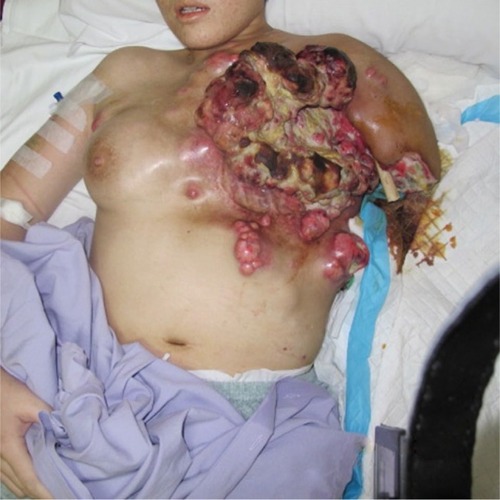
CT of the thorax and abdomen was negative for distant metastases. CT angiogram was not performed, as the patient and family did not agree for her to undergo this invasive investigation. The patient was advised to have another round of palliative chemotherapy and debulking surgery, including a left forequarter amputation. She denied all modalities of intervention. She was given regular subcutaneous morphine and fentanyl patches for analgesia, and her wounds were dressed regularly. The left upper limb tissue gradually became waxy and soft. The flesh over the limb also began to spontaneously separate from the underlying bone, exposing parts of the humerus. The patient eventually requested amputation of the left upper limb, as it was becoming increasingly difficult for her family and nurses to take care of the truncal lesions with the rotting flesh and fragile, non-functioning limb getting in the way. The amputation at the site of the exposed left humerus was performed under intravenous sedation by an experienced orthopedic consultant. The patient succumbed 2 weeks later, slightly more than 6 months from the onset of the disease ( and ). The next of kin has provided written consent for publication of this case report and the accompanying images.
Figure 1 Cancer on the trunk with left humerus exposed and gangrenous arm.
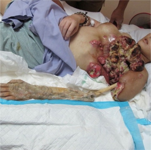
Figure 2 Left humerus post-midshaft amputation.
Discussion
The World Health Organization has recognized metaplastic breast carcinoma (MBC) as a unique pathological entity.Citation1–Citation6 MBC accounts for less than 1% of invasive breast carcinomas, with the mean age at presentation of 50 years. The tumor is usually large, with axillary lymph node metastases in 25% of cases. Fifty percent of cases develop local or distant spread within 5 years, with lung and bone metastases, similar to a sarcoma.Citation7,Citation8 Shahrun et alCitation9 reported six cases that developed tumor recurrence within a year of diagnosis. Immunohistochemistry with broad cytokeratin panel is the best tool to diagnose MBC.Citation10–Citation12 MBC has poor response to neoadjuvant chemotherapy; surgery is the primary management.Citation8 There is no difference in overall survival between patients undergoing mastectomy or breast conserving surgery.Citation13 MBCs frequently express EGFR, and have been shown to demonstrate positive response toward targeted therapy.Citation14,Citation15 Adjuvant radiotherapy has also been reported to improve overall survival.Citation16
Conclusion
Based on our literature review, this is the youngest, and the first case of metaplastic breast carcinoma with extensive angioinvasion leading to upper limb gangrene. In a multi-ethnic country such as Malaysia, traditional medication and alternative therapy is still favored by some over modern medicine, due to deep-rooted cultural beliefs. Such practices, when unregulated, may deny patients of timely access to proper health care, derail them from reality, and dampen the prognosis of salvageable cases. Having said that, much remains unknown regarding this rare subtype of invasive breast cancer. Future direction should be geared toward development of targeted, systemic therapy with improved clinical outcomes.
Author contributions
Norlia Abdullah conceived the idea to report this case, was the primary consultant managing this case, provided the photos, reviewed and edited the write-up. Reynu Rajan wrote the case report and did the literature review. Nik Muhd Aslan Abdullah managed the case and provided the oncological advice. Abdul Yazid Mohd Kassim managed the case and provided the orthopedic advice. All authors contributed toward data analysis, drafting and revising the paper and agree to be accountable for all aspects of the work.
Disclosure
The authors report no conflicts of interest in this work.
References
- TavassoliFADevileePPathology & Genetics Tumours of the Breast and Female Genital OrgansLyonIARC Press2003
- WargotzESNorrisHJMetaplastic carcinomas of the breast: I. Matrix-producing carcinomaHum Pathol19892076286352544506
- WargotzESDeosPHNorrisHJMetaplastic carcinomas of the breast: II. Spindle cell carcinomaHum Pathol19892087327402473024
- WargotzESNorrisHJMetaplastic carcinomas of the breast: III. CarcinosarcomaCancer1989647149014992776108
- WargotzESNorrisHJMetaplastic carcinomas of the breast: IV. Squamous cell carcinoma of ductal originCancer19906522722762153044
- WargotzESNorrisHJMetaplastic carcinomas of the breast: V. Metaplastic carcinoma of the breast with osteoclastic giant cellsHum Pathol19902111114211502227922
- Günhan-BilgenMemişAUstünEEZekiogluOOzdemirNMetaplastic carcinoma of the breast: clinical, mammographic, and sonographic findings with histopathologic correlationAJR Am J Roentgenol200217861421142512034610
- LuiniAAguilarMGattiGMetaplastic carcinoma of the breast, an unusual disease with worse prognosis: the experience of the European Institute of Oncology and review of the literatureBreast Cancer Res Treat2007101334935317009109
- ShahrunNSRohaizakMNaqiyahINurismahMIMetaplastic breast carcinomas: a report of six casesMedicine & Health200942127132
- LuiPCTseGMTanPHFine-needle aspiration cytology of metaplastic carcinoma of the breastJ Clin Pathol200760552953316798932
- AdemCReynoldsCAdlakhaHRochePCNascimentoAGWide spectrum screening keratin as a marker of metaplastic spindle cell carcinoma of the breast: an immunohistochemical study of 24 patientsHistopathology200240655656212047767
- LeiblSGogg-KammererMSommersacherADenkHMoinfarFMetaplastic breast carcinomas: are they of myoepithelial differentiation?: immunohistochemical profile of the sarcomatoid subtype using novel myoepithelial markersAm J Surg Pathol200529334735315725803
- DaveGCosmatosHDoTLodinKVarshneyDMetaplastic carcinoma of the breast: a retrospective reviewInt J Radiat Oncol Biol Phys200664377177516246496
- LeiblSMoinfarFMetaplastic breast carcinomas are negative for Her-2 but frequently express EGFR (Her-1): potential relevance to adjuvant treatment with EGFR tyrosine kinase inhibitors?J Clin Pathol200558770070415976335
- DanceyJEFriedlinBTargeting epidermal growth factor receptor–are we missing the mark?Lancet20033629377626412853203
- TsengWHMartinezSRMetaplastic breast cancer: to radiate or not to radiate?Ann Surg Oncol20111819410320585866