
Abstract
More than half the number of patients with cancer, who are treated with radiotherapy, will have radiodermatitis at some point during their treatment. Radiodermatitis either occurs early on in the treatment period or appears months or up to several years later. Acute radiodermatitis is a burn injury that varies in severity according to both treatment and inherent patient factors. Most acute radiodermatitis reactions resolve after several weeks but some reactions persist and can cause complications. Late-onset radiodermatitis is characterized by telangiectasia that forms on atrophic and fragile skin. These radiodermatitis reactions can have a significant negative impact on concomitant and subsequent therapeutic protocols and most particularly on the patient’s quality of life. Today, treatment of radiodermatitis reactions is in its infancy. Although there is insufficient evidence available to form recommendations that would prevent or reduce radiodermatitis, some advances have been made using low level light therapy (LLLT) or vascular lasers to control the symptoms. Some recent preclinical and clinical research suggests that LLLT has biostimulating properties which allow the tissues to regenerate and heal faster, reduce inflammation, and prevent fibrosis. Also, in late-onset radiodermatitis pulsed dye laser treatment has been shown to be beneficial in clearing radiation-induced telangiectasia. In the absence of evidence-based recommendations, the objective of this paper is to review how to prevent or manage the symptoms of radiodermatitis reactions.
Introduction
International data indicate that 50% of patients diagnosed with cancer will receive some form of radiation therapy. Radiodermatitis is a substantial side effect that arises directly from radiation exposure during cancer treatment, and concerns around 95% of all cancer patients receiving radiation therapy.Citation1,Citation2 It is particularly problematic in cancers of the breast, perineum, and head and neck region, where the skin is part of the target volume.Citation3
Radiodermatitis is referred to as being an acute reaction when it occurs around the time of therapy and either chronic or late onset when it appears 5–10 years after the end of treatment. Symptoms of acute radiodermatitis have been classified into three levels; grade 1 (mild erythema), grade 2 (dry desquamation), and grade 3 (severe moist desquamation).Citation4 In recent years, skin sparing and modern equipment such as intensity-modulated radiation therapy reduced dose intensity on the skin and new equipment has reduced the severity of acute radiodermatitis for many patients.Citation5 Nevertheless, grade 1 skin toxicity still remains a problem for around 90% of patients and grade 2 for 30% of patients.Citation6 While radiodermatitis may resolve over time, it can profoundly affect the patient’s quality of life and may limit the treatment duration and dose delivered.Citation2
Chronic radiodermatitis occurs in a third of all patients and appears up to at least 10 years after radiotherapy treatment.Citation7 Typical symptoms are telangiectasia, pigmentation, cutaneous atrophy (dry papery skin), dermal sclerosis, and keratoses.Citation4 Chronic radiodermatitis is an increasing problem, as today >80% of all women treated for breast cancer are now surviving for ≥10 years.Citation8
Fortunately, there are several solutions available today to prevent and treat radiodermatitis and skin reactions. Dermocosmetic support is recommended to protect and promote tissue repair; light technology, such as biophotomodulation and vascular laser treatments, is becoming established as safe and effective to protect and treat both acute and chronic radiodermatitis.Citation9,Citation10
The objective of this review paper is to provide an overview of the different types of radio and combined chemotherapy-induced radiodermatitis and highlights the evidence for management and treatment.
Radiodermatitis
Radiodermatitis is the result of cutaneous or subcutaneous lesions induced by external beam radiation. The skin is particularly sensitive to radiation damage because it is a continuously renewing organ. In healthy skin tissue, there is a delicate balance between the death and rebirth of each cell type. Acute radiation damage occurs in the dermis with the first radiation dose. A number of basal keratinocytes are destroyed, leaving only the remaining keratinocytes to cornify. Thus, the balance between the normal cell production at the basal layer and cell destruction at the skin surface is disrupted. This process continues with continuing radiation thereby altering the integrity of the epidermis, the skin barrier, and skin healing processes. This leads to structural, histologic, and vasculature changes of the skin and underlying connective tissue.Citation11
With cumulative radiation doses, these acute injuries become apparent. The skin barrier dysfunction manifests as erythema, skin dryness, flaking, folliculitis (skin rash), xerosis, pruritus, and hyperpigmentation. Additionally, the physical barrier and cutaneous immune system are compromised and the skin becomes more sensitive to allergens, ultraviolet radiation, and infection.Citation12
Some cutaneous cell types grow and renew themselves rapidly and in others the life cycle is slower. Radiation affects these two populations differently and the probability of destroying a skin cell increases with the level of radiation. Thus, it is possible to have different types of skin reactions occurring at the same time postradiation depending on which cell types have been affected.Citation13
Supportive dermocosmetic skin care for patients starting radiotherapy
Regular skin care assessment and close collaboration between radiation oncologists and dermatologists to manage skin reactions early and throughout treatment have been repeatedly suggested to improve patient comfort, enhance quality of life, and improve clinical outcome.Citation14–Citation16 However, there is little evidence concerning skin care products to alleviate the severity of skin reactions.Citation17
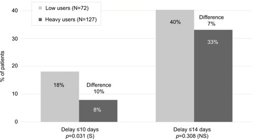
One recent, multinational, real-life study to evaluate a combination of hygiene products was performed in women starting radiotherapy following breast cancer. The objective was to evaluate the tolerance of a specific dermocosmetic regimen on the irradiated area and the effectiveness in delaying or reducing the intensity of acute radiodermatitis. This was measured by erythema, edema, skin dryness, desquamation, physical appreciation, and patient appreciation using the patient benefit index at the start and end of treatment (6±2 weeks).Citation18 Patients were provided with a dermocosmetic kit before the first radiotherapy session. The kit included five products specifically formulated with gentle ingredients that respect skin physiology and tested for use on sensitive skin. In total, 253 women were included in the study, following a tumor excision, partial or full mastectomy. The results revealed two categories of users who were defined by the number (0–5) and frequency of products used (never, sometimes, often, and every day). Low users made up 36% of the study group and 57% were heavy users. This study shows that this skin care regimen was well tolerated on irradiated skin and heavy product users observed less frequent radiodermatitis reactions than low product users (). These results were further supported by a recent conference communication that found that the frequency of severe (grade 3 or 4) radiodermatitis was significantly lower in the intervention group (chi-square =4.61; p=0.03), although the time to onset of skin toxicity was similar among the intervention group compared with a previous cohort (median time to onset, 17 vs 17.5 days),Citation19 thus indicating that patient education can improve adherence to skin care plans and reduce radiation dermatitis. These reports continue to provide support for international recommendations for supportive skin care in radiotherapy.
Figure 1 Time to onset of radiodermatitis from radiotherapy start.
Acute radiodermatitis
Acute radiodermatitis is a burn injury that varies in severity according to both treatment and inherent patient factors.Citation1 Treatment-related factors that influence the degree to which skin tissues are damaged include the total dose, fractionation, type and quality of the beam, and surface area and volume of tissue exposed.Citation11 Also, certain anatomical regions where the target is close to the skin are more susceptible to developing radiodermatitis. These areas include skin folds around the breast and inguinal areas, thin cutaneous regions (clavicle, periauricular area, or axilla), mucosal regions (vulva, anus, and mouth), or altered skin following a previous treatment.
Concomitant radiotherapy with chemotherapy such as epidermal growth factor receptor (EGFR) inhibitors seems to increase the intensity of the EGFR inhibitors-induced rash and delays the appearance by 2–5 weeks after the beginning of treatment.Citation20–Citation22
Typical radiotherapeutic treatment such as a dose intensity of 20 Gy with conventional fractionation (2 Gy/fraction; five fractions/week) results in sustained erythema that usually appears between 10 and 14 days after radiotherapy.Citation23 Although most acute radiodermatitis reactions resolve after 2–3 weeks, some reactions persist and can cause complications such as treatment delays, diminished esthetic appeal, and reduced quality of life.Citation2,Citation24,Citation25
Treatment of acute radiodermatitis
Today, there is insufficient evidence available to form recommendations that would prevent or reduce radiodermatitis. However, several multidisciplinary groups have proposed guidelines that suggest management strategies with the objective to avoid or reduce the severity of radiodermatitis reactions: Association Francophone des Soins Oncologiques de Support,Citation26 Multinational Association of Supportive Care in Cancer,Citation4 European Skin Management in Oncology,Citation16 and consensus guidelines for the management of radiodermatitis and coexisting acne-like rash.Citation21,Citation27 Nevertheless, many centers develop their own protocols.
The general recommendations are presented in . Low level light therapy (LLLT), otherwise known as photobiomodulation (PBM) or “soft laser” (red light or infrared, power <150 mW), is better known in dermatology to treat ulcers.Citation28 However, the biological mechanisms behind the therapeutic effect are currently not well understood. Recent molecular and cellular research suggests that LLLT has biostimulating properties which allow the tissues to regenerate and heal faster.Citation9,Citation10 Animal and clinical studies suggest that LLLT has analgesic properties, reduces inflammation, and prevents fibrosis.Citation29–Citation31
Table 1 General recommendations
Treating radiodermatitis with LLLT is based on previously demonstrated decreased severity and duration of oral mucositis.Citation32–Citation34 However, its preventative and curative use in acute radiodermatitis is currently under evaluation. It was reported that the incidence of radiodermatitis reduced with light emitting diode treatments immediately after radiation therapy for breast cancer. However, a further controlled clinical study was unable to reproduce the results.Citation35 Nevertheless, based on the strong level of evidence available, Bensadoun and Nair proposed a protocol to prevent or treat radiation dermatitis using LLLT. The authors suggest that laser therapy can be started after only a few radiation sessions and before erythema appears or even after erythema has appeared. The treatment was shown to be painless and can be practiced thrice per week, either before or after the radiotherapy session.Citation36 More recently, Censabella et al performed an open, prospective, comparative study and found that skin toxicity improved significantly following multiwave locked system® laser therapy.Citation37
Chronic radiodermatitis
Chronic radiodermatitis, otherwise referred to as late reaction, is characterized by its highly variable, delayed apparition. This late-onset reaction can appear from 6 months up to 20 or 30 years after treatment and can reappear when provoked by the administration of a different treatment such as chemotherapy or antibiotics. The frequency and severity have been shown to be linked with the total dose (+50 Gy to the skin) and favored by short treatment intervals, limited fraction size, large treatment areas, and tumor infiltrate. Other external factors that aggravate chronic radiodermatitis include repetitive trauma, sun exposure, and further irradiation. Chronic radiodermatitis is dominated by telangiectasia that forms on atrophic and fragile skin, associated with areas of hyper or hypopigmentations.Citation38 Less commonly, a more severe form of delayed radionecrosis occurs where the skin sits close to bone or cartilage such as the nose, ears, or scalp.Citation23
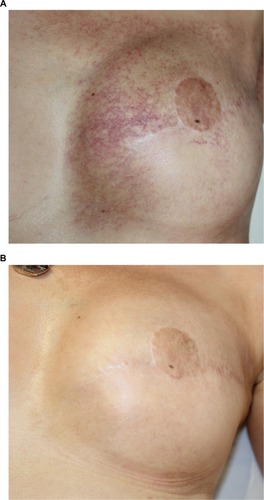
Pulsed dye laser treatment has been shown to be beneficial in clearing radiation-induced telangiectasia. The first prospective study reported by Lanigan et al was performed on eight individuals. This study demonstrated that lesions could be whitened using very short pulse durations (0.45 ms) using a 585 nm pulsed dye laser.Citation38 The study was later supported by a larger comparative study with 13 participants that compared three sessions of either pulsed dye laser or pulsed intense light laser. Patients treated with the pulsed dye laser had a reduction in lesion size of 90%, compared to 50% with the intense pulsed light (IPL). IPL systems are high-intensity light sources, which emit polychromatic light. Unlike laser systems, IPL works with noncoherent light in a broad wavelength spectrum of 515–1200 nm. Both procedures were well tolerated, except for one case of achromia with the IPL.Citation39 Furthermore, over a number of years, Dr JM Mazer performed several open clinical studies on large patient numbers to further refine the number treatment modality ().Citation40 The first study included 110 women, treated with a pulsed dye vascular laser (ScleroPlus laser [Candela] followed by Vbeam laser [Candela]) at 1.5 ms pulse duration, wavelength 595 nm, with a spot size diameter of 7 mm, and fluence of 9–11.5 J/cm2. Between one and five sessions were required to reduce the number of telangiectasias by 80%. A second study was performed with a more recent pulsed dye laser (Vbeam Perfecta laser [Candela]) comparing 1.5 to 6 ms pulse durations, with purpuragenic fluences, for the same spot size diameter and the same wavelength of 595 nm. The number of patients included for post-breast cancer radiotherapy was 176, presenting a total of 234 different lesions, 144 on the presternal area and 90 on the lateral side of the breast. The main criterion of efficacy was the number of needed sessions to obtain 80% reduction in telangiectasias, according to both the patient and the physician. The objective was to investigate if longer pulse durations would be more effective on more dilated vessels, typical of radiodermatitis, according to the rules of selective photothermolysis. However, although the efficacy was satisfactory for both pulse times, the shorter pulse durations were more effective: all patients obtained at least 80% regression in telangiectasia, after two or three sessions for 84% of cases with the 1.5 ms, and in less than five sessions in 95% of cases.Citation41 Pulsed dye laser has been shown to be safe and well tolerated in this population with particularly atrophied skin. Purpura resolved within 10–15 days. No further supportive care is required apart from sunscreen for at least 6 weeks following the procedure. No severe side effects were observed, neither scars nor skin necrosis nor ulceration. Patients reported a significant improvement in quality of life. The physicians noticed an improved skin thickness, probably related to the remodeling effect, known with this laser. The authors suggested that other lasers, particularly fractional ablative lasers or, on this fragile skin, non-ablative lasers, could be effective in reducing atrophy and improving the skin texture.
Figure 2 Example of results obtained after two sessions of vascular laser and two sessions of fractional non-ablative laser.
Conclusion
Today, there is increasing evidence to support various strategies to limit and treat cutaneous reactions to radiotherapy. To prevent acute radiodermatitis, daily dermocosmetic use is useful from the beginning of radiotherapy. There is evidence for the efficacy of PBM to both prevent and cure acute radiodermatitis. In chronic radiodermatitis, treatment with vascular lasers, especially pulsed dye laser, using short pulse durations, has been shown to be effective with an excellent tolerance, inducing a better quality of life for the patients.
Disclosure
SS is an employee of La Roche-Posay Laboratoire Dermatologique, France. The authors report no other conflicts of interest in this work.
Acknowledgments
The authors thank Amanda Whereat of Speak the Speech Consulting for providing assistance to write this article. This article was funded by La Roche-Posay Laboratoire Dermatologique, France.
References
- ChanRJWebsterJChungBMarquartLAhmedMGarantziotisSPrevention and treatment of acute radiation-induced skin reactions: a systematic review and meta-analysis of randomized controlled trialsBMC Cancer2014145324484999
- DengGCassilethBRSkin Injury: Acute Dermatitis and Chronic Skin Changes Supportive Care and Quality of Life5th edPhiladelphia, PALippincott Williams & Wilkins2008
- DeSantisCELinCCMariottoABCancer treatment and survivorship statistics, 2014CA Cancer J Clin201464425227124890451
- WongRKBensadounRJBoers-DoetsCBClinical practice guidelines for the prevention and treatment of acute and late radiation reactions from the MASCC Skin Toxicity Study GroupSupport Care Cancer201321102933294823942595
- PignolJPOlivottoIRakovitchEA multicenter randomized trial of breast intensity-modulated radiation therapy to reduce acute radiation dermatitisJ Clin Oncol200826132085209218285602
- ChanRJLarsenEChanPRe-examining the evidence in radiation dermatitis management literature: an overview and a critical appraisal of systematic reviewsInt J Radiat Oncol Biol Phys2012843e357e36222713836
- WhelanTJPignolJPLevineMNLong-term results of hypofractionated radiation therapy for breast cancerN Engl J Med2010362651352020147717
- American Cancer SocietyCancer Facts & Figures 2017AtlantaAmerican Cancer Society2017 Available from: https://www.cancer.org/Accessed OCtober 12, 2017
- AvciPGuptaASadasivamMLow-level laser (light) therapy (LLLT) in skin: stimulating, healing, restoringSemin Cutan Med Surg2013321415224049929
- HashmiJTHuangYYSharmaSKEffect of pulsing in low-level light therapyLasers Surg Med201042645046620662021
- CoxJDAngKRadiation Oncology: Rationale, Technique, Results9th edPhiladelphia, PAMosby Elsevier2010
- RyanJLIonizing radiation: the good, the bad, and the uglyJ Invest Dermatol20121323 Pt 298599322217743
- SinghMAlaviAWongRAkitaSRadiodermatitis: a review of our current understandingAm J Clin Dermatol201617327729227021652
- McQuestionMEvidence-based skin care management in radiation therapy: clinical updateSemin Oncol Nurs2011272e1e17
- BolderstonALloydNSWongRKHoldenLRobb-BlendermanLSupportive Care Guidelines Group of Cancer Care Ontario Program in Evidence-Based CareThe prevention and management of acute skin reactions related to radiation therapy: a systematic review and practice guidelineSupport Care Cancer200614880281716758176
- BensadounRJHumbertPKrutmannJDaily baseline skin care in the prevention, treatment, and supportive care of skin toxicity in oncology patients: recommendations from a multinational expert panelCancer Manag Res2013540140824353440
- KumarSJuresicEBartonMShafiqJManagement of skin toxicity during radiation therapy: a review of the evidenceJ Med Imaging Radiat Oncol201054326427920598015
- BlomeCAugustinMBehechtnejadJRustenbachSJDimensions of patient needs in dermatology: subscales of the patient benefit indexArch Dermatol Res20113031111720714739
- AndrithsLEducating breast cancer patients reduces radiation dermatitis severityPaper presented at: Oncology Nursing Society (ONS) 42nd Annual Congress2017Denver, CO, USA
- BonnerJAHarariPMGiraltJRadiotherapy plus cetuximab for squamous-cell carcinoma of the head and neckN Engl J Med2006354656757816467544
- BernierJBonnerJVermorkenJBConsensus guidelines for the management of radiation dermatitis and coexisting acne-like rash in patients receiving radiotherapy plus EGFR inhibitors for the treatment of squamous cell carcinoma of the head and neckAnn Oncol200819114214917785763
- TejwaniAWuSJiaYAgulnikMMillenderLLacoutureMEIncreased risk of high-grade dermatologic toxicities with radiation plus epidermal growth factor receptor inhibitor therapyCancer200911561286129919170238
- HymesSRStromEAFifeCRadiation dermatitis: clinical presentation, pathophysiology, and treatment 2006J Am Acad Dermatol2006541284616384753
- JohansenJOvergaardJRoseCCosmetic outcome and breast morbidity in breast-conserving treatment – results from the Danish DBCG-82TM national randomized trial in breast cancerActa Oncol200241436938012234030
- ArenasMSabaterSHernandezVCosmetic outcome of breast conservative treatment for early stage breast cancerClin Transl Oncol20068533433816760008
- AFSOSToxicité cutanée radio-induit. [AFSOS radio-induced cutaneous toxicity]2014 Available from: http://www.afsos.org/fiche-referentiel/toxicite-cutanee-radio-induite/Accessed April 12, 2014 French
- ReguiaiZBachetJBBachmeyerCManagement of cutaneous adverse events induced by anti-EGFR (epidermal growth factor receptor): a French interdisciplinary therapeutic algorithmSupport Care Cancer20122071395140422539049
- MesterALaser biostimulationPhotomed Laser Surg201331623723923741992
- BjordalJMJohnsonMIIversenVAimbireFLopes-MartinsRALow-level laser therapy in acute pain: a systematic review of possible mechanisms of action and clinical effects in randomized placebo-controlled trialsPhotomed Laser Surg20062415816816706694
- LuoLSunZZhangLLiXDongYLiuTCEffects of low-level laser therapy on ROS homeostasis and expression of IGF-1 and TGF-beta1 in skeletal muscle during the repair processLasers Med Sci201328372573422714676
- de OliveiraFASanders-PinheiroHLow-level laser therapy decreases renal interstitial fibrosisPhotomed Laser Surg2013311050750824102169
- ClarksonJEWorthingtonHVFurnessSMcCabeMKhalidTMeyerSInterventions for treating oral mucositis for patients with cancer receiving treatmentCochrane Database Syst Rev20108CD001973
- BjordalJMBensadounRJTunerJFrigoLGjerdeKLopes-MartinsRAA systematic review with meta-analysis of the effect of low-level laser therapy (LLLT) in cancer therapy-induced oral mucositisSupport Care Cancer20111981069107721660670
- LallaRVBowenJBaraschAMASCC/ISOO clinical practice guidelines for the management of mucositis secondary to cancer therapyCancer2014120101453146124615748
- FifeDRayhanDJBehnamSA randomized, controlled, double-blind study of light emitting diode photomodulation for the prevention of radiation dermatitis in patients with breast cancerDermatol Surg201036121921192721070458
- BensadounRJNairRGLow-level laser therapy in the management of mucositis and dermatitis induced by cancer therapyPhotomed Laser Surg2015331048749126447605
- CensabellaSClaesSRobijnsJBulensPMebisJPhotobiomodulation for the management of radiation dermatitis: the DERMIS trial, a pilot study of MLS® laser therapy in breast cancer patientsSupport Care Cancer20162493925393327116013
- LaniganSWJoannidesTPulsed dye laser treatment of telangiectasia after radiotherapy for carcinoma of the breastBr J Dermatol20031481777912534598
- NymannPHedelundLHaedersdalMIntense pulsed light vs. long-pulsed dye laser treatment of telangiectasia after radiotherapy for breast cancer: a randomized split-lesion trial of two different treatmentsBr J Dermatol200916061237124119309367
- MazerJMIndications pour les lasers médicaux en dermatologie. [Indications for medical lasers in dermatology]Presse Med2002315223231 French11878141
- MazerJMCommunication presentationPresented at American Society for Laser Medicine and Surgery, IncApr 05–09, 2017San Diego, CA, USA
- DrenoBBensadounRJHumbertPAlgorithm for dermocosmetic use in the management of cutaneous side-effects associated with targeted therapy in oncologyJ Eur Acad Dermatol Venereol20132791071108023368717
- LacoutureMEMechanisms of cutaneous toxicities to EGFR inhibitorsNat Rev Cancer200661080381216990857