
Abstract
Protein-based therapies hold great promise for treating many diseases. Nevertheless, the challenges of producing therapies with targeted attributes via standardized processes may hinder the development of protein formulations and clinical translation of the advanced therapies. Microfluidics represents a promising technology to develop protein formulations with pre-programmed functional characteristics, including size, morphology, and controlled drug release property. In this review, we discuss some examples of adopting microfluidics for fabricating particle- and fiber/tube-based formulations and highlight the advantages of microfluidics-assisted fabrication.
Introduction
Protein-based therapies have been rapidly developed to treat many diseases, such as cancer and metabolic and autoimmune disorders.Citation1 The delivery of proteins such as growth factor can also have significant impact in the field of regenerative medicine. Globally, the market of protein-based therapy is expanding dramatically, which was valued at around $174.7 billion in 2015 and is expected to reach over $240 billion by 2020.Citation2 Examples of protein products include monoclonal antibodies and fusion protein.Citation3,Citation4
In general, protein drugs show higher binding selectivity and specificity to target molecules with low toxicity compared to small-molecule drugs.Citation5 However, they often suffer from low bioavailability due to degradation, aggregation, adsorption, and denaturation.Citation6 To enhance the performance of protein formulations, functional characteristics can be programmed during the preparation steps. These characteristics include size, morphology, and controlled drug release property.
To effectively deliver drugs to target site, micro-/nanoparticles encapsulating protein drugs have been developed, offering protection against premature degradation as well as serving as drug depot for controlled release of protein.Citation7 Proteins can also be engineered into different configurations, such as fiber and tube, which can be applied in various drug delivery and tissue engineering applications.Citation8,Citation9 Nevertheless, the lack of advanced fabrication approaches that facilitate the development of protein formulations with controllable attributes significantly hinders clinical translation of protein-based formulations.Citation10
The Food and Drug Administration (FDA) has issued the guidelines of current Good Manufacturing Practices (cGMP) for biomanufacturing to ensure products (eg, drugs) are fabricated with the desired characteristics in terms of identity, strength, quality, and purity.Citation11 The use of microfluidics, which refers to the manipulation of fluid flow in tiny scale (micro- or pico-liter), to fabricate biologically relevant materials is a promising approach owing to the multiple advantages it offers.Citation12 Here, we review the rationale and examples of adopting microfluidics for the fabrication of protein formulations with pre-programmed functional characteristics and highlight the role of microfluidics in advancing clinical translation of protein-based therapies.
Introduction of microfluidic technologies
Microfluidics is a technology characterized by the study and manipulation of fluids at the submillimeter scale and has shown great promise in biomedical applications.Citation13 The core of the technology is a device with channels of submillimeter scale and fluid flow in the device is driven by pump or manual injection. Due to the small dimension of the channel, the Reynolds number, which represents the relative importance of inertia to viscous force, is small (typically between 10−6 and 10) and hence laminar flow often results.Citation14 This allows better control over molecules/particles distribution in fluidic system. One distinction between microscale and macroscale fluid phenomena is that the relative effects of various forces are different. Surface tension and capillary forces are dominant at the microscale, whereas the effect of gravity is less important as the dimension becomes smaller. This facilitates operations such as generating monodisperse emulsion droplets and patterning cells on surface. The interfacial flow is especially crucial in the case of droplet generation.Citation14 Emulsion droplets are generated when two or more immiscible streams such as water and oil, in the presence of a surfactant, are injected into a microfluidic channel in various configurations. Some examples of device configuration are T-junction, flow-focusing, and concentric flow.Citation15 These emulsion droplets serve as uniform-sized reactor to compartmentalize the bulk reaction mixture into discrete and controllable units. This can lead to consistent material fabrication. The mode of droplet formation is determined by the ratio of viscous force in the continuous phase to the interfacial tension (ie, capillary number).Citation16 Besides, active techniques such as electrical, magnetic, centrifugal, and mechanical inputs can also be introduced to further manipulate the droplet formation.Citation14 Significant effort has been devoted to study the physical mechanisms of droplet formation, which was reviewed elsewhere.Citation17
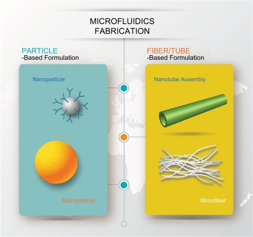
In general, microfluidic system is associated with benefits such as sample volume reduction, device miniaturization, rapid heat and mass transfer, amenability to scaling up and high-throughput screening, and enhanced processing accuracy and efficiency.Citation18–Citation20 The amount of reagent input and device footprint can be reduced in microfluidic operation compared with conventional macroscale bioprocessing.Citation21 This can lead to reduced cost and better controllability. In addition, the physical properties of resulting particle formulation can be easily tuned by adjusting the total flow rate, polymer concentration, aqueous-to-organic flow rate ratio, the use of a co-solvent, and the rate of solvent mixing.Citation22 Protein therapeutics created by microfluidic approaches can be categorized into two main types: particle-based and fiber/tube-based formulations (). We will examine each category and discuss how microfluidics contributes to programing functional characteristics of protein.
Figure 1 Illustration of different forms of protein formulation fabricated using microfluidics system.
Particle-based formulation
Polymeric micro/nanoparticle as protein carrier
Since protein therapeutics are very sensitive to enzymatic degradation, aggregation, adsorption, and denaturation and they usually target specific sites of the body, encapsulating them in carrier can protect them from degradation and delivering them to target site via surface modification (in the case of nanoparticle (NP)). The first US FDA-approved protein delivery therapy was polyethylene glycol(PEG)ylated adenosine deaminase (Adagen®; Leadiant Biosciences, Pomezia, Italy) in 1990. Since then, various classes of carriers for protein delivery have been developed.Citation15 Examples of nanoscale carriers include lipid-based (eg, liposome and exosome) and inorganic (eg, calcium phosphate and mesoporous silica) NPs. The NPs can shield protein from denaturation and prolong circulation in the blood stream.Citation23 Unlike NPs that can be administered systematically, microscale carriers are usually administered locally at the target site or subcutaneously for sustained protein release via diffusion through surface pore or degradation of carrier material.Citation24
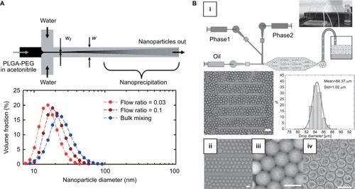
Two common strategies of micro/nanoparticle formation are nanoprecipitation and emulsification. During nanoprecipitation, the reduction of the quality of the solvent in which the ingredient of NPs is present triggers particle formation. The change in solvent quality can be achieved by altering salt concentration, solubility conditions, pH, etc.Citation15 For instance, precursors of NP and protein can be dissolved in an aqueous solvent and added to another solvent with good water miscibility but as a poor solvent for the constituents. NPs will be precipitated out of the mixture. NPs synthesized in typical bulk condition are usually prone to polydispersity and batch-to-batch variation due to longer timescale of solvent exchange (in the order of seconds) than that of precursor nucleation and growth, resulting in uncontrolled NP aggregation. The use of microfluidics can improve the process by expediting solvent exchange via hydrodynamic flow focusing, where the precursor solution is focused into a narrow stream by two streams of anti-solvent flowing in parallel ().Citation25 Rapid diffusion can occur through the interface. Vortex and turbulence created in high speed flow within hydrodynamic focusing can enable even shorter mixing time and increase the production rate.Citation26,Citation27 A recent report described the use of a Staggered Herringbone Mixer for rapid mixing of two streams and channeled to a Tangential Flow Filtration device for subsequent purification, which led to high-throughput liposome production and effective removal of non-entrapped protein (ovalbumin).Citation28
Figure 2 (A) Top: Schematic of formation of nanoparticle via hydrodynamic flow focusing. Bottom: The size distribution of nanoparticles fabricated by different approaches (flow ratio =0.03 and 0.1 refer to ratio of flow rates of PLGA-PEG/water). Reprinted (adapted) with permission from Karnik R, Gu F, Basto P, et al. Microfluidic platform for controlled synthesis of polymeric nanoparticles. Nano Lett. 2008;8(9):2906–2912.Citation25 Copyright (2008) American Chemical Society. (B) (i) Schematic of formation of microfluidic emulsion droplets. Middle: Image of droplets produced and their size distribution. Scale bar is 200 µm. (ii) Images of droplets stored in the device, (iii) collected off-chip, and (iv) forming microparticles via self-assembly after 24 hours. Scale bar is 100 µm. Copyright © 2014. Dove Medical Press. Adapted from Bai S, Debnath S, Gibson K, et al. Biocatalytic self-assembly of nanostructured peptide microparticles using droplet microfluidics. Small. 2014;10(2):285–293.Citation39
Another common way of fabricating micro/nanoparticles is by emulsification followed by solvent depletion, such as evaporation and solvent diffusion.Citation15 Tiny emulsion droplets containing protein and particle precursor are first generated by vigorous agitation or mixing two immiscible phases. A subsequent mixing step can be applied to generate double emulsion for preparing core–shell structures. The droplet content will condensate and form micro/nanoparticles after solvent depletion. The final size of particles is directly related to the size of the emulsion droplet, which can be very heterogeneous in bulk mixing, thus resulting in uncontrollable drug release profile. The benefit of forming monodisperse emulsion droplet in a microfluidic platform can contribute significantly to the formation of monodisperse particles with higher encapsulation efficiency and more controlled drug release rate.Citation29,Citation30 Also, the precise introduction of reagents into the droplets could be realized using pulse width modulation as demonstrated in one report.Citation31 The cell-free synthesis of green fluorescent proteins inside double emulsions was monitored and the influence of DNA concentration on protein produced could be evaluated. In another report, microparticles composed of poly(vinyl alcohol) loaded with bovine serum albumin were fabricated with a size range of 23–47 µm and an encapsulation efficiency of 84%.Citation32 Another report described the fabrication of monodisperse silk fibroin microparticles as a drug carrier using microfluidic platform due to its good biocompatibility, lack of toxicity, and immunogenicity.Citation33
Furthermore, growing evidence has shown that particle shape may influence the particle targeting, drug loading, and release properties.Citation34 For example, more hydrophobic drug molecules could be loaded into worm-like micelles than spherical ones due to the presence of larger core volume.Citation35 Based on the emulsification technique, the shape of the particle could be modulated by changing the geometry of the microchannel.Citation36 Rod- and disc-like particles could be generated via controlled deformation of emulsion droplet inside the channel. Besides, a method that integrates microscope projection photolithography and microfluidics allowed continuous production of particles of distinct shapes.Citation37 The monomer stream was infused through a microchannel above a mask containing desired features. Particles were polymerized when exposed to UV light through the mask and carried along by the stream for subsequent reaction to take place. The versatility of this approach enabled a wider variety of shapes to be produced for various applications.
Self-assembly within emulsion droplet
Self-assembly represents another strategy of formation of protein formulation, which involves the assembly of molecular building blocks into peptide- or protein-based particle.Citation38 In bulk, the process is usually conducted with minimal control over the macroscopic size and shape of the resulting particle. By confining building blocks into discrete, monodisperse emulsion droplets, we can exert better control over the particle properties. In one example, thermolysin and amino acid derivatives Fmoc-S (fluorenylmethyloxycarbony-serine) and F-OMe (phenylalanine methyl ester) were encapsulated in emulsion droplets for catalytic condensation to form microparticles (). The resulting spherulite microparticles were similar to those formed in bulk reaction but were more homogenous in size.Citation39 In another example, emulsion droplet was used to study the coacervation of intrinsically disordered proteins by employing elastin-like polypeptides as model, where different coacervate architectures (ie, multilayer, mixed coacervate, and puncta) could be achieved by introducing a mixture of polypeptides with varying sequence, molecular weight, and concentration into the droplets.Citation40 Overall, the use of emulsion droplet permits the study and control of individual microenvironment for self-assembly to occur.
Fiber/tube-based formulation
Flow-based self-assembly
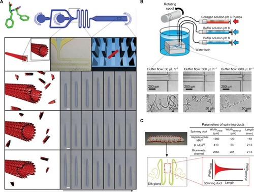
In addition to droplet-based system, self-assembly of peptide/ protein can also be controlled in a flow-based microfluidic device. For instance, the diphenylalanine building block would self-assemble into nanotubes in a spontaneous manner. By compartmentalizing nanotubes in microfluidic platform filled with micron-scale pillars and exposing them to building blocks of various concentrations, it is possible to manipulate the assembly process by controlling the net monomer influx at given time points and visualize the process (elongation and shortening) in real-time ().Citation41 The control over the dimensional alteration will facilitate the design of supramolecular polymers such as protein formulation for various applications.
Figure 3 (A) Top: Diphenylalanine monomer and the design of the microfluidic device for nanotube assembly. Bottom: Illustration and imaging of nanotube formation at supercritical, critical, and subcritical monomer concentrations in flow. Scale bar is 5 µm. Adapted from Arnon ZA, Vitalis A, Levin A, et al. Dynamic microfluidic control of supramolecular peptide self-assembly. Nat Commun. 2016;7:13190. Creative Commons license and disclaimer available from: http://creativecommons.org/licenses/by/4.0/ legalcode.Citation41 (B) Schematic of microfluidic chip for collagen microfiber formation and images of microfibers fabricated with varying buffer flow rates. Reprinted (adapted) with permission from Haynl C, Hofmann E, Pawar K, Förster S, Scheibel T. Microfluidics-produced collagen fibers show extraordinary mechanical properties. Nano Lett. 2016;16(9):5917–5922.Citation43 Copyright (2008) American Chemical Society. (C) Schematic of the microfluidic spinning process of recombinant spider dragline silk and geometries of the spinning ducts of spider and silkworm. A biomimetic, microfluidic channel was designed to mimic the contracting geometry of spinning ducts to form compact and ordered silk protein microfiber. Adapted from Peng Q, Zhang Y, Lu L, et al. Recombinant spider silk from aqueous solutions via a bio-inspired microfluidic chip. Sci Rep. 2016;6:36473. Creative Commons license and disclaimer available from: http://creativecommons.org/licenses/by/4.0/legalcode.Citation45
Microfiber production
Protein fibers are one of the major components of extracellular matrix, and some of them such as silk fiber are also found in nature. Micro- and nano-scale fibers hold great promise as implantable scaffold. They possess attributes such as short diffusion distance and high surface-to-volume ratio for mass exchange, thus they are suitable as cell culture platform and localized drug delivery vehicle. There are various ways to manufacture fibers, but not many of them are applicable to produce protein formulation.Citation42 For example, melt spinning requires heating polymer above its melting point which can cause protein denaturation. Wet spinning, which is achieved by extruding material into a coagulation bath for solvent to diffuse out, often involves the use of harsh chemical that may not be optimal for protein application as well as for producing fiber with relatively a large diameter (20–200 µm).Citation43 Electrospinning is another technique that is commonly used to fabricate fibers, but the use of high voltage to draw the charged solution may preclude the loading of sensitive biological materials.
By using microfluidic flow focusing, microscale fibers can be generated with predictable and controllable properties. Through adjusting the flow rates of protein and surrounding buffer, fibers of various diameters can be fabricated. One study reported that collagen fibers with diameters as small as 3 µm could be produced ().Citation43 Meanwhile, it has been observed that the silk gland in the silkworm and spider function similarly as a microfluidic system.Citation44 The concentrated spinning dope would flow along a contracted gland and is exposed to solution with ionic gradients which regulate crystallization and protein aggregation. A biomimetic microfluidic device that mimics the specific geometry found in silkworm or spider or by means of flow focusing can reproduce the silk spinning process ().Citation45 In addition, the shearing and elongational forces supplied by microfluidics also direct the alignment of β-sheets during fiber formation, resulting in fibers of tunable diameter, stiffness, and toughness.Citation46,Citation47 Compared with traditional wet spinning, microfluidic-based approach is more economical and nontoxic, which will facilitate the rapid and inexpensive fabrication of fiber of protein formulation.
Future perspectives
The demand for protein-based therapies is constantly rising, but fabricating protein formulations with controllable and pre-programmed functional characteristics is still a challenge. To comply with cGMP, technologies that enable controlled fabrication of protein formulation with pre-programmed functional characteristics must be developed. These characteristics include size, morphology, and controlled drug release property. Microfluidics has emerged as a potential platform to advance biofabrication of particle- and fiber/tube-based protein formulation. Here, we have reviewed several technologies that can not only improve the quality of protein formulations but also facilitate the design of supramolecular polymers for various protein applications. However, most of the previous reports were proof-of-concept studies and the translation of microfluidic-based formulation to preclinical and clinical application is still a challenge. One limitation is that microfluidic platform generally has a low production rate. To address this, efforts are devoted to scaling up the microfluidics technologies, such as by utilizing a microfluidic module with 128 cross-junctions that can produce droplets at a rate of 5.3 mL/min.Citation48 Inspiration can also be drawn from the computer chip industry where massively parallel and miniaturized processing systems are developed to analyze big data. We envision that increased adoption of microfluidic technologies should pave the way for more effective protein-based therapy in the near future.
Acknowledgments
This work was supported by the Chinese University of Hong Kong start-up fund.
Disclosure
The authors report no conflicts of interest in this work.
References
- CraikDJFairlieDPLirasSPriceDThe future of peptide-based drugsChem Biol Drug Des201381113614723253135
- DewanSSGlobal Markets and Manufacturing Technologies for Protein Drugs (BIO021E)2016 Available from: https://www.bccresearch.com/report/download/report/bio021eAccessedNovember 19, 2018
- WeidleUHSchneiderBGeorgesGBrinkmannUGenetically engineered fusion proteins for treatment of cancerCancer Genom Proteom201296357372
- ChamesPvan RegenmortelMWeissEBatyDTherapeutic antibodies: successes, limitations and hopes for the futureBr J Pharmacol2009157222023319459844
- TiwariGTiwariRSriwastawaBDrug delivery systems: an updated reviewInt J Pharm Invest201221211
- FixJAOral controlled release technology for peptides: status and future prospectsPharm Res19961312176017648987068
- LangerRDrug delivery and targetingNature19983926679 Suppl5109579855
- XuWYangYDrug sorption onto and release from soy protein fibersJ Mater Sci Mater Med200920122477248619609653
- BrunoBJMillerGDLimCSBasics and recent advances in peptide and protein drug deliveryTher Deliv20134111443146724228993
- KobsaSSaltzmanWMBioengineering approaches to controlled protein deliveryPediatr Res200863551351918427296
- Federal Register DivisionThe Code of Federal Regulations of the United States of America1999 Available from: https://www.gpo.gov/fdsys/pkg/FR-1999-01-08/content-detail.htmlAccessed November 19, 2018
- ChanHFMaSLeongKWCan microfluidics address biomanufacturing challenges in drug/gene/cell therapies?Regen Biomater201632879827047674
- WhitesidesGMThe origins and the future of microfluidicsNature2006442710136837316871203
- ZhuPWangLPassive and active droplet generation with microfluidics: a reviewLab Chip2016171347527841886
- ZhangYChanHFLeongKWAdvanced materials and processing for drug delivery: the past and the futureAdv Drug Deliv Rev201365110412023088863
- ZhuPKongTLeiLTianXKangZWangLDroplet breakup in expansion-contraction microchannelsSci Rep201662152726899018
- TehSYLinRHungLHLeeAPDroplet microfluidicsLab Chip20088219822018231657
- OttinoJMWigginsSIntroduction: mixing in microfluidicsPhilos Trans A Math Phys Eng Sci2004362181892393515306477
- ChanHFZhangYHoYPChiuYLJungYLeongKWRapid formation of multicellular spheroids in double-emulsion droplets with controllable microenvironmentSci Rep20133346224322507
- ChanHFZhangYLeongKWEfficient one-step production of micro-encapsulated hepatocyte spheroids with enhanced functionsSmall201612202720273027038291
- SongHChenDLIsmagilovRFReactions in droplets in microfluidic channelsAngew Chem Int Ed Engl200645447336735617086584
- KhanIUSerraCAAntonNVandammeTFProduction of nanoparticle drug delivery systems with microfluidics toolsExpert Opin Drug Deliv201512454756225345543
- de JongWHBormPJDrug delivery and nanoparticles: applications and hazardsInt J Nanomedicine20083213314918686775
- KohaneDSMicroparticles and nanoparticles for drug deliveryBiotechnol Bioeng200796220320917191251
- KarnikRGuFBastoPMicrofluidic platform for controlled synthesis of polymeric nanoparticlesNano Lett2008892906291218656990
- ValenciaPMBastoPAZhangLSingle-step assembly of homogenous lipid-polymeric and lipid-quantum dot nanoparticles enabled by microfluidic rapid mixingACS Nano2010431671167920166699
- KimYLee ChungBMaMMass production and size control of lipid-polymer hybrid nanoparticles through controlled microvorticesNano Lett20121273587359122716029
- DimovNKastnerEHussainMPerrieYSzitaNFormation and purification of tailored liposomes for drug delivery using a module-based micro continuous-flow systemSci Rep2017711204528935923
- DamiatiSKompellaUBDamiatiSAKodziusRMicrofluidic devices for drug delivery systems and drug screeningGenes201892103
- XuQHashimotoMDangTTPreparation of monodisperse biodegradable polymer microparticles using a microfluidic flow-focusing device for controlled drug deliverySmall20095131575158119296563
- ChangJCSwankZKeiserOMaerklSJAmstadEMicrofluidic device for real-time formulation of reagents and their subsequent encapsulation into double emulsionsSci Rep201881814329802303
- PessiJSantosHAMiroshnykIJoukoYliruusiWeitzDAMirzaSMicrofluidics-assisted engineering of polymeric microcapsules with high encapsulation efficiency for protein drug deliveryInt J Pharm20144721-2828724928131
- MitropoulosANPerottoGKimSMarelliBKaplanDLOmenettoFGSynthesis of silk fibroin micro- and submicron spheres using a co-flow capillary deviceAdv Mater20142671105111024339048
- ChenJClayNKongHNon-spherical particles for targeted drug deliveryChem Eng Sci2015125202425838583
- GengYDischerDEHydrolytic degradation of poly(ethylene oxide)-block-polycaprolactone worm micellesJ Am Chem Soc200512737127801278116159254
- DendukuriDTsoiKHattonTADoylePSControlled synthesis of nonspherical microparticles using microfluidicsLangmuir: the ACS Journal of Surfaces and Colloids20052162113211615751995
- DendukuriDPregibonDCCollinsJHattonTADoylePSContinuous-flow lithography for high-throughput microparticle synthesisNat Mater20065536536916604080
- HabibiNKamalyNMemicAShafieeHSelf-assembled peptide-based nanostructures: smart nanomaterials toward targeted drug deliveryNano Today2016111416027103939
- BaiSDebnathSGibsonKBiocatalytic self-assembly of nano-structured peptide microparticles using droplet microfluidicsSmall201410228529323913836
- SimonJRCarrollNJRubinsteinMChilkotiALópezGPProgramming molecular self-assembly of intrinsically disordered proteins containing sequences of low complexityNat Chem20179650951528537592
- ArnonZAVitalisALevinADynamic microfluidic control of supramolecular peptide self-assemblyNat Commun201671319027779182
- TamayolAAkbariMAnnabiNPaulAKhademhosseiniAJunckerDFiber-based tissue engineering: progress, challenges, and opportunitiesBiotechnol Adv201331566968723195284
- HaynlCHofmannEPawarKFörsterSScheibelTMicrofluidics-produced collagen fibers show extraordinary mechanical propertiesNano Lett20161695917592227513098
- KonwarhRGuptaPMandalBBSilk-microfluidics for advanced biotechnological applications: a progressive reviewBiotechnol Adv201634584585827165254
- PengQZhangYLuLRecombinant spider silk from aqueous solutions via a bio-inspired microfluidic chipSci Rep201663647327819339
- KinahanMEFilippidiEKösterSTunable silk: using microfluidics to fabricate silk fibers with controllable propertiesBiomacromolecules20111251504151121438624
- LuoJZhangLPengQTough silk fibers prepared in air using a biomimetic microfluidic chipInt J Biol Macromol20146631932424613677
- NisisakoTToriiTMicrofluidic large-scale integration on a chip for mass production of monodisperse droplets and particlesLab Chip20088228729318231668