
Abstract
The novel coronavirus disease 2019 (COVID-19) pandemic is severely challenging the healthcare systems and economies of the world, which urgently demand vaccine and therapy development to combat severe acute respiratory syndrome coronavirus 2 (SARS-CoV-2). Hence, advancing our understanding of the comprehensive entry mechanisms of SARS-CoV-2, especially the host factors that facilitate viral infection, is crucial for the discovery of effective vaccines and antiviral drugs. SARS-CoV-2 has previously been documented to reach cells by binding with ACE2 and CD147 receptors in host cells that interact with the spike (S) protein of SARS-CoV-2. A novel entry factor, called neuropilin 1(NRP1), has recently been discovered as a co-receptor facilitating the entry of SARS-CoV-2. NRP1 is a single-pass transmembrane glycoprotein widely distributed throughout the tissues of the body and acts as a multifunctional co-receptor to bind with different ligand proteins and play diverse physiological roles as well as pathological and therapeutic roles in different clinical conditions/diseases, including COVID-19. The current review, therefore, briefly provides the overview of SARS-CoV-2 entry mechanisms, the structure of NRP1, and their roles in health and various diseases, as well as extensively discusses the current understanding of the potential implication of NRP1 in SARS-CoV-2 entry and COVID-19 treatment.
Introduction
Neuropilin 1 (NRP1) is one of two homologous neuropilins (NRP) expressed in all vertebrates that has important physiological and pathological roles.Citation1 It was identified first in 1987 by Takagi and his coworkers as a neuronal receptor in developing Chick nervous system.Citation2 NRP1 can exist in two isoforms, namely, secretory and transmembrane isoforms.Citation3,Citation4 The former, also known as truncated or soluble NRP1, circulates freely in the body fluid, whereas the latter isoform, transmembrane NRP1, is a highly conserved single-pass transmembrane protein that interacts with different ligands and has multifaceted functions, including mediating of varieties of physiological and pathological processes; as a result, it is commonly called NRP1.Citation1,Citation3,Citation4
Since its emergence in December 2019, the 2019 coronavirus disease (COVID-19) pandemic, caused by novel severe acute respiratory syndrome coronavirus 2 (SARS-CoV-2), has been severely challenging the global healthcare systems, economies, and social life.Citation5 COVID-19 is primarily a disease of the respiratory tract typically transmitted from person to person through respiratory droplets and direct contact with SARS-CoV-2 infected individuals or inanimate objects. It is highly infective and transmissive compared to SARS-CoV, as it usually spreads rapidly via active pharyngeal viral shedding.Citation6–Citation8
The global threat of COVID-19 urgently demands extensive efforts to develop vaccines and antiviral therapies that curb the global spread and impact of COVID-19. Thus, growing our extensive knowledge regarding SARS-CoV-2 entry mechanisms is central for designing COVID-19 therapies and vaccines. The entry of SARS-CoV-2 to the host cells requires binding with the host receptors via the viral protein called spike (S) protein.Citation9 Besides ACE2 and CD147, recently, a novel receptor called NRP1 was identified for SARS-CoV-2 entry to the host cell.Citation10,Citation11 Hence, this minireview presents the role of NRP1 in SARS-COV-2 entry and as a possible therapeutic target.
Overview of SARS-CoV-2 Entry Mechanism
It is the virus–host cell interactions that determine the cellular entry of SARS-CoV-2 and its dissemination across the tissues.Citation12 To infect humans, like SARS-CoV, the S protein of SARS-CoV-2 must first bind to surface receptors of human cells lining the respiratory or intestinal tracts. Once attached, the virus invades the cell then replicates multiple copies of itself. The copies of viruses are then released and lead to the SARS-CoV-2 transmission.Citation12,Citation13
The process of viral attachment to and invasion of human cells occurred using different cellular receptors. Several studies on SARS-CoV-2 entry have so far been carried out mainly on ACE2 and have confirmed that, like SARS-CoV, the S protein of SARS-CoV-2 uses ACE2 as a host surface receptor to enable the virus to enter and infect the cell.Citation10,Citation14–Citation19 In addition to ACE2, CD147 protein has been identified as a co-receptor in host cells to enhance the ability of SARS-CoV-2 to enter human cells, and cause COVID-19 disease.Citation11,Citation14,Citation16,Citation17,Citation20,Citation21
In spite of these entry mechanisms, it was not clearly understood why the tissue tropism of SARS-CoV-2 varies from what is expected from virus–host cell interaction via ACE2 receptor, and why SARS-CoV-2 readily infects tissues other than the respiratory system, such as the brain, heart, and other tissues with no or low ACE2 expression. These raised the possibility that other host factors are required to facilitate virus–host cell interactions in cells with low ACE2 protein level.Citation6,Citation22 Thus, some studies were recently conducted to reveal these unclear mechanisms, and intriguingly, a major breakthrough study by Daly et al as well as a genetic based study by Cantuti-Castelvetri et al and Li and Buck identified a novel receptor called NRP1 that potentially answer the puzzling question what makes SARS-CoV-2 highly infectious and capable of rapidly spreading in human cells.Citation6,Citation22,Citation23 They concluded that NRP1 may serve as an alternative or independent doorway for SARS-CoV-2 entry to and invasion of the human cells.
The Structure and Ligands of Neuropilin 1
NRP1, formerly known as A5 protein, is a 120–140 kDa non-tyrosine kinase multifunctional transmembrane heptameric protein with an ATWLPPR sequence consisting of 923 amino acids.Citation1 It is encoded by the NRP1 gene located on the 10p11.22 human chromosome locus and shares 44% amino acid sequence identity with its homology, NRP2.Citation3,Citation4,Citation24
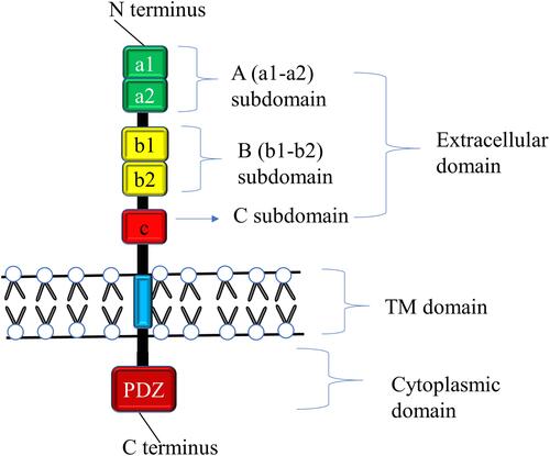
NRP1 comprises a large N-terminus extracellular domain, a relatively very small plasma membrane spanning transmembrane domain, and a short cytoplasmic tail in the inner side of the cell membrane. The extracellular domain in turn comprised of three different subdomains, namely A, B, and C subdomains ().Citation1,Citation3 The A or a1-a2 subdomains, also called semaphorin (SEMA) binding subdomains, are located on amino end and contains two complement binding motifs (CUB), namely the complement binding components (C1r and C1s), Uegf (urchin embryonic growth factor), and bone morphogenetic protein 1 (BMP1), whereas the B subdomain involving b1-b2 subdomains are found in the middle of the extracellular domain and are characteristics of clotting factor V and VIII, and discoidin proteins.Citation25–Citation27 This domain is also known as the vascular endothelial growth factors (VEGF) binding subdomain as it serves as a binding site for VEGF.Citation1 The third domain, called C domain, has similarity with MAM (Meprin, A5/NRP1, protein tyrosine phosphatase μ and К) and contributes for homo- or hetero-dimerization of other receptors with the transmembrane domain, thereby affecting distinct downstream signaling cascades.Citation3 The transmembrane domain is a single-pass protein consisting of a conserved GXXXG repeat crossing the cell membrane.Citation27–Citation29 A short domain of NRP1 that contains 43–44 amino acids but lacks tyrosine kinase activity is the cytosolic tail. This domain possesses a PDZ-binding motif that can interact with various proteins such RGS-GAIP-interacting protein (GIPC) and synectin that are essential for via VEGF receptor 2 (VEGFR2) signaling, arterial morphogenesis as well as in maintaining the structural integrity of transmembrane proteins.Citation1,Citation3,Citation27
Figure 1 Schematic diagram of NRP1 Structure. NRP1 contains a large N-terminus extracellular domain comprising A (a1-a2), B (b1-b2), and C subdomains, a very small single-pass plasma membrane spanning TM domain, and a short cytoplasmic domain in the inner side of the cell membrane possessing PDZ-binding motif that can interact with various proteins. The a1/a2/b1 segment binds with SEMA3s, VEGFs, and other proteins, and the C domain involved in receptor dimerization with the TM domain. The b1 of NRP1 binds with S protein of SARS-CoV-2 and facilitate infection.
Although it lacks a direct cellular signaling role, NRP1 is most widely known for its crucial role as a multifunctional co-receptor by forming a complex with other membrane receptors to form holoreceptors.Citation1 Numerous previous studies have shown that NRP1 is a membrane protein that serves as a surface receptor that can bind with wide varieties of protein families, including heparin-binding members of the VEGF family, class 3 members of the SEMA family (SEMA3s) such as SEMA3A, 3C, and 3F, transforming growth factor-β1 (TGF-β1).Citation28,Citation30,Citation31 The SEMA3s are well known to bind to the a1/a2/b1 portion, whereas the VEGFs bind to b1/b2 segments of NRP1. In addition, NRP1 has recently been demonstrated to act as a receptor for extracellular microRNAs, fibroblast growth factor 2 (FGF-2), galectin-1, hepatocyte growth factor (HGF), plexin, β1 integrin, epidermal growth factor (EGF), platelet-derived growth factor (PDGF), hedgehog (Hh), and other growth factors, albeit their binding sites are not well characterized.Citation32,Citation33
Furthermore, a small portion of S protein is currently recognized to be complementary with the b1 domain of human NRP1 protein and allows the binding of SARS-CoV-2 to host cells and hence facilitate infection.Citation6,Citation22
The Role of Neuropilin 1 in Health and Disease
Many studies have pointed out that NRP1 is broadly distributed in the tissues of the human body, with a predominant expression in blood endothelial cells, vascular smooth muscle cells, mesenchymal stem cells, retinal vasculature, neurons, and epithelial cells lining the respiratory and gastrointestinal tracts.Citation1,Citation3,Citation6,Citation22,Citation24 NRP1 has been established to play a myriad of physiological, pathological, and therapeutic roles. As indicated by the plethora of evidence, NRP1 has versatile functions in regulating a wide array of biological processes, such as axon guidance within the central and peripheral nervous systems, angiogenesis, vascular permeability, and cell survival, proliferation, differentiation, migration, and invasion.Citation2,Citation3,Citation30,Citation32
In recent years, NRP1 has also been shown to be expressed by various immune cells, such as macrophages, including alveolar, adipose tissue, bronchial, and vascular macrophages, dendritic cells, T-cells, particularly CD8 cells, regulatory T-cells, B-cells, and mast cells where it controls a multitude of functions, including development, migration, and recruitment, communication between different immune cells, as well as immune system regulation under normal physiological condition.Citation3,Citation4 NRP1 has also been detected in bone cells like osteoclasts and osteoblasts where it has an important role in regulating bone remodeling, for instance osteo-protection through its binding with SEMA3A.Citation3,Citation34,Citation35
Besides, many other studies have improved the understanding of the roles of NRP1 in different pathological conditions, including cancer, immunological disorders, and bone diseases though the molecular mechanisms behind these functions are still to be elucidated.Citation3,Citation33,Citation34 NRP1 is generally overexpressed in various clinical disorders, including malignancies, where it upregulates the oncogenic activities of malignant cells by enhancing cell survival and proliferation, and angiogenesis, as well as by contributing therapeutic resistance.Citation1,Citation3,Citation33
Moreover, NRP1 has been investigated to have a potential role as a therapeutic target in various pathological disorders, for instance in cancer it serves as an antiangiogenic target as well as in cancer and autoimmune diseases it acts as a site for immunotherapies but it needs selective targeting of NRP1 under a particular clinical setting.Citation3,Citation33
Intriguingly, recent encroaching studies have unveiled an additional role of NRP1 in COVID-19 infection, which was found to be a cofactor and facilitator of SARS-CoV-2 entry and could pave the way to a new possible target of intervention for COVID-19.Citation6,Citation22 More recently, an NRP1 receptor was reported in one study as a potential new target for pain inhibitors to treat chronic pain.Citation36 This discovery is based on the grounds of the pain-relieving activity of SARS-CoV-2, which is suggested to mediate the interactions of NRP1 with S-protein by preventing the normal binding of a protein called VEGF-A to NRP1 and blocking pain signals to give pain relief. However, before developing analgesics, more research is required on how NRP1 contributes to pain signaling.
Neuropilin 1 and COVID-19
Neuropilin 1 Mediated SARS-CoV-2 Entry Mechanism
According to recent studies, NRP1 is identified as a novel co-receptor as well as a potentiating factor for SARS-CoV-2 entry process by enhancing the interaction of the virus with ACE2 ().Citation6,Citation22,Citation23,Citation37 Daly et al suggested that it may act as an alternative doorway for SARS-CoV-2 to enter and infect human cells.Citation22 Consistently, the study by Cantuti-Castelvetri et al using tissues from human autopsies revealed that NRP1 significantly potentiates SARS-CoV-2 infectivity.Citation6 Furthermore, this study demonstrated that the NRP1 is expressed strongly in respiratory and olfactory epithelia, while the ACE2 is absent or expressed at low level and hence it may provide an independent gateway for viral entry and invasion of the host cells. Thus, the NRP1 may mediate SARS‑CoV‑2 entry into the brain via the olfactory bulb.Citation6 In agreement with this, a study by Davies et al showed NRP1 is expression in the CNS, involving olfactory‑related regions such as the olfactory tubercles and para-olfactory gyri, suggesting the potential role of NRP1 as an additional mediator of SARS‑CoV‑2 infection implicated in the neurologic manifestations of COVID-19.Citation37 Moreover, one review article has also suggested that the brainstem has a relatively high expression of ACE2 receptor, and possibly NRP1, that SARS-CoV-2 exploits for cell infection. Thus, the respiratory, cardiovascular, gastrointestinal, and neurological functions of the brainstem may be compromised indefinitely as a result of brainstem damage manifested with neurological symptoms even in mild cases of COVID-19 and may results in long-lasting consequences.Citation38 Their abundant expression on epithelia exposed to the external environment as well as their multifaceted functions are the possible reasons that make NRP1 an ideal entry factor for SARS-CoV-2 as well as a critical contributing factor for multisystem involvement of SARS-CoV-2 infection.Citation4,Citation6,Citation22
Table 1 A Summary Table on the Role of NRP1 in SARS-CoV-2 Infection
SARS-CoV-2 uses a small piece of S protein located on the outer surface of the virus to attach to a complementary region of NRP1 receptors on human cells and hence to penetrate the host cells ().Citation6 The cell entry phase of SARS-CoV-2 depends on priming of S protein by host cell proteases.Citation39,Citation40 At the S1/S2 boundary, there is a multi-basic sequence motif called RRAR containing arginine (R) and alanine (A) amino acids, with a sequence of Arg-Arg-Ala-Arg. The RRAR amino acid sequence is a unique feature of SARS-CoV-2 that provides a cleavage site for a host proprotein convertase (furin) and possibly form additional cell surface receptor binding sites and thus enhances pathogenicity by priming the fusion activity.Citation39–Citation41 This is confirmed by another study demonstrating that SARSCoV-2 virus with a natural deletion of the S1/S2 furin cleavage site is associated to attenuated pathogenicity in hamster models.Citation42
Figure 2 The potential NRP1 mediated SARS-CoV-2 entry mechanism into human cells. The trimeric S protein of SARS-CoV-2 binds to host ACE2 via RBM of RBD. Furin mediated cleavage of S protein at S1/S2 site exposes the CendR motif of S1 and enables binding to the b1 subdomain of NRP1. Further processing of S protein by TMPRSS2 on the cell surface (early entry pathway) and CTSL in endolysosome (late entry pathway) exposes the FP and triggers membrane fusion, and the viral RNA get into the host cytoplasm. The genomic RNA undergoes replication and translation to form new SARS-CoV-2 virions after assembly in ERGIC and the new viruses finally released into the outside of the cell.
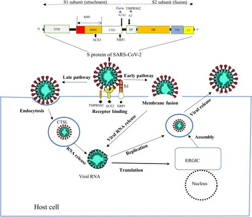
The furin protease cuts the full-length S protein into S1 and S2 functional polypeptides and forms a multi-basic RRAR sequence on the carboxyl-terminal of S1 polypeptide. Studies based on x-ray crystallography and biochemical approaches have shown that the S1 C-end rule (CendR) motif is known to directly interact to b1 domain of NRP1 by electrostatic attraction and activate the cell surface receptors.Citation4,Citation6,Citation22 However, even though the CendR peptide of S1 domain is a major recognition site for NRP1, it is not yet known which other segments of NRP1 and the S protein may interact. In addition, a comparable binding between the carboxy-terminal sequence of S1 subunit and NRP1 homolog, NRP2, was also demonstrated.Citation22
A more recent study by Zhenlu and Matthias also indicated that NRP1 facilitate SARS-CoV-2 infection by stimulating the separation of S1 and S2 subunits.Citation23 This study modeled the structures of NRP1 a2/b1/b2 binding to S protein of SARS-CoV-2 and showed NRP1 on binding with S protein trimer bound to an ACE2 dimer. The S1 binds more strongly to the host membrane in the presence of NRP1 and destabilizes the S1/S2 interface and hence increases the likelihood of the S2 subunit to be pulled out rather than S1 being stretched. Thus, NRP1 attachment may stimulate the easier dissociation of S2 from the S1 subunit that triggers membrane fusion and, thus, increase virus infectivity.Citation23
Following the binding of ACE2 and NRP1 with S1 of SARS-CoV-2, further processing of S protein by transmembrane serine protease 2 (TMPRSS2) on the cell surface (early entry pathway) and cathepsin L(CTSL) in endolysosome (late entry pathway) occur. This exposes the fusion peptide (FP) that triggers membrane fusion, and the viral RNA get into the host cytoplasm. Then the genomic RNA undergoes replication and translation to form new virions after assembly in ERGIC, and the new viruses then are released into the outside of the cell.Citation43,Citation44 Previous studies showed that NRPs are known to mediate the internalization of CendR ligands through an endocytosis resembling micropinocytosis but it is unclear whether NRP1 allows attachment and receptor-mediated endocytosis in SARS-CoV-2 infected patients.Citation6,Citation22,Citation45,Citation46 Thus, further studies need to be done to clearly explain the role of NRP1 in the entry and infection mechanisms of SARS-CoV-2.
Neuropilin 1 Expression and Correlation with COVID-19
A significant proportion of COVID-19 carriers remain asymptomatic or presentwith mild symptoms such as respiratory symptoms, fever, cough, shortness of breath, and myalgia, malaise, arthralgia, chest pain, nasal congestion, runny nose, headache, sore throat, and diarrhea. In some patients, however, COVID-19 becomes severe and results in serious complications such as severe pneumonia, acute respiratory distress syndrome, pulmonary edema, coagulopathy, systemic inflammation, sepsis, septic shock or multiple organ failure, and even death.Citation5
A number of studies have reported that the SARS-CoV-2 infected tissues of the body as well as the severity of COVID-19 is correlated with the expression or activity of ACE2 and CD147 in that tissues.Citation47 However, the tissue expressing such proteins and the COVID-19 targeted tissues differs and so COVID-19 may involve and complicate tissues/organs with low ACE2 protein expression. Besides its role in the viral entry, recent studies also proposed that NRP1 may be the potential factor for the involvement or complicating organs other than the respiratory tracts.Citation6,Citation22 Daly et al observed a significantly enhanced SARS-CoV-2 infection in cells expressing NRP1 wt-GFP compared to GFP control, whereas it was not increased in cells expressing the T316R mutant.Citation22 The overexpression of both NRP1 and NRP2 in SARS-CoV-2 infected lung tissue compared to adjacent bronchoalveolar lavage fluid (BALF) cells in serious patients with COVID-19 was further noted in this report. But the study added that the NRP1 isoform is more likely to function as a co-factor for SARS-CoV-2 that displays limited viral infectivity when expressed alone, though markedly increases viral infectivity when co-expressed with ACE2.
Unlike ACE2, NRP1 is abundant in the respiratory and olfactory epithelium, which is expressed in almost every type of cell in the nasal passages and is even expressed in olfactory neurons. Interestingly, this may explain the infectivity of SARS-CoV-2 in these epithelia as well as the potential route of spread that provides a direct path to SARS-CoV-2 to reach the cells through the olfactory bulb and to the infiltration of the virus into the CNS, disrupting olfaction and causing anosmia associated with COVID-19.Citation4,Citation6 However, unlike in other respiratory infections, the anosmia in the context of COVID-19 is unusually presented with no nasal inflammation and discharge, albeit the reason remains unanswered.Citation4
The morbidity and mortality rate of COVID-19 is highest among elderly and those patients with comorbidities including obesity, diabetes, cardiovascular diseases, and polycystic ovarian syndrome (PCOS).Citation48,Citation49 These high morbidity and mortality levels of COVID-19 in comorbid conditions are suggested to be associated with NRP1 expression level.
In a study of the cryopreserved human diabetic kidney single RNA sequencing dataset, NRP1 was significantly up-regulated unlike ACE2, proposing the possible explanation for the increased risk of COVID-19 in diabetic patients.Citation6 This however contradicts with the previous several lines of evidence that demonstrated suppressed NRP1 expression in cultured differentiated podocytes, podocytes from diabetic db/db mice as well as in diabetic patients diagnosed with diabetic nephropathy.Citation50,Citation51 Treatment with Epoetin-β or continuous erythropoietin receptor activator (CERA) of diabetic db/db mice correlated with overexpression of NRP1 in podocytes of treated animals compared with the untreated, indicating the role of NRP1 in diabetic disease, particularly in the development of diabetic nephropathy.Citation51,Citation52 Hence, further studies should be done regarding the increased risk of COVID-19 in diabetic patients and correlation with NRP1 expression level.
Vascular pathologies, such as arterial injury, coagulopathy, and sepsis have also been suggested to be associated with severe COVID-19, possibly due to NRP1 mediated SARS-CoV-2 vascular dysfunction and coagulopathy. The binding and outcompeting of typical angiogenic ligands at the b1 domain of NRP1 with S protein of SARS-CoV-2 may promote such abnormalities during severe COVID-19.Citation4,Citation53
More recently, however, a contrasting result was documented in PCOS women by Moin et al and indicated a lowered level of soluble NRP1 and increased level of RAS-related proteins in PCOS women, suggesting that lower plasma soluble NRP1 levels may indicate increased risk of COVID-19 disease.Citation49 Though the NRP1 isoform in this study was soluble NRP1, to reconcile these contrasting findings and to arrive at a conclusion, further extensive studies need to conductand demonstrate whether the up-regulation and participation of NRP-1 in COVID-19 could transform into long-term complication.
Potential Implications of Neuropilin 1 in COVID-19 Treatment
To date, multiple clinical trials have been performed to improve the vaccine and drugs of SARS-CoV-2 infection by targeting viral entry mechanisms. Thus, human recombinant soluble ACE2 (hrsACE2) and ACE2 activator targeting ACE2 have been demonstrated to have a potential role in the therapeutic strategy of COVID-19, though they have associated untoward effects.Citation8 Besides, the discovery of CD147 receptor provides a key target for the development of specific antiviral drugs against SARS-CoV-2.Citation54 Currently, a clinical trial aimed at blocking CD147 protein by monoclonal antibodies to inhibit the binding of S protein of SARS-CoV-2 and subsequent infection is underway in China.
Furthermore, targeting the interaction between the novel entry receptor, NRP1, and SARS-CoV-2 may provide a new route that may have important implications in COVID-19 therapies. A recent study finding by researchers at the University of Bristol highlighted that inhibiting the interaction between S1 protein and NRP1 through a RNA interference or selective inhibitors was observed to reduce the entry and infectivity of SARSCoV-2 in cell culture.Citation22 A small molecule EG00229 selectively blocked the direct binding between b1 and the CendR peptide of S1 in addition to VEGF-A binding. This suggests that blocking the SARS-CoV-2 interaction with NRP1 may serve as a valuable therapeutic intervention in the treatment of COVID-19.
The therapeutic approach of COVID-19 has to be considered in combination with the immunosuppressive role of NRP1 as it has multitude functions in the immune system and may give an interesting potential target for immunotherapies such as monoclonal antibodies for possible use against COVID-19, similar to autoimmune diseases and cancers.Citation3,Citation6 Thus, targeting NRP1 may also potentially provide a new avenue for future antiviral treatment to avert the current COVID-19 pandemic. Perhaps this may speed up vaccine research that may have important implication for the development of vaccines against viral S protein in order to prevent SARS-CoV-2 infection. However, this needs further study, whether blocking NRP1 could be effective for the treatment and prevention of SARS-CoV-2 infection. Moreover, targeting NRP1 needs to be closely monitored as it has widespread tissue distribution, multisystem involvement as well as its potential dose-dependent effects.
Concluding Remarks
NRP1 is a non-tyrosine kinase single-pass transmembrane glycoprotein that is highly expressed throughout the tissues of all vertebrates and functions as a cell surface receptor to interact with different ligands and to mediate varieties of physiological and pathological processes. It has myriads of physiological, pathological and therapeutic roles in humans. There have been several previous studies showing the biological roles of NRP1 in axonal guidance, immune regulation and bone hemostasis. Besides, NRP1 plays an important role in various pathological conditions including malignancies, autoimmune and bone diseases, as well as it serves as a therapeutic target in treatment of disorders.
Currently, NRP1 has been observed to play an important pathological role and potentially therapeutic role in COVID-19. In addition to ACE and CD147, NRP1 has recently been reported to be an essential host factor that drives SARS-CoV-2 infectivity using a four amino acid sequence, RRAR, attached to the carboxy-terminal of S1 subunit of S protein, called the CendR motif, to bind with NRP1 and enable viral entry into the host cells. NRP1 is now identified to serve as a novel potentiating factor that provides an alternative and independent gateway for SARS-CoV-2 entry into the human cells, especially in those cells that paradoxically express low levels of ACE2.
The discovery of NRP-1 as an entry or potentiating factor of SARS-CoV-2 infection may pave the way for potential NRP1 targeted antiviral therapies. Nevertheless, while it is reasonable to suggest NRP1 as a therapeutic target, it is too early to think about antiviral drug candidates at this stage of basic research, and hence more intensive research is required to demonstrate the precise role of NRP1 in the SARS-CoV-2 infection and transmission.
Abbreviations
ACE2, angiotensin converting enzyme 2; BALF, bronchoalveolar lavage fluid; BMP1, bone morphogenetic protein 1; CendR, C-end rule; COVID-19, coronavirus disease 2019; CUB, complement C1r/C1s, Uegf, BMP1; EGF, epidermal growth factor; FGF-2, fibroblast growth factor 2; HGF, hepatocyte growth factor (HGF); GIPC, RGS-GAIP-interacting protein; Hh-hedgehog; MAM, meprin, A5/NRP1, protein tyrosine phosphatase μ and К; NRP, neuropilin; NRP1, neuropilin 1; NRP2, neuropilin 2; PCOS, polycystic ovarian syndrome; PDGF, platelet-derived growth factor; RRAR, arginine-arginine-alanine-arginine; S protein, spike protein; SARS-CoV, severe acute respiratory syndrome coronavirus; SARS-CoV-2, severe acute respiratory syndrome coronavirus 2; SEMA, semaphorin; TGF-β1, transforming growth factor-β1; Uegf, urchin embryonic growth factor; VEGF, vascular endothelial growth factor; VEGFR2, vascular endothelial growth factor receptor 2.
Author Contributions
All authors made a significant contribution to the work reported, whether that is in the conception, study design, execution, acquisition of data, analysis and interpretation, or in all these areas; took part in drafting, revising or critically reviewing the article; gave final approval of the version to be published; have agreed on the journal to which the article has been submitted; and agreed to be accountable for all aspects of the work.
Disclosure
The authors report no conflict of interests for this work.
Additional information
Funding
References
- Elpek GÖ. Neuropilins and liver. World J Gastroenterol. 2015;21(23):7065. doi:10.3748/wjg.v21.i23.7065
- Takagi S, Kasuya Y, Shimizu M, et al. Expression of a cell adhesion molecule, neuropilin, in the developing chick nervous system. Dev Biol. 1995;170(1):207–222. doi:10.1006/dbio.1995.1208
- Roy S, Bag AK, Singh RK, Talmadge JE, Batra SK, Datta K. Multifaceted role of neuropilins in the immune system: potential targets for immunotherapy. Front Immunol. 2017;8:1228. doi:10.3389/fimmu.2017.01228
- Mayi BS, Leibowitz JA, Woods AT, Ammon KA, Liu AE, Raja A. The role of Neuropilin-1 in COVID-19. PLoS Pathog. 2021;17(1):e1009153. doi:10.1371/journal.ppat.1009153
- Abebe EC, Dejenie TA, Shiferaw MY, Malik T. The newly emerged COVID-19 disease: a systemic review. Virol J. 2020;17(1):1–8. doi:10.1186/s12985-020-01363-5
- Cantuti-Castelvetri L, Ojha R, Pedro LD, et al. Neuropilin-1 facilitates SARS-CoV-2 cell entry and infectivity. Science. 2020;370(6518):856–860. doi:10.1126/science.abd2985
- Shi J, Wen Z, Zhong G, et al. Susceptibility of ferrets, cats, dogs, and other domesticated animals to SARS–coronavirus 2. Science. 2020;368(6494):1016–1020. doi:10.1126/science.abb7015
- Li Y, Zhou W, Yang L, You R. Physiological and pathological regulation of ACE2, the SARS-CoV-2 receptor. Pharmacol Res. 2020;157:104833. doi:10.1016/j.phrs.2020.104833
- Lu R, Zhao X, Li J, et al. Genomic characterisation and epidemiology of 2019 novel coronavirus: implications for virus origins and receptor binding. Lancet. 2020;395(10224):565–574. doi:10.1016/S0140-6736(20)30251-8
- Dimitrov DS. The secret life of ACE2 as a receptor for the SARS virus. Cell. 2003;115(6):652–653. doi:10.1016/S0092-8674(03)00976-0
- Wang K, Chen W, Zhang Z, et al. CD147-spike protein is a novel route for SARS-CoV-2 infection to host cells. Sign Transduct Target Ther. 2020;5(1):1–10. doi:10.1038/s41392-020-00426-x
- Synowiec A, Szczepański A, Barreto-Duran E, Lie LK, Pyrc K. Severe acute respiratory syndrome coronavirus 2 (SARS-CoV-2): a systemic infection. Clin Microbiol Rev. 2021;34(2).
- Belouzard S, Millet JK, Licitra BN, Whittaker GR. Mechanisms of coronavirus cell entry mediated by the viral spike protein. Viruses. 2012;4(6):1011–1033. doi:10.3390/v4061011
- Essahib W, Verheyen G, Tournaye H, Van de Velde H. SARS-CoV-2 host receptors ACE2 and CD147 (BSG) are present on human oocytes and blastocysts. J Assist Reprod Genet. 2020;37(11):2657–2660. doi:10.1007/s10815-020-01952-x
- Lukassen S, Chua RL, Trefzer T, et al. SARS‐CoV‐2 receptor ACE 2 and TMPRSS 2 are primarily expressed in bronchial transient secretory cells. EMBO J. 2020;39(10):e105114. doi:10.15252/embj.2020105114
- Radzikowska U, Ding M, Tan G, et al. Distribution of ACE2, CD147, CD26, and other SARS‐CoV‐2 associated molecules in tissues and immune cells in health and in asthma, COPD, obesity, hypertension, and COVID‐19 risk factors. Allergy. 2020;75(11):2829–2845. doi:10.1111/all.14429
- Radzikowska U, Ding M, Tan G, et al. Distribution of ACE2, CD147, cyclophilins, CD26 and other SARS-CoV-2 associated molecules in human tissues and immune cells in health and disease. bioRxiv. 2020.
- Samavati L, Uhal BD. ACE2, much more than just a receptor for SARS-COV-2. Front Cell Infect Microbiol. 2020;10:317. doi:10.3389/fcimb.2020.00317
- Zhao Y, Zhao Z, Wang Y, Zhou Y, Ma Y, Zuo W. Single-cell RNA expression profiling of ACE2, the receptor of SARS-CoV-2. Am J Respir Crit Care Med. 2020;202(5):756–759. doi:10.1164/rccm.202001-0179LE
- Helal MA, Shouman S, Abdelwaly A, et al. Molecular basis of the potential interaction of SARS-CoV-2 spike protein to CD147 in COVID-19 associated-lymphopenia. J Biomol Struct Dyn. 2020;1–11. doi:10.1080/07391102.2020.1822208
- Shilts J, Crozier TW, Greenwood EJ, Lehner PJ, Wright GJ. No evidence for basigin/CD147 as a direct SARS-CoV-2 spike binding receptor. Sci Rep. 2021;11(1):1–10. doi:10.1038/s41598-020-80464-1
- Daly JL, Simonetti B, Klein K, et al. Neuropilin-1 is a host factor for SARS-CoV-2 infection. Science. 2020;370(6518):861–865. doi:10.1126/science.abd3072
- Li Z, Buck M. Neuropilin-1 assists SARS-CoV-2 infection by stimulating the separation of spike protein domains S1 and S2. bioRxiv. 2021. doi:10.1101/2021.01.06.425627
- Soker S, Takashima S, Miao HQ, Neufeld G, Klagsbrun M. Neuropilin-1 is expressed by endothelial and tumor cells as an isoform-specific receptor for vascular endothelial growth factor. Cell. 1998;92(6):735–745. doi:10.1016/S0092-8674(00)81402-6
- Gaboriaud C, Gregory-Pauron L, Teillet F, Thielens NM, Bally I, Arlaud GJ. Structure and properties of the Ca2+-binding CUB domain, a widespread ligand-recognition unit involved in major biological functions. Biochem J. 2011;439(2):185–193. doi:10.1042/BJ20111027
- Fuentes-Prior P, Fujikawa K, Pratt KP. New insights into binding interfaces of coagulation factors V and VIII and their homologues-lessons from high resolution crystal structures. Curr Protein Pept Sci. 2002;3(3):313–339. doi:10.2174/1389203023380639
- Kiedzierska A, Smietana K, Czepczynska H, Otlewski J. Structural similarities and functional diversity of eukaryotic discoidin-like domains. Biochimica et Biophysica Acta. 2007;1774(9):1069–1078. doi:10.1016/j.bbapap.2007.07.007
- Gu C, Limberg BJ, Whitaker GB, et al. Characterization of neuropilin-1 structural features that confer binding to semaphorin 3A and vascular endothelial growth factor 165. J Biol Chem. 2002;277(20):18069–18076. doi:10.1074/jbc.M201681200
- Roth L, Nasarre C, Dirrig-Grosch S, et al. Transmembrane domain interactions control biological functions of neuropilin-1. Mol Biol Cell. 2008;19(2):646–654. doi:10.1091/mbc.e07-06-0625
- Kofler N, Simons M. The expanding role of neuropilin: regulation of transforming growth factor-β and platelet-derived growth factor signaling in the vasculature. Curr Opin Hematol. 2016;23(3):260–267. doi:10.1097/MOH.0000000000000233
- Mercurio AM. VEGF/neuropilin signaling in cancer stem cells. Int J Mol Sci. 2019;20(3):490. doi:10.3390/ijms20030490
- Guo H-F, Vander Kooi CW. Neuropilin functions as an essential cell surface receptor. J Biol Chem. 2015;290(49):29120–29126. doi:10.1074/jbc.R115.687327
- Nasarre C, Roth M, Jacob L, et al. Peptide-based interference of the transmembrane domain of neuropilin-1 inhibits glioma growth in vivo. Oncogene. 2010;29(16):2381–2392. doi:10.1038/onc.2010.9
- Hayashi M, Nakashima T, Taniguchi M, Kodama T, Kumanogoh A, Takayanagi H. Osteoprotection by semaphorin 3A. Nature. 2012;485(7396):69–74. doi:10.1038/nature11000
- Gomez C, Burt‐Pichat B, Mallein‐Gerin F, et al. Expression of Semaphorin‐3A and its receptors in endochondral ossification: potential role in skeletal development and innervation. Dev Dynam. 2005;234(2):393–403. doi:10.1002/dvdy.20512
- Moutal A, Martin LF, Boinon L, et al. SARS-CoV-2 Spike protein co-opts VEGF-A/Neuropilin-1 receptor signaling to induce analgesia. Pain. 2021;162(1):243. doi:10.1097/j.pain.0000000000002097
- Davies J, Randeva HS, Chatha K, et al. Neuropilin‑1 as a new potential SARS‑CoV‑2 infection mediator implicated in the neurologic features and central nervous system involvement of COVID‑19. Mol Med Rep. 2020;22(5):4221–4226. doi:10.3892/mmr.2020.11510
- Yong SJ. Persistent brainstem dysfunction in long-COVID: a hypothesis. ACS Chem Neurosci. 2021;12(4):573–580. doi:10.1021/acschemneuro.0c00793
- Hoffmann M, Kleine-Weber H, Pöhlmann S. A multibasic cleavage site in the spike protein of SARS-CoV-2 is essential for infection of human lung cells. Mol Cell. 2020;78(4):779–84. e5. doi:10.1016/j.molcel.2020.04.022
- Millet JK, Whittaker GR. Physiological and molecular triggers for SARS-CoV membrane fusion and entry into host cells. Virology. 2018;517:3–8. doi:10.1016/j.virol.2017.12.015
- Wrapp D, Wang N, Corbett KS, et al. Cryo-EM structure of the 2019-nCoV spike in the prefusion conformation. Science. 2020;367(6483):1260–1263. doi:10.1126/science.abb2507
- Lau S-Y, Wang P, Mok BW-Y, et al. Attenuated SARS-CoV-2 variants with deletions at the S1/S2 junction. Emerg Microb Infect. 2020;9(1):837–842. doi:10.1080/22221751.2020.1756700
- Kielian M. Enhancing host cell infection by SARS-CoV-2. Science. 2020;370(6518):765–766. doi:10.1126/science.abf0732
- Murgolo N, Therien AG, Howell B, et al. SARS-CoV-2 tropism, entry, replication, and propagation: considerations for drug discovery and development. PLoS Pathog. 2021;17(2):e1009225. doi:10.1371/journal.ppat.1009225
- Simons M, Gordon E, Claesson-Welsh L. Mechanisms and regulation of endothelial VEGF receptor signalling. Nat Rev Mol Cell Biol. 2016;17(10):611. doi:10.1038/nrm.2016.87
- Teesalu T, Sugahara KN, Kotamraju VR, Ruoslahti E. C-end rule peptides mediate neuropilin-1-dependent cell, vascular, and tissue penetration. Proc Natl Acad Sci. 2009;106(38):16157–16162. doi:10.1073/pnas.0908201106
- Qiao J, Li W, Bao J, et al. The expression of SARS-CoV-2 receptor ACE2 and CD147, and protease TMPRSS2 in human and mouse brain cells and mouse brain tissues. Biochem Biophys Res Commun. 2020;533(4):867–871. doi:10.1016/j.bbrc.2020.09.042
- Ssentongo P, Ssentongo AE, Heilbrunn ES, Ba DM, Chinchilli VM. Association of cardiovascular disease and 10 other pre-existing comorbidities with COVID-19 mortality: a systematic review and meta-analysis. PLoS One. 2020;15(8):e0238215. doi:10.1371/journal.pone.0238215
- Moin ASM, Sathyapalan T, Atkin SL, Butler AE. The relationship of soluble neuropilin-1 to severe COVID-19 risk factors in polycystic ovary syndrome. Metabolism Open. 2021;9:100079. doi:10.1016/j.metop.2021.100079
- Bondeva T, Rüster C, Franke S, et al. Advanced glycation end-products suppress neuropilin-1 expression in podocytes. Kidney Int. 2009;75(6):605–616. doi:10.1038/ki.2008.603
- Loeffler I, Rüster C, Franke S, Liebisch M, Wolf G. Erythropoietin ameliorates podocyte injury in advanced diabetic nephropathy in the db/db mouse. Am J Physiol Renal Physiol. 2013;305(6):F911–F8. doi:10.1152/ajprenal.00643.2012
- Bondeva T, Wolf G. Role of neuropilin-1 in diabetic nephropathy. J Clin Med. 2015;4(6):1293–1311. doi:10.3390/jcm4061293
- Machhi J, Herskovitz J, Senan AM, et al. The natural history, pathobiology, and clinical manifestations of SARS-CoV-2 infections. J Neuroimmune Pharmacol. 2020;1–28.
- Ulrich H, Pillat MM. CD147 as a target for COVID-19 treatment: suggested effects of azithromycin and stem cell engagement. Stem Cell Rev Rep. 2020;16(3):434–440. doi:10.1007/s12015-020-09976-7