
Abstract
Background
The effects of pollution on health have received increasing attention in recent years. Extrinsic skin aging occurs via multiple processes, and pollution is now recognized as a major component, causing increased pigmentation and wrinkles via oxidative mechanisms. We tested the antipollution efficacy of a cosmetic facial cream (FC) by assessing its effects on carbon particle adhesion to skin and on oxidative and inflammatory pathways in the skin.
Methods
In an in vivo study, FC was applied once to the forearms of healthy subjects. Carbon E153 powder was applied, and the skin was washed under standardized conditions. Images were taken using a dermoscope to determine the area of particle adherence. Each participant served as their own control, with the contralateral forearm being untreated with the FC but otherwise following the same protocol. In a 5-day ex vivo study, skin explants were treated with the FC daily and exposed to vaporized pollutants on day 2 and day 4 via a closed system. Explants were sampled at baseline and day 5 and culture media on day 5. The parameters evaluated were cellular viability on microscopy, Nrf2 immunostaining, malondialdehyde (MDA) levels in culture, melanin levels, and gene expression profile (TYR, IL6, and CYP1A1).
Results
In the in vivo adhesion study, after standardized washing, carbon particle deposition on skin treated with the FC was significantly lower than that on untreated skin. In the ex vivo study, samples treated with the FC had reduced Nrf2 staining and MDA levels vs polluted controls. Melanin did not change significantly. The FC modulated pollution-induced increases in CYP1A1, IL-6, and TYR.
Conclusion
This FC reduces particle adhesion to skin after a single application and protects against pollution-induced oxidative and inflammatory pathways in the skin.
Introduction
Recent years have witnessed a greater awareness of the damage human activity has wreaked on the environment. More than ever, attention, in both the lay and scientific community, is focused on the role of environmental pollution in various disease conditions.Citation1–Citation3 The effect of air pollution on skin, while seeming a logical corollary to skin being the barrier between environment and the body, has only recently begun to be supported by scientific evidence. Pollution has been shown to have multiple deleterious effects on the skin including hyperpigmentation, wrinkles, sallow appearance of the skin, and exacerbation of acne,Citation4 skin cancer,Citation5 atopic dermatitis,Citation6 and psoriasis.Citation7
The etiology of skin aging is multifactorial with numerous intrinsic and extrinsic factors colluding to produce visible changes in phenotype. Pollution has been recognized as one of the driving factors that accelerate skin aging, besides solar radiation, as proposed by the exposome theory.Citation8 Protection of skin from air pollution is considered an essential part of a multipronged approach to slowing skin aging.
Air pollution varies by region and level of rural or industrial activity. However, some of the universal components include polycyclic aromatic hydrocarbons (PAHs), fine and very fine particulate matter (PM2.5 and PM10), heavy metals, and gases such as sulfur dioxide, nitrogen oxide, and carbon monoxide.Citation3,Citation7 The interaction of one or more of these components with skin results in the generation of reactive oxygen species (ROS) that react with cellular proteins, lipids, and DNA, triggering downstream events.Citation9 An inflammatory cycle is triggered, with the release of cytokines, causing inflammatory cell migration and infiltration and further generation of free radicals.Citation7 Changes in membrane lipids and proteins damage cell structure and function and, in particular, the skin’s barrier function.Citation10 Epidermal vitamins C and E – nonenzymatic antioxidants – are depleted,Citation11 as are multiple enzymatic antioxidants.Citation9
Protecting skin against such damage may be approached in two ways: by the formation of a physical barrier on the skin surface to limit pollution contact with the skin and by counteracting the subsequent downstream events – namely oxidative stress. A novel facial cream (FC) was designed to act in both ways against pollution: it contained a film-forming exopolysaccharide (EPS) capable of adsorbing heavy metals and preventing particulate matter adhesionCitation12,Citation13 and carnosine and niacinamide to minimize the oxidative and inflammatory downstream effects of pollution. We report the results from an in vivo study to determine the effects of topical application of the cream on inhibiting carbon powder adhesion to healthy skin as well as an ex vivo study to determine the effects of the cream against pollution-induced markers of oxidation and inflammation in skin explants.
Methods
Particle adhesion study
Subjects
Twenty healthy adult female subjects were enrolled (inclusion criteria: 18–70 years). Individuals who were pregnant, breastfeeding, tattooed on the inner forearms, or recently exposed to sun or psoralen and ultraviolet A (PUVA) therapy were excluded. All subjects provided written informed con sent. The study was performed in accordance with the 1964 Declaration of Helsinki and its successive updates and the recommendations on Good Clinical Practice ICH E6 (R2) 2016. The study was conducted in France, and as the nature of the study did not fall within the classification of medical research, as described in decree number 2017-884,Citation14 but rather of a cosmetic product, it did not require ethics committee approval. Prior to the current study, the product had previously undergone safety testing as recommended in the 2008 Colipa guidelines for the evaluation of the efficacy of cosmetic productsCitation15 and had been determined to be safe for use as a cosmetic product.
Protocol
A 5 cm × 5 cm square was marked on both inner forearms, with a smaller 4 cm × 4 cm square inside this, using a dermographic pencil. Participants were instructed to avoid touching, wetting, or rubbing their forearms throughout the study.
Subjects were randomized to have the FC applied to either the left or right forearm; the arm with no FC applied served as a control. A technician applied 50 mL (2 µL/cm2) of the cream to the larger square on the treated arm and left it to dry for 15 minutes. Then, the powder of carbon particles (carbon E153 powder [Colorey SAS, Lozanne, France], granulometry diameter [D]: D10: 2 µm, D50: 5 µm, D90: 55 µm) was applied to the inner square on both forearms: 4 mg on each arm, using a brush and fingertip. The test areas were rinsed with 30 mL of water; after that, 75 µL of neutral liquid soap was applied and massaged for 30 seconds, and then rinsed under running water for 5 seconds. The skin was dried by placing a tissue on the skin without rubbing.
Imaging
Images were taken with a C-Cube Dermoscope Clinical Research Edition camera (Pixience SAS, Toulouse, France) before FC application (T0), immediately after the application of carbon E153 powder (T1), and immediately after washing (T2). This camera uses a probe with an embedded high-resolution camera (10-megapixel complementary metal oxide semiconductor sensor) coupled with a patented lighting technique to avoid glare. Images were analyzed with Kalliste imaging software (Microvision, Evry Cedex, France). For each subject, a region of interest was delimited on the photograph. To establish the surface area covered with carbon E153 powder, the photograph was processed using adaptive thresholding to convert the image to a binary image of clean vs carbon-covered skin.
Statistical analyses
Results are shown as mean ± SD. For each area – treated and control – the efficacy coefficient was calculated according to the following formula: ([T2–T0]–[T1–T0])/(T1–T0). The Shapiro–Wilk test was used to assess the normality of distribution (1%), which confirmed normal distribution; therefore, the Student’s t-test was used. A P-value of <0.05 was considered statistically significant.
Ex vivo study
Experimental groups
Human skin explants were obtained from subjects undergoing esthetic surgery in accordance with French Law number 2011-814 of July 7, 2011, on bioethics.Citation16 Skin was cut into 14 mm disks and maintained in survival medium at 37°C in a humid 5% CO2 atmosphere. There were four experimental groups, each containing seven explants, which are given in .
Table 1 Experimental groups in the antipollution activity: ex vivo study
Pollution exposure and FC application
The FC was applied on days 0, 1, 2, 3, 4, and 5 with a small spatula (2 µL/cm2). Explants were exposed to pollution on days 2 and 4, in a closed system that exposes the stratum corneum to vaporized solution introduced using a nebulizer. The explants were placed in 900 µL/well of Hanks’ balanced salt solution (HBSS), exposed to the pollutants for 1.5 hours, and then returned to the incubator.
The pollutant mixture used was composed of diesel particles (PM2.5 and PM10) 0.01%, benzene 1 µL/mL, benzo[a] pyrene 1 mg/mL, and heavy metals (National Institute of Standards and Technology standard reference material, 1650b). The pollutant concentration in the chamber was chosen to be reflective of the levels found in a polluted city.
Sampling
At day 0 and day 5, three explants from each group were cut into three parts each: one was fixed in buffered formalin, one was frozen at –80°C, and one was used to extract RNA for subsequent genomic study. A further three explants from each group were frozen for subsequent melanin quantification. Culture medium was harvested on day 5 and stored at –20°C for malondialdehyde (MDA) quantification.
Histological processing
Skin samples were fixed in buffered formalin, dehydrated, and impregnated in paraffin using a Leica PEARL dehydration automat and Leica EG1160 embedding station (Leica Microsystems, Wetzlar, Germany). Five-micrometer sections were cut with a Leica RM2125 Minot microtome. Frozen samples were cut into 7 µm-thick sections using a Leica CM 3050 cryostat. Sections were then mounted on Superfrost plus silanized glass slides, and a Leica DMLB or Olympus BX43 microscope was used for viewing. Pictures were digitized with a numeric DP72 Olympus camera with CellD storing software.
Skin viability
Paraffinized sections were stained with Masson’s trichrome (Goldner variant) to assess general morphology and skin viability.
Nuclear factor (erythroid-derived 2)-like 2 (Nrf2) immunostaining
Nrf2 immunostaining was conducted on deparaffinized sections using a monoclonal anti-Nrf2 (phospho S40) antibody (reference ab76026, clone EP1809Y; Abcam, Cambridge, UK) diluted at 1:200 in 0.3% BSA in PBS and 0.05% Tween 20. The antibody is specific for the serine-40 phosphorylated nuclear form of Nrf2. Sections were incubated for 1 hour at room temperature with an avidin–biotin amplifier system and revealed using a peroxidase kit (ref. SK-4600; Vector laboratories, Burlingame, CA, USA). Sections were evaluated by a histopathologist, and the level of staining was categorized as follows: very weak, weak, moderate, quite clear, clear, very clear, or strong.
MDA
MDA in culture medium on day 5 was measured for the four explants per batch with an enhanced thiobarbituric acid reagent (TBAR) method. MDA was assayed in HBSS medium with the addition of TBAR solution (thiobarbituric acid, hydrochloric acid, and trichloroacetic acid) and placed in a water bath at 80°C for 15 minutes followed by a liquid/liquid extraction of MDA with butanol. MDA was measured by spectrofluorimetry using a Tecan Infinite M200 Pro microplate reader (excitation: 515 nm, emission: 550 nm; Tecan Group Ltd., Männedorf, Switzerland).
Melanin quantification
Epidermal melanin levels were determined using the method described previously by Costin and Raab.Citation17 Briefly, skin samples were incubated in 1 N sodium hydroxide solution for 1 hour at 80°C. The quantity of melanin in NaOH of each sample was measured by absorbance at 330 nm using an Infinite M200 Pro microplate reader (Tecan Group Ltd.).
RNA extraction and evaluation
Tissue was disrupted and homogenized and total RNA was extracted using the ReliaPrep RNA Tissue Miniprep System for fibrous tissue (Promega Corporation, Fitchburg, WI, USA). The RNA was purified with DNase digestion and quantified using BioDrop spectrophotometer (BioDrop, Cambridge, UK) and evaluated for quality by capillary electrophoresis with Experion (Bio-Rad, Irvine, CA, USA).
Reverse transcription of RNA and quantitative PCR (qPCR)
To generate cDNA, 100 ng of each RNA sample was reverse transcribed using the iScript kit (Bio-Rad). For qPCR reactions, cDNA was diluted 1/10 in RNase/DNase-free water and amplified with primer pairs for the tested genes and two housekeeping genes, GAPDH and B2M. Three samples were generated, and repeated experiments were performed on all cDNAs, such that each test condition was sampled six times. Appropriate controls were included to discount nonspecific signal generated by amplification of contaminating DNA fragments. For quantification of all genes, the number of cycles was normalized to the reference gene (GAPDH or B2M) and different treatments were evaluated by comparison with the control values on day 5. A fold change of ≥1.45 or ≤0.6 was considered upregulated or repressed, respectively.
Results
Particle adhesion in vivo study
Subjects
Twenty-three subjects were enrolled; three were excluded because the images could not be assessed, leaving 20 subjects in the final analysis. Subject characteristics are summarized in .
Table 2 Characteristics of the study subjects
Carbon E153 powder adhesion
The mean (± SD) areas of skin with carbon E153 powder deposition at baseline were 0.178 (±0.222) and 0.141 (±0.246) mm2 in the FC-treated and control areas, respectively. At T1 (immediately after the application of carbon E153 powder), these areas were 59.039 (±7.120) and 61.114 (±5.474) mm2, and at T2 (immediately after washing), they were 3.978 (±3.937) mm2 and 8.532 (±6.176) ().
Table 3 Areas of skin with carbon E153 powder adhesion (mean [SD])
Thus, the efficacy coefficient, that is, the reduction in the area with carbon particle deposition following washing, was –0.931 (±0.073; 95% CI –0.963 to –0.899) in the treated area and –0.862 (±0.098; 95% CI –0.908 to –0.816) in the control area. This represented a 93.1% reduction in carbon E153 powder deposition in the treated area vs only 86.2% on untreated skin under the same conditions of cleansing. Thus, the application of FC resulted in a statistically significant (P=0.004) reduction in carbon E153 powder adherence to skin.
Ex vivo study
Tissue viability
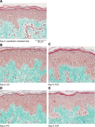
shows the histological images of skin tissue showing general morphology at baseline and day 5. There were no appreciable changes in general morphology in any of the treatment groups during the course of the study, suggesting that skin explants were viable throughout the study.
Figure 1 Viability study: general morphology at day 0 and day 5 in all sample groups.
Nrf2 immunostaining
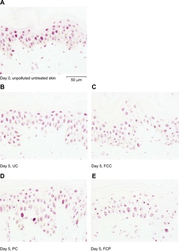
Day 0 skin sections showed clear staining of Nrf2 in the living epidermis. At day 5, FC application led to a decrease in the level of staining compared to the control group. Exposure to pollution led to an increase in the level of staining for Nrf2. However, the group with FC application prior to pollution exposure showed levels of staining that were similar to the control group ( and ).
Figure 2 Nrf2 immunostaining levels at day 0 and day 5 in living epidermis.
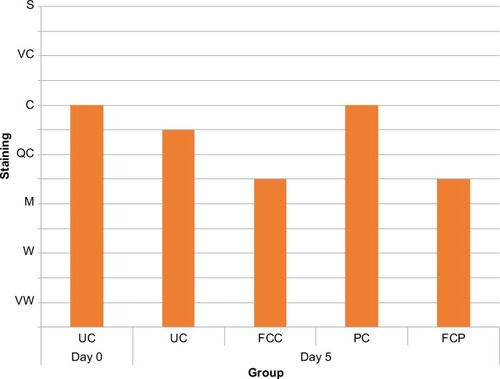
Figure 3 Nrf2 immunostaining at day 0 and day 5 in all sample groups.
MDA
Mean MDA concentrations in survival culture medium on day 5 for all groups are presented in . Exposure of skin to pollution for 4 days led to a statistically significant increase of 38% in MDA concentrations in the culture media (108.5 vs 149 nmol/L in untreated control [UC] and pollution-exposed control [PC] groups, respectively, P<0.01). Topical application of the FC produced a nonsignificant reduction in MDA concentration in the PC group (7% decrease). MDA levels were significantly lower in the FC-treated and exposed to pollution (FCP) group compared to the PC group (27% decrease, P<0.01).
Table 4 MDA levels in culture medium at day 5 (mean [SD])
Melanin
Melanin levels in the different treatment groups are reported in . There were no statistically significant differences in the values among groups (P>0.05).
Table 5 Epidermal melanin levels at day 0 and day 5 (mean [SD])
Gene expression
Application of the FC to skin did not in itself lead to the upregulation or downregulation of the genes studied. Exposure to pollution led to a change in the expression of three genes in skin explants, and the FC modulated this expression ().
Table 6 Gene expression at day 5 of ex vivo antipollution activity study
CYP1A1: in the PC group, exposure to pollution led to a 5.74-fold increase in CYP1A1 expression compared to the UC group. In the FCP group, this overexpression was limited to 2.22-fold. IL6: in the PC group, exposure to pollution led to a 1.83-fold increase in IL6 expression; in the FCP group, this overexpression was limited to 1.34-fold (<1.45 fold, hence not considered upregulated). TYR: in the PC group, exposure to pollution led to a 1.53-fold increase in TYR expression compared to the FCP group, where this gene was not overexpressed.
Discussion
We present in this study two levels of evidence for the protective effects of topical application of a novel FC against pollution. In vivo, the application of the FC resulted in a statistically significant decrease in the adherence of carbon E153 powder to the skin after washing compared to nude skin (control area). At the cellular level, exposure to pollution led to changes indicative of increased oxidative stress, inflammation, and pigmentation: increased staining for the active form of Nrf2, increased MDA levels in culture medium, and upregulation of CYP1A1, IL6, and TYR genes in human skin explants. Prior application of the FC was able to block to a certain degree all of these changes induced in the skin by exposure to pollution. These results are consistent with certain key ingredients of the FC, which were included to minimize fine particle adhesion to skin and control oxidation and inflammation to ameliorate the downstream effects of such exposure.
In our study, the pollution-induced increase in MDA concentration and in the active form of Nrf2 indicates oxidative stress,Citation18 which is reported in the literature to be the principle deleterious action of pollution on skin.Citation19,Citation20 The reduction in MDA levels and staining for the active nuclear form of Nrf2 in the skin explants pretreated with FC demonstrates the protective effect against the cellular changes induced by pollution. This can partly be explained by the presence of niacinamide which has a known anti-inflammatory actionCitation21 and both niacinamide and carnosine with known antioxidantCitation22 action in the skin. Pollution also induced upregulation of IL6 and CYP1A1 suggesting an inflammatory response to oxidative injuryCitation23,Citation24 and a protective effect of the FC as seen by the modulation of this response with its application. IL-6 plays a key role in local and systemic inflammation, and CYP1A1 codes for a CYP/CYP450-related detoxification enzyme whose expression is induced by some PAHs, a component of urban pollution.Citation25
Other authors have described the effects of pollution on pigment spot formation in the skin.Citation26 In our study, after only two periods of exposure to pollution, we observed an upregulation of the TYR gene, which codes for tyrosinase, a key enzyme in melanogenesis.Citation27 We did not observe an increase in melanin content in pollution-exposed skin. This may be due to the short duration of the study (5 days) that was not long enough to see changes in melanin levels and represents a potential area for future investigation. The finding that TYR was upregulated by pollution and modulated by the FC suggests an antipollution protective effect of the product.
A film-forming EPS called as alteromonas ferment extract was included in the formulation for its anti-adhesion effect. Previous in vitro and clinical studies showed that a product containing the same EPS, in skin exposed to heavy metals, hydrocarbons, and particulate matter, significantly reduced particle adhesion to skin and protected keratinocyte membranes from lipid peroxidation, preserved cell integrity, and normalized the collagen network.Citation12,Citation13
Strengths and limitations
The strength of this study is that under controlled, standardized, replicable conditions, we provided two levels of evidence on the protective effect of this FC against pollution. The exposure of skin explants to vaporized pollution particles in a closed environment was chosen to be a close approximation of real-life conditions, although ozone, a known component of air pollution, was not included, a point that may be considered a limitation. Other limitations include the absence of a placebo control group and unblinded nature of the clinical study, although a standardized method of powder application and washing was used to minimize interindividual and intergroup variability. The particle adhesion study was conducted on forearm skin, which differs from facial skin, the main target of pollution effects, in characteristics such as density of sebocytes present. The forearm area was chosen for the ease of assessment and the lower risk of inadvertent introduction of carbon particles to the subjects’ eyes or upper airways. However, this is a drawback of the in vivo study as facial sebum might affect the adhesion of carbon particles on the skin. A study on facial skin would provide more directly-applicable results. In the clinical study, we report a statistically significant decrease in particle binding in treated skin, yet this study does not allow us to establish a clinical cutoff value beyond which particle binding is known to induce or not skin aging as this is clearly complex and was not the aim of this study. However, when taken together with results from the ex vivo study, clearly this level of mitigation of adhesion of particles has a relevant benefit in terms of markers of oxidative stress.
Increasing understanding of the effects of pollution on skin health suggests that protecting skin against pollution should be a key area of improvement of skincare and antiaging regimens. The effects of ultraviolet exposure on skin aging have been well documented, and applying sun protection is now a standard recommendation in antiaging skincare. As understanding and awareness of the effects of pollution on skin continue to increase, everyday-use products that provide this protection are of increasing relevance especially in highly polluted urban or industrial nuclei.
Conclusion
Application of FC containing an EPS, carnosine, and niacinamide prior to exposure to carbon particles significantly decreased particle adherence to skin in human subjects. In human skin explants, exposure to urban dust led to an increase in indicators of oxidative stress, while the daily application of the cream over 5 days had a protective effect against pollution-induced changes. These results suggest that this novel FC can help to reduce the harmful oxidative effects of pollution on skin.
Acknowledgments
J. Marshall provided medical writing assistance in drafting the manuscript.
Disclosure
MN, GB, PV, and CG are employees of ISDIN S.A, the manufacturer of the cosmetic product tested in this study. The authors report no other conflicts of interest in this work.
References
- PilzVWolfKBreitnerSC-reactive protein (CRP) and long-term air pollution with a focus on ultrafine particlesInt J Hyg Environ Health2018221351051829428699
- PhungVUedaKKasaokaSAcute Effects of Ambient PM2.5 on All-Cause and Cause-Specific Emergency Ambulance Dispatches in JapanInt J Environ Res Public Health2018152307
- WHO [homepage on the Internet]Air Quality Guidelines for Particulate Matter, Ozone, Nitrogen Dioxide and Sulfur Dioxide, Global Update 2005Summary of Risk Assessment2006 Available from: http://www.who.int/phe/health_topics/outdoorair/outdoorair_aqg/en/Accessed September 18, 2018
- KrutmannJMoyalDLiuWPollution and acne: is there a link?Clin Cosmet Investig Dermatol201710199204
- BaudouinCCharveronMTarrouxRGallYEnvironmental pollutants and skin cancerCell Biol Toxicol200218534134812240965
- KimJKimEHOhISymptoms of atopic dermatitis are influenced by outdoor air pollutionJ Allergy Clin Immunol2013132249549823763977
- PuriPNandarSKKathuriaSRameshVEffects of air pollution on the skin: A reviewIndian J Dermatol Venereol Leprol201783441542328195077
- KrutmannJBoulocASoreGBernardBAPasseronTThe skin aging exposomeJ Dermatol Sci201785315216127720464
- HalliwellBCrossCEOxygen-derived species: their relation to human disease and environmental stressEnviron Health Perspect1994102Suppl 10512
- BaroniABuomminoEde GregorioVRuoccoERuoccoVWolfRStructure and function of the epidermis related to barrier propertiesClin Dermatol201230325726222507037
- ThieleJJTraberMGTsangKCrossCEPackerLIn vivo exposure to ozone depletes vitamins C and E and induces lipid peroxidation in epidermal layers of murine skinFree Radic Biol Med19972333853919214574
- BorelMLamarqueELoingEUnique natural exopolysaccharides for biomimetic protective effect against urban pollutionJ Cosmet Sci201768112613229465393
- ZhangZCaiRZhangWFuYJiaoNA Novel Exopolysaccharide with Metal Adsorption Capacity Produced by a Marine Bacterium Alteromonas sp. JL2810Mar Drugs2017156175
- Decree no 2017-884 of 9 May 2017. Journal Officiel de la Republique Française (JORF) Available from: https://www.legifrance.gouv.fr/jo_pdf.do?id=JORFTEXT000034634217Accessed July 10, 2018
- Colipa, The European Cosmetics AssociationGuidelines for the evaluation of the efficacy of cosmetic products2008 Available from: https://www.cosmeticseurope.eu/files/4214/6407/6830/Guidelines_for_the_Evaluation_of_the_Efficacy_of_Cosmetic_Products_-_2008.pdf
- Law no. 2011-814 of 7 July onbioethics (article 18). JORF 157 of 8 July 2011 page 11826 text 1NOR: ETSX1117652L Available from: https://www.legifrance.gouv.fr/affichTexte.do?cidTexte=JORFTEXT000024323102&dateTexte=20180508Accessed July 10, 2018
- CostinGRaabHOptimized In Vitro Pigmentation Screening Assay Using A Reconstructed Three Dimensional Human Skin ModelRom J Biochem20135011527
- OsburnWOKenslerTWNrf2 signaling: an adaptive response pathway for protection against environmental toxic insultsMutat Res20086591–2313918164232
- ValacchiGSticozziCPecorelliACervellatiFCervellatiCMaioliECutaneous responses to environmental stressorsAnn N Y Acad Sci20121271758123050967
- VierkötterASchikowskiTRanftUAirborne particle exposure and extrinsic skin agingJ Invest Dermatol2010130122719272620664556
- GehringWNicotinic acid/niacinamide and the skinJ Cosmet Dermatol200432889317147561
- QuinnPJBoldyrevAAFormazuykVECarnosine: its properties, functions and potential therapeutic applicationsMol Aspects Med19921353794449765790
- PaquetPPiérardGEInterleukin-6 and the skinInt Arch Allergy Immunol199610943083178634514
- ShimadaTFujii-KuriyamaYMetabolic activation of polycyclic aromatic hydrocarbons to carcinogens by cytochromes P450 1A1 and 1B1Cancer Sci20049511614720319
- Gene [Internet]Bethesda (MD)National Library of Medicine (US), National Center for Biotechnology Information2018 Available from: https://www.ncbi.nlm.nih.gov/gene/1543Accessed 10 May, 2018
- HülsAVierkötterAGaoWTraffic-related air pollution contributes to development of facial lentigines: further epidemiological evidence from Caucasians and AsiansJ Invest Dermatol201613651053105626868871
- JimbowKParkJSKatoFAssembly, target-signaling and intracellular transport of tyrosinase gene family proteins in the initial stage of melanosome biogenesisPigment Cell Res200013422222910952389