Abstract
Josef Jadassohn (1863–1936) and his assistant, Felix Lewandowsky (1879–1921), were eminent German dermatologists who had several dermatology conditions linked eponymously. One such condition is Jadassohn–Lewandowsky syndrome, which is a type of pachyonychia congenita – a disease that is characterized by severe thickening of the nail due to massive nail hyperkeratosis. This report describes Jadassohn–Lewandowsky syndrome and the men behind this disease.
Introduction
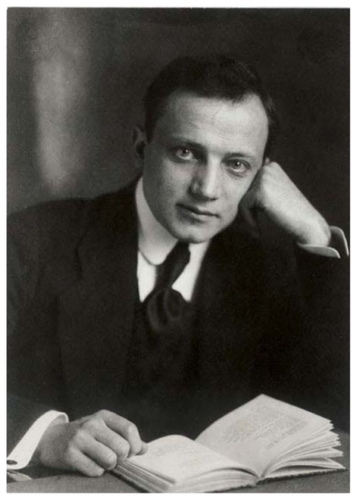
Josef Jadassohn (1863–1936) () was a pioneering German dermatologistCitation1–Citation4 who, with another great German dermatologist, Felix Lewandowsky (1879–1921)Citation5,Citation6 (), described a syndrome,Citation7 later known as Jadassohn–Lewandowsky syndrome.Citation8–Citation15 This report describes Jadassohn–Lewandowsky syndrome and the men for whom it is named.
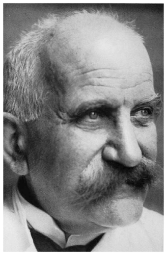
Figure 2 Felix Lewandowsky (1879–1927).
Josef Jadassohn (1863–1936)
Joseph (Josef) Jadassohn was a German dermatologist. Among his many contributions to medicine, he is credited for describing several medical conditions,Citation1–Citation4 many of which were named after him and his assistants, including nevus sebaceous of Jadassohn, Jadassohn–Lewandowsky syndrome, Jadassohn–Dössekker disease (myxoedema tuberosum), and Naegeli–Franceschetti–Jadassohn syndrome.Citation2
Jadassohn was born in Liegnitz, Germany on September 10, 1863.Citation1–Citation4 He studied medicine in Göttingen, Heidelberg, Leipzig, and Breslau, obtaining his doctorate in 1887 at Breslau.Citation2 He was an assistant of Albert Neisser at Allerheiligen Hospital in Breslau until 1892, the director of the university skin clinic in Bern (1896–1917), and a professor of dermatology at Breslau University (1917–1932).Citation1
Jadassohn was a pioneer in the field of allergology and was among the first to take an immunological approach in the research of dermatological disorders, contributing to the understanding of the immunopathology of tuberculosis and trichophytosis.Citation1 Jadassohn is credited for introducing patch testing to diagnose contact dermatitis, and in 1901, he described a rare childhood dermatological disorder, known as granulosis rubra nasi (a papular red lesion of the nose associated with increased sweating).Citation1
He was particularly interested in drug reactions, leprosy, eczema, tuberculosis, syphilis, and mycotic infections. His awareness of the social aspects of venereal diseases led to his appointment to the Committee on Hygiene of the League of Nations.Citation3 He was a corresponding member of the British Association of Dermatology, and in the year prior to his death, he was made an honorary fellow of the Royal Society of Medicine. Josef Jadassohn died in Zurich on March 24, 1936.Citation1,Citation2
Felix Lewandowsky (1879–1921)
Felix Lewandowsky was a Germany dermatologist who is credited for describing several medical conditionsCitation5,Citation6 that were later named after him, including Jadassohn–Lewandowsky syndrome, Lewandowsky’s syndrome, and Lewandowsky–Lutz dysplasia.
Lewandowsky was born on October 1, 1879, and in 1902, he earned his doctorate at Strassburg.Citation5,Citation6 From 1903 until 1907 he worked at a dermatology clinic in Bern, where he was an assistant to Jadassohn. On returning to Hamburg, he worked in Eduard Arning’s (1855–1936) department at St George’s Hospital. In 1917, he was appointed director of the dermatological clinic at Basel.Citation5,Citation6
Lewandowsky specialized in tuberculosis and infantile skin diseases.Citation5 In 1916, he published an important study on tuberculosis of the skin, entitled Die Tuberkulose der Haut.Citation5,Citation6 Prior to his death, he described epidermodysplasia verruciformis, a rare skin disease, sometimes known as Lewandowsky–Lutz dysplasia (named for him and dermatologist Wilhelm Lutz).Citation5 Lewandowsky died on October 31, 1921.
Jadassohn–Lewandowsky syndrome
Pachyonychia congenita (PC) is characterized by onychogryposis, hyperkeratosis of the palms, soles, knees, and elbows, tiny cutaneous horns, and leukoplakia of the oral mucous membranes. Hyperhidrosis of the hands and feet is usually present.Citation12 The hallmark of PC is severe nail thickening due to massive nail hyperkeratosis. The Online Mendelian Inheritance in Man® (OMIM®) database contains three entries for this disease: PC1 (OMIM #167200), PC2 (OMIM#167210), and PC recessive (OMIM 260130).
PC1 is known as Jadassohn–Lewandowsky type, and PC2 is called Jackson–Lawler type. PC1 is caused by a mutation in KRT6A (located on 12q13.13) and KRT16 (17q21.2). PC2 is caused by a mutation in KRT6B (12q13.13) and KRT16 (17q21.2).
In PC, the fingernails and toenails become thickened, yellow- gray, and transversely overcurved with marked subungual hyperkeratosis, most notably at the distal end. Nail trimming is impossible, because the nails are extremely hard.Citation14 These abnormalities usually develop during childhood, but late-onset PC has been described. Focal palmoplantar keratoderma is associated with these nail changes in 60% of patients or can result from mutations without detectable nail changes.Citation14 Up to 75% of patients have oral leukokeratosis; hyperhidrosis of the palms and soles, follicular keratosis, and acral bullae (especially during warmer months) are common.
PC that is associated with late symptom onset has been described by several groups and is called pachyonychia congenita tarda, type 1.Citation12
PC1 is considered as a type of keratoderma that principally involves the plantar surfaces. Onset of plantar calluses can be delayed up to 7 years of age. Nail changes may be evident at birth but more commonly develop within the first few months of life. They are characterized by upward angulation of the distal end of the nail due to subungual hyperkeratosis, giving the characteristic “door wedge” distal hyperkeratosis. Calluses develop over the plantar pressure points; palmar calluses are not seen except in manual workers. Blistering can occur on walking. Hair abnormalities and oral, laryngeal, and follicular hyperkeratosis have also been described in PC1.Citation14
Jadassohn and Lewandowsky’s original patient was a 15-year-old female who was admitted with fungating tuberculosis of the skin. Abnormalities had been present since birth, and she also had unusual keratinization of the skin and tongue. In their case description, Jadassohn and Lewandowsky stated, “The nail plates of all the fingers and toes are extremely thickened, and so hard that they cannot be cut with scissors; the father has to trim them with a hammer and chisel.”Citation8
The familial nature of this condition was established in 1921, when Murray documented seven affected persons in three generations of a family.Citation8 In a similar report, Kumer and Loos described 24 cases in a five-generation family in 1935.Citation12
Patients with PC2 tend to have cystic lesions (steatocystoma multiplex, vellus hair cysts), which reflects the expression of K6b and K17 in adnexal structures. The cystic lesions form during childhood and appear progressively. Coarse or twisted hair, early primary tooth loss, and natal teeth are other features of PC2, reflecting the expression of the affected genes in the hair follicle apparatus and teeth.Citation14 Approximately 60% of patients have oral keratosis, 16% have natal or neonatal teeth, and 10% have angular stomatitis. Some patients also develop chronic intraoral candidosis. The keratosis requires no treatment, but dental advice should be sought regarding natal or neonatal teeth. Laryngeal changes that require a tracheostomy for respiratory distress during childhood have been reported.Citation12
The incidence of cyst formation and natal teeth is higher among PC2 patients, although cysts are more commonly seen in PC1 patients (25%–33%). Previously unreported clinical features of PC include the development of painful oral and nipple lesions during breastfeeding, copious production of waxy material in ears, and the inability to walk without an ambulatory aid (50%).Citation12
There are several clinical differences between PC1 and PC2. PC1 patients develop oral leukokeratosis, whereas those with PC2 have natal teeth and epidermoid cysts (cylindromas) but no oral leukoplakia. Corneal dystrophy is exclusively a feature of PC2. Both disorders are clearly autosomal dominant.Citation12 In PC2, the nail thickening and keratosis are less severe than in type 1.
Thickened nails can be treated surgically in some instances, most frequently warranted in PC1. The localization of the four implicated keratins in the nail bed suggests that tissue is the chief site of pathology. Affected nails that are viewed end-on appear almost normal but have a dense wedge of subungual keratin, constituting the focus of nail bed disease.Citation13 Treatments for PC are based on focal complaints, such as ingrowth and paronychia, but there are no established therapies.Citation13
The Pachyonychia Congenita Project is an initiative that was established to serve PC patients and their families. It provides several services to health care providers that are interested in PC. Further information about this project is available at http://www.pachyonychia.org/homeScientist.php.
Disclosure
The authors report no conflicts of interest in this work.
References
- ObituaryBr J Dermatology193648323324
- Whonamedit? Josef Jadassohn [web page on the Internet]February 12002 Available from: http://www.whonamedit.com/doctor.cfm/743.htmlAccessed April 5, 2011
- [No authors listed]Josef Jadassohn (1863–1936) dermatologistJAMA19692078151115124884744
- SulzbergerMBJosef Jadassohn. A personal appreciationAm J Dermatopathol19857131363883835
- HäusermannPLutterSMeigelWRufliTZur Person Felix LewandowskyH&G Zeitschrift für Hautkrankheiten200277182183 German
- Whonamedit? Felix Lewandowsky [web page on the Internet]January 312002 Available from: http://www.whonamedit.com/doctor.cfm/1397.htmlAccessed April 5, 2011
- JadassohnJLewandowskyFPachyonychia congenita. Keratosis disseminata circumscripta (follicularis). Tylomata. Leucokeratosis linguaeNeisserAJacobiEIkonographia DermatologicaBerlinUrbach and Schwarzenberg19062931
- MurrayFACongenital anomalies of the nails. Four cases of hereditary hypertrophy of the nail bed associated with a history of erupted teeth at birthBr J Dermatol192133409412
- KanskyABasta-JuzbasicAVidenicNIvankovicDStanimirovicAPachyonychia congenita (Jadassohn–Lewandowsky syndrome) – evaluation of symptoms in 36 patientsArch Dermatol Res19932851–236378470932
- MurugeshSBReddySRagunathaSMohammed FaizalMMShashikalaPAcro-osteolysis: a complication of Jadassohn– Lewandowsky syndromeInt J Dermatol200746220220517269978
- ScullyCLangdonJEvansJMarathon of eponyms: 10 Jadassohn– Lewandowsky syndrome (Pachyonychia congenita)Oral Dis201016331031120374515
- OMIM®: Online Mendelian Inheritance in Man®Pachyonychia congenita, type 1; PC1 [web page on the Internet] Updated April 30, 2010. Available from: http://omim.org/entry/167200Accessed April 5, 2011
- DawberRPRBaranRDe BerkerDDisorders of nailsChampionRHBurtonJLBurnsTBreathnachSTextbook of Dermatology46th edOxfordBlackwell Science Ltd199828492850
- TostiAPiracciniBMBiology of nails and nail disordersWolffKGoldsmithLAKatzSIGilchrestBAPallerASLeffellDJFitzpatrick’s Dermatology in General Medicine7th edNew YorkMcGraw-Hill2008778794
- LeachmanSAKasparRLFleckmanPClinical and pathological features of pachyonychia congenitaJ Investig Dermatol Symp Proc2005101317