
Abstract
Iatrogenic bile duct injuries (BDIs) after laparoscopic cholecystectomy, being one of the most common performed surgical procedures, remain a substantial problem in gastrointestinal surgery with a significant impact on patient’s quality of life. The primary aim of this review was to discuss the classification of BDIs, the proposed methods to prevent biliary lesions, the associated risk factors, and the management challenges depending on the timing of recognition of the injury, its extension, the patient’s clinical condition, and the availability of experienced hepatobiliary surgeons. Early recognition of BDI is of paramount importance and limiting the diagnosis delay is crucial for an optimal postoperative outcome. The therapeutic management depends on the type and gravity of the biliary lesion, and includes endoscopic, radiologic, and surgical approaches.
Introduction
Laparoscopic cholecystectomy (LC) is one of the most frequently performed procedures worldwide in general surgery.Citation1 Annually, more than 750,000 procedures are performed in the US.Citation2 Bile duct injury (BDI) represents the most serious complication of LC, with an incidence of 0.3%–0.7%, resulting in a significant impact on quality of life (QoL), overall survival, and frequent medico-legal liabilities.Citation1–Citation4 The incidence of iatrogenic BDI has significantly gone up since LC became the “gold standard” in the treatment of symptomatic cholelithiasis. It has been demonstrated that the primary cause of BDI is the misinterpretation of biliary anatomy in 71%–97% of all cases.Citation1 Over the years, various classifications of biliary injuries have been proposed and different methods have been described to prevent iatrogenic biliary tract lesions. Early recognition of BDI is of paramount importance. The optimal treatment is influenced by the timing of recognition of the injury, the extent of BDI, the patient’s clinical condition, and the availability of experienced hepatobiliary surgeons. This article aims to discuss the classification and management challenges of iatrogenic BDIs after cholecystectomy.
Classification of BDIs
Before the advent of LC, the most frequent lesion to biliary tract was represented by bile duct stricture, and biliary injuries were classified using the “Bismuth classification”Citation5 according to the level of biliary injury. This classification includes five types of bile duct lesions according to the distance from the hepatic hilus, the level of injury, the involvement of bile duct bifurcation, and individual right sectoral duct. Few years later, Strasberg et alCitation6 proposed a new classification, similar to the Bismuth, but incorporating additional biliary injuries seen more commonly in the laparoscopic era, in particular biliary leaks. In the “Strasberg classification”, biliary injuries were stratified form type A to type E, as summarized in . McMahon et alCitation7 proposed another classification by distinguishing major and minor biliary injuries: lacerations under 25% of the common bile duct (CBD) diameter or cystic duct-common hepatic duct (CD-CHD) junction were considered as minor injury, whereas transection or laceration over 25% of CBD diameter and postoperative bile duct stricture were classified as major injury. The “Stewart-Way classification”Citation8 involved four classes based on the mechanism and anatomy of biliary injury, as shown in . This classification arose from the analysis of operative reports, providing the human mistakes and cognitive processes involved in the mechanisms of BDIs. Class I injury occurs in two ways: 1) either the CBD is mistaken for the cystic duct, but the error is recognized before CBD is divided in 72% of cases, or 2) an incision made in the cystic duct for the cholangiogram catheter is unintentionally extended into the CBD (28% of cases). Class II injuries consist of lateral damage to the CHD with resultant stricture and/or leak. This type of injury occurs when clips or electro-cautery are used too close to the CBD, mainly in cases where visibility is limited due to severe inflammation or excessive bleeding. Class III injuries are the most common (about 60% of cases) and involve complete transection of the main bile duct, which always includes the CD-CHD junction. Class IV injuries include transection or leak of the right hepatic duct (RHD) or posterolateral sectoral duct, often combined with injury to the right hepatic artery.
Figure 1 Stewart-Way classification of bile duct injuries.
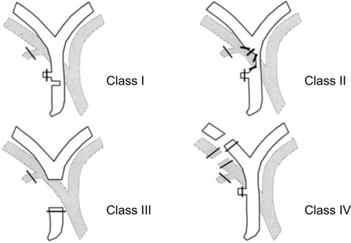
Table 1 Bismuth and Strasberg classification
The “Amsterdam classification” proposed by Bergman et al in 1996Citation9 is also very commonly used in many articles, especially among endoscopists. This classification includes four types of ductal injury: type A with leakage from cystic ducts or peripheral hepatic radicles, type B presenting as major bile duct leakage, type C showing an isolated ductal stricture, and type D with complete transection of the bile duct.
For an easy reading, we have chosen the Stewart-Way classification system in this manuscript, as it better describes the mechanisms of biliary lesion during LC, including also concomitant vascular injuries.
Prevention of iatrogenic BDI
Over the years, various methods have been proposed and described in the scientific literature to prevent iatrogenic biliary tract lesions. They are summarized in . The “critical view of safety (CVS)” technique was introduced by Strasberg in 1995Citation10 and it is considered the gold standard to perform a safe cholecystectomy with identification of biliary structures during dissection. Three criteria are required to achieve the CVS: 1) the hepatocystic triangle must be cleared of adipose and fibrotic tissues; the CBD and CHD must not be exposed; 2) the lower third of the gallbladder must be separated from the liver bed to expose the cystic plate; and 3) two and only two structures should be seen entering the gallbladder. CVS was conceived not as a way to do LC but as a way to avoid biliary injury.
Table 2 Summary of proposed methods to prevent bile duct injuries
In such cases, some surgeons prefer performing the infundibular method in order to work very close to gallbladder infundibulum, reducing the risk of biliary injuries, but care should be taken with “hidden cystic duct” syndrome which presents a deceptive appearance of a false infundibulum that misleads the surgeon into identifying the CBD as the cystic duct.Citation11 The “antegrade dissection or fundus first/dome-down technique” represents a way of dissection from the gallbladder fundus up to the infundibulum away from Calot’s triangle.Citation12 In this way, the gallbladder is left pedunculated by the cystic artery and cystic duct, which can be clipped and divided in turn, reducing the risk of biliary lesions. Another easy, safe, and definitive operation for the “difficult gallbladder” is described and has been termed “subtotal or partial cholecystectomy”,Citation13 consisting in leaving the posterior wall of the gallbladder attached to the liver and securing the cystic duct at its origin from within the gallbladder.
Different anatomic landmarks have been described: among them, in 1924, Henri Rouvière, a French surgeon, described a 2–5 cm sulcus running to the right of liver hilum, anterior to the caudate lobe, and usually containing the right portal triad or its branches.Citation14 It can be considered a useful landmark site to start dissection of the hepatocystic triangle during LC.Citation15 The “Rouvière’s sulcus” is present in the majority of patients, ranging from 68% to 90%,Citation16 and is clearly visible during laparoscopy, being visible in one form or the other (a sulcus, a slit, or a scar). Another anatomic landmark to guide gallbladder dissection is represented by the “cystic lymph node” or Mascagni’s node, which always lies lateral to the biliary tree and should form the medial end point of dissection.Citation17
Some authors proposed a mnemonic “B-SAFE method”, by using five anatomic landmarks (B, bile duct; S, sulcus of Rouvière; A, hepatic artery; F, umbilical fissure; E, enteric/duodenum) to correctly place their cognitive map during dissection.Citation18
The routine use of “intra-operative cholangiography (IOC)” has been proposed for the better declaration of biliary anatomy, detection of silent CBD stones, and reduction of incidence of BDIs, but it is burdened by morbidity and mortality and hence should be performed in facilities where the necessary equipment and experience are available.Citation1,Citation19,Citation20 The opinions about the “routine” or “selective” use of IOC still represent a matter of debate.Citation1,Citation19
“Laparoscopic ultrasound (LUS)” was shown to provide highly sensitive mapping of the extra-hepatic biliary anatomy but the difficult learning curve and the lack of randomized controlled trials have reduced its use in clinical practice.Citation21,Citation22
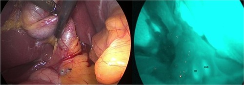
One of the latest innovations in minimally invasive technology is fluorescence image-guided surgery; “near-infrared fluorescent cholangiography (NIRF-C)” represents a novel intra-operative imaging technique that allows a real-time enhanced visualization of the extrahepatic biliary tree by fluorescence.Citation23–Citation25 NIRF-C well represents a useful method to identify the CVS with the aid of real-time fluorescent vision with a perfect combination between surgical anatomy and technical innovation, as shown in .Citation26
Figure 2 Intra-operative real-time identification of biliary structures with visible light (VL) on left and by fluorescence (NIRF-C) on right.
Abbreviation: NIRF-C, near-infrared fluorescent cholangiography.
In some difficult situations, the opinion of a surgical colleague in vicinity during operation may also represent a valid alternative, the so-called “colleaguography”, in place of IOC or other methods, as proposed by some authors.Citation27
Despite the plethora of publications and debates, there is still no consensus as to which method is the best to prevent BDIs during LC. Recently, highest consensus was achieved on the importance of the CVS and NIRF-C, as well as ante-grade dissection or fundus first/dome-down technique and partial cholecystectomy as alternative techniques.Citation28,Citation29
A general recommendation is that in all cases of complicated cholecystectomies, the surgeon must not hesitate when considering conversion to open approach because there is no substitute for experience and caution in biliary surgery.
Risk factors
There are several risk factors which can contribute to the iatrogenic injury of the biliary tract: anatomical factors; patient-related factors; and factors related to the gallbladder disease, the surgical technique, and the surgeon. Among the anatomical factors, the numerous anatomical variants of the biliary tract represent a possible cause of iatrogenic lesion, for example, the different variants of the cystic duct, such as short cystic duct, cystic duct running parallel to the CBD, anomalies of the CD-CHD junction, presence of the hepatocystic duct, accessory cystic duct, the existence of aberrant bile ducts (eg, Luschka duct).Citation30 Among the patient-related factors, severe obesity, previous surgery on the biliary tract, and underlying liver disease seem to be predisposing factors for peri-operative complications.
However, acute cholecystitis causes a series of modifications of the local anatomy (adhesions, thickening of the tissues, inflammation, bleeding) that are associated with an increased risk of iatrogenic lesion.Citation31,Citation32 Among the factors related to the technique, the laparoscopic approach itself represents a risk factor, hence the importance of a correct “learning curve” for young surgeons.Citation33,Citation34 The human factors also play a crucial role in the pathophysiology of the iatrogenic bile lesion: the excessive safety of the surgeon, the rush to finish the intervention, the fatigue and personal concerns of the surgeon, the performance anxiety, the superficiality of the surgical act, and the lack of humility in converting to open surgery in doubtful cases may determine a relevant damage to the biliary tree.Citation8–Citation35
LC can be seen as a trivial intervention but it could hide many difficulties with dramatic consequences. For these reasons, caution and experience are essential in biliary surgery.
Clinical presentation
Clinical presentation depends essentially on the extent and type of iatrogenic lesion, if this is not discovered intra-operatively. The presence of a possible biliary leak can start with non-specific symptoms, such as nausea, vomiting, bloating, widespread abdominal pain, general discomfort, and anorexia. Rarely, the patient presents the clinical picture of biliary peritonitis, despite large amounts of bile in the abdomen. The evolution of biliary leak is very subtle, because of the presence of these vague symptoms. Patients with stricture at level of the CBD, in cases where the CBD is mistaken for the cystic duct, will present the classic Charcot’s triad with pain in the right hypochondrium, fever, and jaundice. A general recommendation is that any alteration in the normal postoperative course after LC must suggest a possible damage to the biliary tract. For these reasons, a careful clinical evaluation of patients is essential. A delay in the diagnosis is crucial for the postoperative outcome.
Timing of recognition
Early recognition of BDI is of paramount importance. Intra-operative recognition should typically prompt immediate surgical repair as this confers the best outcomes; however, <25%–30% of BDIs are recognized during operation and not all surgeons perform an intra-operative cholangiography.Citation36,Citation37 If the surgeon is inexperienced, an intra-abdominal drain should be placed, and the patient should be immediately transferred to a high-volume hepatobiliary center. There are several data suggesting that these injuries are best managed by a surgeon with expertise in biliary reconstruction with better outcomes.Citation37–Citation39 There are several factors that lead to a recognition of intra-operative lesion, but the most important is the surgeon’s awareness to suspect and/or evaluate for a BDI. Diagnosis often occurs in the immediate postoperative period (within 6 weeks post-intervention) or, in some cases, later (over 6 weeks). A careful clinical evaluation helps for a correct diagnosis. The appearance of persistent or abnormal abdominal pain in the right hypochondrium, the leakage of bile from drainage (if placed during surgery), a picture of biliary peritonitis, and the appearance of fever and jaundice with increased liver function tests are all elements supporting a diagnosis of any iatrogenic lesion of the biliary tree. Radiologic investigations should be obtained for the correct identification of the damage, its extension, and gravity and to plan therapeutic strategies. Abdominal ultrasound represents the first diagnostic tool aimed at finding any liver collections, dilatation of the CBD, and any associated vascular lesions.Citation40 The abdominal computed tomography (CT) scan is the principal investigation that allows to define the possible presence of focal intra- or peri-hepatic fluid collections, ascites, biliary obstruction with upstream dilatation, or long-term sequelae of a long-standing bile stricture, such as lobar hepatic atrophy or signs of secondary biliary cirrhosis. The CT scan also allows to identify any associated vascular lesions, such as to the right hepatic artery.
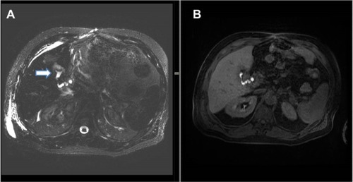
Magnetic resonance cholangio-pancreatography (MRCP) represents the “gold standard” for the complete morphological evaluation of the biliary tree as it offers detailed information about the integrity of the biliary tract.Citation41,Citation42 MRCP can also be done with a contrast agent (a gadolinium-based contrast agent, the gadoxetic acid and its salt, gadoxetate disodium), and it is much more informative in bile leaks than plain MRCP.Citation42 The use of a contrast agent during MRCP allows detection of active bile leakage by direct visualization of contrast material extravasation into fluid collections in addition to demonstrating the anatomical site of the leakage and the type of BDI. For example, in cystic duct leak was demonstrated at MRCP after gadoxetic acid injection. In the same way, hepatobiliary scintigraphy may confirm the presence of an active biliary leak with the progressive accumulation of the radiotracer inside the peritoneal cavity, but this examination is much less reliable. However, if fluid collection is found in ultrasound, CT, or MRCP, an abdominal drain should be placed right away under radiological guidance in order to improve patient’s clinical condition (peritonitis, sepsis, tissue repairing). If there is a delay of more than a week in operative treatment of Stewart-Way Class III or IV injuries, there should be a timeout for 2–3 months before operation. In this time frame, the patient needs to be stabilized and optimized for surgery. An optimal control of any intra-abdominal fluid collection, inflammation, and infection is essential, and is best achieved with percutaneous drainage.Citation37
Figure 3 (A) Axial three-dimensional MRCP sequence showing a small fluid collection in the gallbladder fossa (white arrow). (B) T1-weighted hepatobiliary acquisition (after gadoxetic acid injection) demonstrates opacification of the small fluid collection suggesting a biliary leak from the cystic duct.
In the postoperative period, re-laparoscopy can identify the biliary injury, drainage of any intra-abdominal collection, and exclusion of injury to other surrounding organs, which can lead to the decision between conservative management and early or late definitive surgical repair.Citation43
There is no role for explorative laparotomy, as this is associated with increased morbidity and mortality.
Therapeutic management
The endoscopic retrograde cholangio-pancreatography (ERCP) examination has an important therapeutic role in cases where a conservative therapeutic approach is preferred based on the type and extent of the biliary lesion (cystic duct leaks, Stewart-Way Class I injuries, and most Class II injuries).
Cystic duct leaks are well managed conservatively; the endoscopic procedure allows to perform a sphincterotomy with the aim of reducing the pressure in the biliary tree and to place a plastic stent or a naso-biliary tube to limit or exclude the fistula (depending on the location of the biliary leak). After the right patient selection, the success rate of endoscopic treatment can approach 100% for cystic duct or peripheral radical leaks. Endoscopic sphincterotomy is an effective and cost-effective single procedure with success rate similar to endoscopic sphincterotomy and biliary stenting, as proposed by Rainio et al.Citation44 Self-expanding biodegradable biliary stents have only recently become available in order to avoid second endoscopy with good results.Citation45 The limits of the endoscopic treatment concern the lack of visualization of aberrant or sectioned bile ducts which do not communicate with the CBD (eg, an aberrant RHD), the cases of complete transection of the bile duct, and the difficulty in visualization of possible intra-hepatic proximal leaks. In this specific case, the percutaneous transhepatic cholangiography (PTC) is superior in the definition of the lesion and in the biliary drainage.
Stewart-Way Class I injuries, which are by definition recognized intra-operatively, can be immediately repaired by using fine monofilament absorbable suture. Insertion of a T-tube catheter is rather contraindicated, as extension of the laceration to facilitate T-tube insertion results in worsening and progression of the injury and an increased likelihood of stricture.Citation37,Citation46
Stewart-Way Class II injuries consist of lateral damage to the CHD with resultant stricture and/or leak. The management of biliary leak requires a multidisciplinary approach with endoscopy and radiological-guided drainage as the first therapeutic options. Stewart-Way Class II injuries with stricture can be treated with multiple plastic stents and self-expanding covered metallic or biodegradable stents, depending on the gravity of biliary lesion.Citation45
Stewart-Way Class III and IV treatment is mostly operative, but even in such conditions Stewart-Way Class III lesions can also be treated mini-invasively by endoscopic and radiologic extra-anatomical rendezvous reconstruction.Citation47,Citation48 Fiocca et alCitation48 proposed a combined endoscopic–radiologic rendezvous technique to treat the complete transection of the main bile duct, as it is possible to avoid difficult surgical reintervention that presents some morbidity and mortality.
Class IV injuries that involve a sectoral bile duct without transection of the duct can often be managed non-operatively, with drainage and stenting via either ERCP or PTC, with good outcomes in many cases.Citation49–Citation53 Class IV injuries involving transection of the bile ducts require reconstruction of the duct (either the RHD or a right sectoral duct) into a defunctionalized Roux limb.
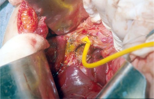
With regard to reparative surgery, there are some basic rules, and the difficulty in repairing damage derives from both the size and caliber of the bile duct and from local inflammatory and sclerotic changes. The timing of the operation is an extremely important factor in the repair of the lesion; therefore, an early identification of the lesion plays a key role. In case of partial or complete transection of the CBD, it is possible to perform a direct end-to-end suture if the distal choledochus is free and without tension. It is also preferable to protect the suture by inserting a Kehr T-tube, as shown in . There is also a second type of T-tube available for biliary drainage, the so-called internal Y-drainage,Citation54 which involves the insertion of the two short branches into both the right and left hepatic ducts, splinting of the anastomosis, and conducting of its long branch into the duodenum by the papilla of Vater. This tube can be removed endoscopically after healing of the end-to-end ductal anastomosis. Moreover, it was suggested as the drainage of choice in end-to-end ductal anastomosis.Citation55–Citation58 In case of impossibility to perform a direct suture or in cases of severe biliary strictures, a biliodigestive anastomosis is recommended, a Roux-en-Y hepaticojejunostomy in most cases, as demonstrated in . If a hepaticojejunostomy cannot be performed due to the presence of dense adhesions with infected and friable tissues, some authors have proposed a combination of endoscopic biliary stenting and pedicled omental patch repair of the bile duct to control bile leak and sepsis as a bridging procedure to definitive hepaticojejunostomy.Citation59 In case of strictures involving the bifurcation or left or RHD, bilateral hepaticojejunostomies may be necessary. For many years, biliary repair has been the prerogative of open surgery. Nowadays, the improvement in surgical technology has allowed to perform reconstruction of iatrogenic biliary lesions even by laparoscopic or robotic approaches with good results.Citation60–Citation62 In a recent paper, Cuendis-Velázquez et alCitation61 have demonstrated that the robotic approach is similar to the laparoscopic technique regarding safety and efficacy in attaining primary patency of hepaticojejunostomy for bile duct repair.
Figure 4 Trans-Kehr cholangiography after the positioning of T-tube to protect biliary anastomotic suture.
Abbreviation: CBD, common bile duct.
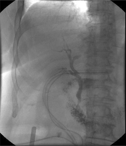
Figure 5 A hepaticojejunostomy was performed for a stricture at biliary confluence after the positioning of T-tube.
Liver resection is very rarely indicated in cases of extensive hepatic necrosis. In a small subset of patients with iatrogenic BDIs, failure of surgical or non-surgical management might lead to acute or chronic liver failure necessitating liver transplantation.Citation63
Bismuth and Strasberg classifications do not include concomitant vascular injuries during LC. In the Stewart-Way classification system, the Class IV biliary injuries are frequently associated with concurrent right hepatic artery lesions. The right hepatic artery injuries may present with hepatic abscess, hemobilia, and right hepatic lobe ischemia.Citation64 Many of these patients require angioembolization for treatment of hemobilia and drainage of hepatic abscesses, and some require hepatectomy.Citation65–Citation68
Furthermore, some authors have also analyzed the consequences of biliary injuries in terms of patient’s QoL. Flores-Rangel et alCitation69 reported that the QoL of the patients undergoing a bilioenteric derivation due to a biliary injury, independently by surgeon’s experience, is reduced significantly when compared to patients undergoing uncomplicated cholecystectomy. Moreover, Booij et alCitation70 have analyzed the long-term impact of BDIs on QoL and found that BDIs are associated with a significant decrease in patient’s QoL and work-related limitations.
Conclusion
Iatrogenic BDIs represent a serious complication which can be brought on by cholecystectomy. The errors leading to laparoscopic bile duct lesions stem principally from misperception of the biliary anatomy. Any effort toward the reduction of the risk profile of everyday cholecystectomy is appreciated. The key points to successful treatment are characterized by early recognition, control of any intra-abdominal fluid collection and infection, nutritional balance, multidisciplinary approach, and surgical repair by an experienced surgeon in biliary reconstruction.
Disclosure
The authors report no conflicts of interest in this work.
References
- PesceAPortaleTRMinutoloVScillettaRLi DestriGPuleoSBile duct injury during laparoscopic cholecystectomy without intraoperative cholangiography: a retrospective study on 1,100 selected patientsDig Surg201229431031422986956
- ArcherSBBrownDWSmithCDBranumGDHunterJGBile duct injury during laparoscopic cholecystectomy: results of a national surveyAnn Surg2001234454955811573048
- WoodsMSEstimated costs of biliary tract complications in laparoscopic cholecystectomy based upon Medicare cost/charge ratios. A case-control studySurg Endosc19961010100410078864095
- CarrollBJBirthMPhillipsEHCommon bile duct injuries during laparoscopic cholecystectomy that result in litigationSurg Endosc19981243103149543519
- BismuthHMajnoPEBiliary strictures: classification based on the principles of surgical treatmentWorld J Surg200125101241124411596882
- StrasbergSMHertlMSoperNJAn analysis of the problem of biliary injury during laparoscopic cholecystectomyJ Am Coll Surg199518011011258000648
- McMahonAJFullartonGBaxterJNO’DwyerPJBile duct injury and bile leakage in laparoscopic cholecystectomyBr J Surg19958233073137795992
- WayLWStewartLGantertWCauses and prevention of laparoscopic bile duct injuries: analysis of 252 cases from a human factors and cognitive psychology perspectiveAnn Surg2003237446046912677139
- BergmanJJvan den BrinkGRRauwsEATreatment of bile duct lesions after laparoscopic cholecystectomyGut19963811411478566842
- StrasbergSMBruntLMRationale and use of the critical view of safety in laparoscopic cholecystectomyJ Am Coll Surg2010211113213820610259
- DalySCDezielDJLiXCurrent practices in biliary surgery: do we practice what we teach?Surg Endosc20163083345335026541721
- NeriVAmbrosiAFersiniATartagliaNValentinoTPAntegrade dissection in laparoscopic cholecystectomyJSLS200711222522817761085
- BornmanPCTerblancheJSubtotal cholecystectomy: for the difficult gallbladder in portal hypertension and cholecystitisSurgery1985981163892743
- RouvièreHOn the configuration and the significance of the groove of the caudate processNewsletter and Memoirs of The Anatomical Society of Paris192494355358
- HughTBKellyMDMekisicARouvière’s sulcus: a useful landmark in laparoscopic cholecystectomyBr J Surg1997849125312549313706
- SinghMPrasadNThe anatomy of Rouviere’s sulcus as seen during laparoscopic cholecystectomy: a proposed classificationJ Minim Access Surg2017132899528281470
- FerzliGTimoneyMNazirSSwedlerDFingerhutAImportance of the node of Calot in gallbladder neck dissection: an important landmark in the standardized approach to the laparoscopic cholecystectomyJ Laparoendosc Adv Surg Tech A2015251283225559890
- SutherlandFDixonEThe importance of cognitive MAP placement in bile duct injuriesCan J Surg201760642442529173261
- FlumDRDellingerEPCheadleAChanLKoepsellTIntraoperative cholangiography and risk of common bile duct injury during cholecystectomyJAMA2003289131639164412672731
- NickkholghASoltaniyektaSKalbasiHRoutine versus selective intraoperative cholangiography during laparoscopic cholecystectomy: a survey of 2,130 patients undergoing laparoscopic cholecystectomySurg Endosc200620686887416738972
- DiliABertrandCLaparoscopic ultrasonography as an alternative to intraoperative cholangiography during laparoscopic cholecystectomyWorld J Gastroenterol201723295438545028839445
- PesceAPortaleTRdi StefanoBCostaSCammisuliFClinical value of intra-operative ultrasonography during laparoscopic cholecystectomyLaparosc Surg201827
- IshizawaTBandaiYKokudoNFluorescent cholangiography using indocyanine green for laparoscopic cholecystectomy: an initial experienceArch Surg2009144438138219380655
- PesceAPiccoloGLa GrecaGPuleoSUtility of fluorescent cholangiography during laparoscopic cholecystectomy: a systematic reviewWorld J Gastroenterol201521257877788326167088
- PesceALatteriSBarchittaMNear-infrared fluorescent cholangiography – real-time visualization of the biliary tree during elective laparoscopic cholecystectomyHPB (Oxford)201820653854529292071
- PesceADianaMCritical view of safety during laparoscopic cholecystectomy: from the surgeon’s eye to fluorescent visionSurg Innov201825319719829557253
- KapoorVK“Colleaguography” in place of cholangiography, to prevent bile duct injury during laparoscopic cholecystectomyJ Minim Access Surg Epub 2018 Aug 7
- ConradCWakabayashiGAsbunHJIRCAD recommendation on safe laparoscopic cholecystectomyJ Hepatobiliary Pancreat Sci2017241160361529076265
- van de GraafFWZaïmiIStassenLPSLangeJFSafe laparoscopic cholecystectomy: a systematic review of bile duct injury preventionInt J Surg20186016417230439536
- NuzzoGGiulianteFGiovanniniIBile duct injury during laparoscopic cholecystectomy: results of an Italian national survey on 56 591 cholecystectomiesArch Surg20051401098699216230550
- KumCKEypaschELeferingRPaulANeugebauerETroidlHLaparoscopic cholecystectomy for acute cholecystitis: is it really safe?World J Surg19962014349 discussion 48-98588411
- GeorgiadesCPMavromatisTNKourlabaGCIs inflammation a significant predictor of bile duct injury during laparoscopic cholecystectomy?Surg Endosc20082291959196418443865
- GigotJEtienneJAertsRThe dramatic reality of biliary tract injury during laparoscopic cholecystectomy. An anonimous multicenter Belgian survey of 65 patientsSurg Endosc199711117111789373288
- MooreMJBennettCLThe learning curve for laparoscopic cholecystectomy. The Southern Surgeons ClubAm J Surg199517055597793496
- HughTBNew strategies to prevent laparoscopic bile duct injury – surgeons can learn from pilotsSurgery2002132582683512464867
- SeerasKKalaniADBile Duct RepairTreasure Island, FLStatPearls Publishing LLC2018 Available at: https://www.ncbi.nlm.nih.gov/books/NBK525989/
- StewartLIatrogenic biliary injuries: identification, classification, and managementSurg Clin North Am201494229731024679422
- StewartLWayLWBile duct injuries during laparoscopic cholecystectomy. Factors that influence the results of treatmentArch Surg19951301011231128 discussion: 11297575127
- de ReuverPRGrossmannIBuschORObertopHvan GulikTMGoumaDJReferral pattern and timing of repair are risk factors for complications after reconstructive surgery for bile duct injuryAnn Surg2007245576377017457169
- LatteriSMalaguarneraGManninoMUltrasound as point of care in management of polytrauma and its complicationJ Ultrasound201720217117728593009
- PalmucciSMauroLACoppolinoMEvaluation of the biliary and pancreatic system with 2D SSFSE, breathhold 3D FRFSE and respiratory-triggered 3D FRFSE sequencesRadiol Med2010115346748220077045
- PalmucciSMauroLALa ScolaSMagnetic resonance cholangiopancreatography and contrast-enhanced magnetic resonance cholangiopancreatography versus endoscopic ultrasonography in the diagnosis of extrahepatic biliary pathologyRadiol Med2010115573274620177983
- GuptaVJayaramanSRole for laparoscopy in the management of bile duct injuriesCan J Surg201760530030428930036
- RainioMLindströmOUddMHaapamäkiCNordinAKylänpääLEndoscopic therapy of biliary injury after cholecystectomyDig Dis Sci201863247448028948425
- SiikiAVaalavuoYAntilaABiodegradable biliary stents preferable to plastic stent therapy in post-cholecystectomy bile leak and avoid second endoscopyScand J Gastroenterol2018515
- PoddaMPolignanoFMLuhmannAWilsonMSKulliCTaitISSystematic review with meta-analysis of studies comparing primary duct closure and T-tube drainage after laparoscopic common bile duct exploration for choledocholithiasisSurg Endosc201630384586126092024
- DonatelliGVergeauBMDerhySCombined endoscopic and radiologic approach for complex bile duct injuries (with video)Gastrointest Endosc201479585586424556053
- FioccaFSalvatoriFMFanelliFComplete transection of the main bile duct: minimally invasive treatment with an endoscopic-radiologic rendezvousGastrointest Endosc20117461393139821963262
- SchizasDPapaconstantinouDMorisDManagement of segmental bile duct injuries after cholecystectomy: a systematic reviewJ Gastrointest Surg Epub 2018 Nov 6
- LiJFrillingANadalinSSurgical management of segmental and sectoral bile duct injury after laparoscopic cholecystectomy: a challenging situationJ Gastrointest Surg201014234435119911237
- MazerLMTapperEBSarmientoJMNon-operative management of right posterior sectoral duct injury following laparoscopic cholecystectomyJ Gastrointest Surg20111571237124221347873
- PereraMTPRMonacoASilvaMALaparoscopic posterior sectoral bile duct injury: the emerging role of nonoperative management with improved long-term results after delayed diagnosisSurg Endosc20112582684269121416174
- PeriniRFUflackerRCunninghamJTSelbyJBAdamsDIsolated right segmental hepatic duct injury following laparoscopic cholecystectomyCardiovasc Intervent Radiol200528218519515770390
- JabłonskaBEnd-to-end ductal anastomosis in biliary reconstruction: indications and limitationsCan J Surg201457427127725078933
- KapoorBSMauriGLorenzJMManagement of biliary strictures: state-of-the-art reviewRadiology2018289359060330351249
- RenzBWBöschFAngeleMKBile duct injury after cholecystectomy: surgical therapyVisc Med201733318419028785565
- JabłońskaBLampePOlakowskiMHepaticojejunostomy vs end-to-end biliary reconstructions in the treatment of iatrogenic bile duct injuriesJ Gastrointest Surg20091361084109319266245
- GazzanigaGMFilauroMMoriLSurgical treatment of iatrogenic lesions of the proximal common bile ductWorld J Surg200125101254125911596885
- NgJJKowAWCPedicled omental patch as a bridging procedure for iatrogenic bile duct injuryWorld J Gastroenterol201723366741674629085219
- Cuendis-VelázquezAMorales-ChávezCAguirre-OlmedoILaparoscopic hepaticojejunostomy after bile duct injurySurg Endosc201630387688226092013
- Cuendis-VelázquezATrejo-ÁvilaMBada-YllánOA new era of bile duct repair: robotic-assisted versus laparoscopic hepaticojejunostomyJ Gastrointest Surg Epub 2018 Nov 6
- GiulianottiPCQuadriPDurgamSBiancoFMReconstruction/repair of iatrogenic biliary injuries: is the robot offering a new option? short clinical reportAnn Surg20182671e7e928657946
- ParrillaPRoblesRVaroELiver transplantation for bile duct injury after open and laparoscopic cholecystectomyBr J Surg20141012636824318962
- HalaszNACholecystectomy and hepatic artery injuriesArch Surg199112621371381992990
- BuellJFCroninDCFunakiBDevastating and fatal complications associated with combined vascular and bile duct injuries during cholecystectomyArch Surg2002137670371012049542
- TzovarasGDervenisCVascular injuries in laparoscopic cholecystectomy: an underestimated problemDig Surg2006235–637037417164583
- SchmidtSCSettmacherULangrehrJMNeuhausPManagement and outcome of patients with combined bile duct and hepatic arterial injuries after laparoscopic cholecystectomySurgery2004135661361815179367
- LiJFrillingANadalinSManagement of concomitant hepatic artery injury in patients with iatrogenic major bile duct injury after laparoscopic cholecystectomyBr J Surg200895446046518161898
- Flores-RangelGAChapa-AzuelaORosalesAJRoca-VasquezCBöhm-GonzálezSTQuality of life in patients with background of iatrogenic bile duct injuryWorld J Surg20184292987299129520485
- BooijKACde ReuverPRvan DierenSLong-term impact of bile duct injury on morbidity, mortality, quality of life, and work related limitationsAnn Surg2018268114315028426479