 ?Mathematical formulae have been encoded as MathML and are displayed in this HTML version using MathJax in order to improve their display. Uncheck the box to turn MathJax off. This feature requires Javascript. Click on a formula to zoom.
Abstract
Background
Bone mass density is an important parameter used in the estimation of the severity and depth of lesions in osteoporosis. Estimation of bone density using existing methods in experimental models has its advantages as well as drawbacks.
Materials and methods
In this study, the X-ray histogram edge detection technique was used to estimate the bone mass density in ovariectomized rats treated orally with germinated brown rice (GBR) bioactives, and the results were compared with estimated results obtained using Archimede’s principle. New bone cell proliferation was assessed by histology and immunohistochemical reaction using polyclonal nuclear antigen. Additionally, serum alkaline phosphatase activity, serum and bone calcium and zinc concentrations were detected using a chemistry analyzer and atomic absorption spectroscopy. Rats were divided into groups of six as follows: sham (nonovariectomized, nontreated); ovariectomized, nontreated; and ovariectomized and treated with estrogen, or Remifemin®, GBR-phenolics, acylated steryl glucosides, gamma oryzanol, and gamma amino-butyric acid extracted from GBR at different doses.
Results
Our results indicate a significant increase in alkaline phosphatase activity, serum and bone calcium, and zinc and ash content in the treated groups compared with the ovariectomized nontreated group (P < 0.05). Bone density increased significantly (P < 0.05) in groups treated with estrogen, GBR, Remifemin®, and gamma oryzanol compared to the ovariectomized nontreated group. Histological sections revealed more osteoblasts in the treated groups when compared with the untreated groups. A polyclonal nuclear antigen reaction showing proliferating new cells was observed in groups treated with estrogen, Remifemin®, GBR, acylated steryl glucosides, and gamma oryzanol. There was a good correlation between bone mass densities estimated using Archimede’s principle and the edge detection technique between the treated groups (r2 = 0.737, P = 0.004).
Conclusion
Our study shows that GBR bioactives increase bone density, which might be via the activation of zinc formation and increased calcium content, and that X-ray edge detection technique is effective in the measurement of bone density and can be employed effectively in this respect.
Introduction
Traditionally, dual X-ray absorptiometry and Archimede’s principle are employed in the measurement of bone mass density (BMD) in biomedical research. The use of dual X-ray absorptiometry to measure bone density has become very popular over the years, and although a significant correlation was obtained when comparing BMD using dual X-ray absorptiometry and Archimede’s principle in rats, the precision, accuracy, and sensitivity of this method in bone density determination remains a topic of concern.Citation1
Previous research has revealed that a progressive decline in BMD occurs with estrogen deficiency at menopause, exposing women to a greater risk for the development of osteoporosis.Citation2,Citation3 Postmenopausal osteoporosis is now a worldwide problem, and the conventional treatment of estrogen replacement and other related chemotherapy are reported to be associated with side effects ranging from alterations in physiology to therapy-related cancers.Citation4–Citation6 Fisher et alCitation7 described osteoporotic fracture as an important public health issue with increasing morbidity, mortality, and prevalence. In the United States alone, osteoporosis affects an estimated 10–28 million Americans over the age of 50, with 33 million people, mostly women, having low bone density and close to 1.2 million bone fractures being related to osteoporosis.Citation8,Citation9 In view of this, natural products, especially phytoestrogens, are being explored as an alternative to hormone therapy for the management of osteoporosis and other related bone degenerative diseases. In this study, germinated brown rice (GBR), a rice with increased levels of bioactive compounds compared to polished rice and brown rice, with potential benefits in the management of chronic disease, was administered to rats with the aim of studying its ability to protect bone from osteoporosis. Bone ash weight and calcium and zinc content were quantified, and bone histology and polyclonal nuclear antigen staining were used to detect new bone formation. BMD was estimated using Archimede’s principle and was compared with edge detection X-ray morphometry in order to explore its applicability in measuring bone density in biomedical research.
Materials and methods
BERNAS Rice Company (Sri Tiram Jaya, Malaysia) supplied the brown rice. Cimicifuga racemosa (Remifemin® 20 mg/tab) was procured from Schaper and Brümmer GmbH (Salzgitter, Germany). Conjugated estrogen (Premarin® 0.625 mg/tab) was obtained from Wyeth Medica Ireland (Newbridge, Ireland). Xylazine HCl 20 mg/mL and ketamine HCl were obtained from Troy Laboratories (Smithfield, Australia).
Animals, grouping, dosing, and frequency
A total of 78 mature 12-week-old Sprague Dawley rats were procured from the Faculty of Veterinary Medicine, University Putra Malaysia (Selangor, Malaysia). They were acclimatized for 2 weeks before ovariectomy or sham operation. Six rats were assigned to each of the 13 treatment groups: group 1: nonovariectomized, nontreated (sham) negative control; group 2: ovariectomized, nontreated positive control; group 3: ovariectomized and treated with estrogen (0.2 mg/kg); groups 4 and 5: ovariectomized and treated with Remifemin® (10 and 20 mg/kg); groups 6–13: ovariectomized and treated with GBR, acylated steryl glucosides (ASG), gamma amino-butyric acid (GABA) or gamma oryzanol (ORZ) at two different doses (100 and 200 mg/kg). Extracts were administered by oral gavage once a day for 8 weeks. The entire animal study was carried out at the animal facility, Faculty of Medicine and Health Science, University Putra Malaysia. Rats were individually housed in plastic cages in a temperature and humidity controlled air-conditioned room (25°C–30°C) with exposure to a 12/12-hour light/dark cycle. The study was carried out according to the guidelines for the use of animals and was approved by the Animal Care and Use Committee (ACUC) of the Faculty of Medicine and Health Sciences, University Putra Malaysia.
Bone mass density and diameter of the femur bone
Femurs were isolated and freed from the surrounding muscles, and bone diameters (diameter of the femur in the distal ¼ and diameter of the femur in the distal ½) were determined using a KERNN 150 mm digital caliper (Kern and Sohn, Balingen, Germany). The bone mass density was estimated using both Archimede’s principle and the X-ray edge detection technique.
Archimede’s principle
The BMD of the left femur was calculated by measuring the mass of the bone in air and measuring the weight again after submerging in a specific volume of distilled water. From these two masses, the density was then calculated using the formula,
where d is density, w is weight, w1 is weight in air, w2 is weight in water, and P is density of distilled water at a given temperature and is expressed as g/cm3.
X-ray morphometry
The bones were placed on a grid view specimen container (CIRS, Norfolk, VA, USA) at the 3, 6, 9, and 12 o’clock positions, and exposed to X-rays using a Philips Mammo diagnostic 3000 X-ray machine (Philips Healthcare, Andover, MA, USA) at controlled exposures of 62 kVp, 4 mAs.
Automatic classification of bone structure
We designed an algorithm that had the ability to classify the bone structure automatically as summarized in . This algorithm was designed with MATLAB software (MathWorks, Natick, MA, USA) and runs on a PC with a HP Pavilion Notebook dv6 computer (Hewlett-Packard, Palo Alto, CA, USA) with the following specifications: core i5, 2.4 GHz processor speed, 520 M cache. According to the proposed algorithm, the original image obtained from the X-ray machine () was filtered through a Wiener filter to remove the noise in the image. The filtered image was then divided into different subimages (). The local range filter and local entropy were applied to extract the features of this subimage. The local range filter was used to specify the magnitude of the gradient. Local range filtering was determined by calculating the difference between the maximum and minimum range values of the filtered window. Local range filtering has a short calculation time, as it operates on only a small number of inputs for each output pixel.Citation10 On the other hand, using local entropy will create a textured image wherein the local entropy is related to the variance in the grayscale in the neighborhood. The local entropy is larger for a heterogeneous region but smaller for a homogeneous neighborhood. Accordingly, the transition region will have larger local entropy values than those in nontransition regions of the image.Citation11 The weighted sum (v and w) of the magnitude of the gradient and the local entropy was used to calculate the pixels’ importance and can be defined as follows,
Figure 1 Flowchart summarizing the proposed algorithm.
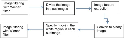
Figure 2 Stages involved in the measurement of bone mass density using X-ray morphometry and edge detection technique in ovariectomized rats treated with germinated brown rice and its bioactives, estrogen, or Remifemin® for a period of 8 weeks.
Abbreviations: X1, osteoporotic; X2, osteopenia; X3, normal bone density; X4, increase bone density; X5–X7, high bone density.
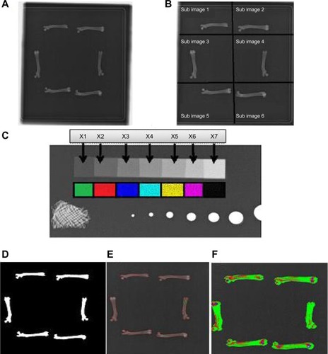
where gra (x, y) and entr (x, y) are the magnitude of the gradient and the local entropy for pixel (x, y), respectively, and v and w are the weight for balancing the local entropy and the magnitude of gradient features, respectively. At this stage, the grayscale subimage was converted to a binary image to obtain an image with two colors: white for the bone and black for the background of the subimage. To enable the classification process, the original grayscale X1–X7 (where X1 denotes lower density and X7 denotes higher density) was converted to RBG (red, blue, green) color for easy identification, as shown in . The range of intensity in each block on the original grayscale was used as a training set for k-nearest neighbor, which is a powerful method of nonparametric analysis.Citation12 There are three key elements of this approach: (1) a set of labeled objects, (2) a distance or similarity metric to compute distance between objects, and (3) the value of the number of nearest neighbor’s k. To classify an unlabeled object, the distance of this object to the labeled objects is computed, and its k-nearest neighbors are identified; the nearest-neighbor list is thus obtained, the test object is classified based on the majority class of its nearest neighbors, and the class labels of these nearest neighbors are then used to determine the class label of the object.Citation12–Citation14 The fractional percentage for the bone was calculated by taking each pixel in the bone and classifying it with k-nearest neighbor to specify each pixel’s structure according to the classification range (X1–X7). – show the segmentation process, the edge detection method, and classification, respectively.
Measurement of serum zinc and calcium concentrations
Blood was obtained from rats at 2, 4, and 8 weeks by cardiac puncture under anesthesia. Serum was collected by centrifugation at 3,000 rpm for 15 minutes and stored at −20°C. Zinc and calcium concentrations were analyzed using Randox analytical kits (Randox, Crumlin, UK) on a Selectra XL automated chemistry analyzer (Vita Scientific, Dieren, The Netherlands) according to the manufacturer’s instructions.
Bone ash weight, calcium and zinc content
Bone calcium was determined by atomic absorption spectrophotometry (S series GE712405 v1.30; Thermo Fisher Scientific, Waltham, MA, USA) using a procedure reported by Harrison et al,15 with some modifications. Briefly, bone samples were dried at 100°C for 24 hours before they were ashed in a muffle furnace at 550°C–600°C for another 24 hours and the ash weighed. The ash was pulverized into powder and hydrolyzed with 6 M HCl. The hydrolysate was then used to determine the calcium content at a wavelength of 422.7 nm, lamp current 100%, and a band pass and measurement time of 0.5 nm and 4 seconds, respectively. Zinc was determined at a wavelength of 213.9 nm, lamp current 75%, and a band pass and measurement time of 0.2 nm and 4 minutes, respectively. Both calcium and zinc concentrations were expressed in mg/L.
Histology
Bones were fixed in 10% neutral formalin for 3 days, during which the formalin was changed every 24 hours. The bones were transferred to 80% formic acid for decalcification for 1 week, then embedded in paraffin and cut into longitudinal sections of 5 μm thickness. The sections were stained with hematoxylin and eosin and later viewed under a light microscope.
Immunohistochemistry
Immunohistochemistry to detect the presence of a nuclear antigen was performed following a previously reported procedure,Citation16 with some modifications. Bone sections were mounted on gelatin-coated glass slides, deparaffinized in three changes of xylol, dehydrated in graded alcohol, and then washed with distilled water. The sections were placed in 10 mM citrate buffer, pH 6.0, for 10 minutes at 50 W in a microwave, then cooled at room temperature for 5 minutes. Nonspecific binding was covered using 5% bovine serum albumin (BSA). Sections were then incubated using hydrogen peroxide (3%) for 30 minutes to block endogenous peroxidase activity and were then washed in phosphate buffered saline containing 0.2% solution of Tween-20 and distilled water. PC10 monoclonal antibody (DakoCytomation, Copenhagen, Denmark) was used as the primary antibody for 1 hour at a ratio of 1:200 then rinsed in phosphate buffered saline and reacted with polyclonal rabbit anti-mouse secondary antibody for 10 minutes at room temperature. The peroxidase reactions were developed in 3,3 diaminobenzidine in chromagen solution (DakoCytomation, Copenhagen, Denmark), counterstained with methylene blue for 2 minutes, and finally, the sections were cleared in xylene and coverslipped for examination under the light microscope. Images were captured at strategic locations on the slide using an image analyzer (Analysis LS Research) attached to the microscope (Olympus BX51, Japan).
Statistical analysis
Data are presented as mean ± standard deviation. Differences were determined by one-way analysis of variance (ANOVA) and mean comparison by Tukey–Kramer post hoc test, using JMP10 statistical software (SAS, Cary, NC, USA). Differences were considered significant at P < 0.05.
Results
The diameter of the femur in the distal ½ in the sham nonovariectomized group, and the ovariectomized groups treated with 0.2 mg/kg estrogen, 200 mg/kg ASG, GBR, or ORZ was significantly higher compared to the ovariectomized, nontreated group (P < 0.05). The diameter of the femur in the distal ¼ in the ovariectomized, nontreated group and groups treated with ORZ 200 mg/kg and estrogen 0.2 mg/kg was significantly higher (P < 0.05) compared to the nontreated group ().
Table 1 Bone mass density of the femur in g/cm3 and diameter of the shaft in the distal 20% (F20) and central 50% (F50) in ovariectomized rats treated with GBR and its bioactives, estrogen, or Remifemin® for a period of 8 weeks
The bone densities of the sham nonovariectomized group and groups treated with estrogen 0.2 mg/kg, and ORZ, ASG, GABA each at 200 mg/kg, and Remifemin® 20 mg/kg were significantly higher compared to the ovariectomized, nontreated group (P < 0.002, P < 0.03, and P < 0.01, respectively). The other treated groups expressed a higher density than the nontreated group although not significantly (P > 0.05) when using Archimede’s principle (). The X-ray edge detection technique yielded the following percentage increase in bone formation in these groups: ASG 200 mg/kg, 0.38%; estrogen 0.2 mg/kg, 1.55%; GABA 200 mg/kg, 0.02%; ORZ 200 mg/kg, 0.6%; Remifemin® 20 mg/kg, 0.04%; and SHAM, 0.28%. These treatment groups showed an increase in BMD and fell under the X4 scale of classification (). A significant correlation was observed between densities obtained using Archimede’s principle and those obtained in X3 (bone density values in class 3 grading using X-ray morphometry) (R2 = 0.737, P = 0.004); a nonsignificant correlation was observed between the two methods in groups that showed a significant increase in bone formation (R2 = 0.710, P = 0.179) ().
Table 2 X-ray edge detection showing density grading and fractional composition of bones in the image after 8 weeks of oral intervention with GBR and its bioactives, estrogen, or Remifemin® in ovariectomized rats
Table 3 Pearson correlation between the results of bone density measurements using Archimede’s principle versus X-ray morphometry and edge detection technique
The weight of the femur bone immediately after sacrifice of rats (wet weight) in the ovariectomized, nontreated group was significantly lower (P < 0.05) than in the sham and other treated groups (). Bone ash weight was higher in the sham group and groups treated with 0.2 mg/kg estrogen, ASG 200 mg/kg, and ORZ 100 mg/kg and 200 mg/kg, and significantly different (P < 0.05) from that of the ovariectomized, nontreated group and the other treatment groups, as shown in .
Table 4 Wet and ash weight, calcium and zinc concentrations of femur bone of ovariectomized rats treated with estrogen, Remifemin®, and germinated brown rice and its bioactives
Bone calcium content in the ovariectomized, nontreated group was significantly lower (P < 0.05) than that in sham and all ovariectomized treated groups. No significant difference in terms of calcium content was observed in groups treated with estrogen 0.2 mg/kg, GBR 200 mg/kg, ASG 100 mg/kg and 200 mg/kg, ORZ 100 mg/kg and 200 mg/kg, GABA 200 mg/kg, and the sham nonovariectomized group (P < 0.05) ().
Bone zinc concentration was significantly lower in the ovariectomized, nontreated group when compared to the sham and all treated groups (P < 0.05). The concentration increased significantly (P < 0.05) in groups treated with estrogen 0.2 mg/kg, Remifemin® 10 mg/kg, ASG 200 mg/kg, GABA 200 mg/kg, ORZ 100 mg/kg and 200 mg/kg compared to the sham nonovariectomized group, as shown in .
There was significant difference in the levels of serum alkaline phosphatase (ALP) between 2 weeks and 8 weeks after treatment in the group treated with ASG 100 mg/kg compared to the ovariectomized, nontreated group and other treatment groups: P < 0.05. During the fourth week after treatment, a significant difference was observed between sham nonovariectomized rats and the group treated with Remifemin® 10 mg/kg (P < 0.0451) ().
Table 5 Serum alkaline phosphatase (ALP) and calcium and zinc concentration in ovariectomized rats after 8 weeks of intervention with germinated brown rice phenolics, ASG, GABA, ORZ, estrogen, and Remifemin® at various oral doses
Serum calcium concentrations were significantly different between treated groups in the eighth week only after commencement of treatment (P = 0.0001). There was no significant difference between the treated groups in the second and fourth week (P = 0.9390 and P = 0.8948, respectively) ().
Serum zinc concentrations showed a significant increase in the sham nontreated group, ORZ 200 mg/kg, and GBR 200 mg/kg 2 weeks after treatment compared to ovariectomized, nontreated and other treated groups (P < 0.0001) (). After 4 weeks of treatment, serum zinc concentration in groups treated with GABA 200 mg/kg, estrogen 0.2 mg/kg, and GBR 200 mg/kg were significantly different compared to other treatment groups (P < 0.0001), as shown in . Zinc concentrations in the nonovariectomized (sham) group and groups treated with GABA 200 mg/kg and estrogen 0.2 mg/kg in the eighth week differed significantly from those of the other treatment groups (P < 0.0001) ().
Bone histology
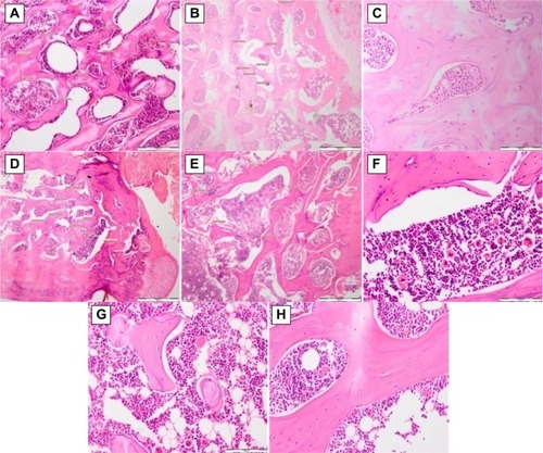
Histologically, the sham nonovariectomized group showed a normal bone cell configuration with more osteocytes and minimal osteoblastic activity compared to ovariectomized, nontreated group (). The ovariectomized, nontreated rats showed vacuolation in the bone marrow, reduced marrow and osteogenic activity, complete absence of osteoblasts, reduced bone density, and marked degenerative changes (), while the group treated with estrogen showed an increase in bone formation with minimal marrow activity and osteoblast entrapment at the margins (). Similarly, the Remifemin®-treated group showed evidence of an increase in new bone formation (). The GBR-treated group showed active proliferation of new bone cells () and the ASG-treated group showed bone marrow with lymphoblastic cells with some proteinaceous fluid. Osteocytes were entrapped at the margin. Active osteoblasts at the margin were in the process of converting to osteocytes, showing active bone formation and increased bone density (). Hematopoietic cells, mature osteocytes, and osteoblasts were present. The GABA-treated group also showed evidence of active bone formation (), and the ORZ-treated group showed an increase in osteoblastic activity and an increase in new bone formation ().
Figure 3 Histological slides showing hematoxylin and eosin staining of bone tissue.
Abbreviations: ASG, acylated steryl glucosides; EST, estrogen; GABA, gamma amino-butyric acid; GBR, germinated brown rice; ORZ, gamma oryzanol; REM, Remifemin®.
Immunohistochemistry
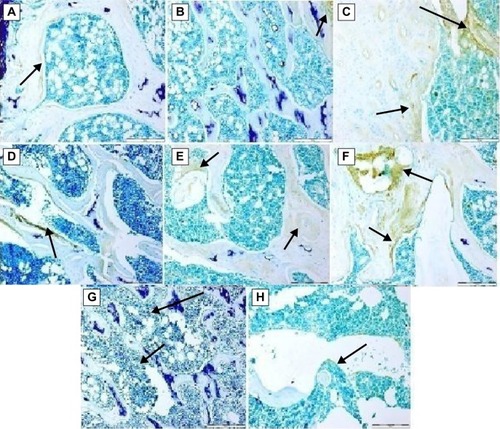
Immunohistochemistry of the bone tissue gave weak positive staining, indicating new bone formation in all treated groups, and the reaction was more prominent in the groups treated with estrogen, remifemin, and ASG (). While groups treated with GBR, GABA and ORZ showed a milder reaction (). The sham and OVX-untreated groups both showed a very little or no reaction ().
Figure 4 Immunohistochemical staining of polyclonal nuclear antigen-positive cells in bone tissue.
Abbreviations: ASG, acylated steryl glucosides; EST, estrogen; GABA, gamma amino-butyric acid; GBR, germinated brown rice; ORZ, gamma oryzanol.
Discussion
It was evident in this study that GBR bioactives increased bone density. To the best of our knowledge this is the first report on the effects of GBR on BMD and the application of the X-ray edge detection technique in the measurement of bone density.
Our results using the edge detection technique quantified the increase in percentage of bone formation in the individual treatments, and the results were highly correlated with those obtained using the standard Archimede’s principle.
The ovariectomized, nontreated rats showed a decrease in BMD. Ovariectomy is known to stimulate oxidative stress and interfere with the antioxidant system in rats.Citation17 This leads to an increase in the level of oxidative stress markers such as hydrogen peroxide, which is highly indicative of bone loss in estrogen shortageCitation18 and also tumor necrosis factor alpha, which is generated as a result of the low level of thiol antioxidants within bone cells.Citation19–Citation21 Our data show that GBR bioactives, specifically ASG, GABA, and ORZ, increased the BMD to a level almost the same as that of the sham nonovariectomized group. GABA and ORZ from GBR have been shown to upregulate genes related to bone formation in ovariectomized rats.Citation22 Serum ALP activity increased in the ovariectomized, nontreated group and other treatment groups especially during the fourth week after ovariectomy. This might be due to an increase in the rate of bone metabolism, especially a few days after ovariectomy, and an increase in osteogenic activity in the treated groups. An increase in ALP was also reported in ovariectomized rats treated with soy isoflavones.Citation23,Citation24 ALP is known as a prominent marker of bone formation.Citation25 Bone remodeling involves both bone formation and resumption in a coupling effect,Citation26 which might explain the increase in the activity of ALP in ovariectomized rats. Serum and bone calcium level, bone wet weight, and the ash content of the bone decreased in the ovariectomized, nontreated group due to a decrease in estrogenic activity, which in turn affected the bone mass and constituency, calcium content, and zinc concentration. Treatment with ORZ, GABA, and ASG at 200 mg/kg restored the values of these parameters to the same level or higher than in the sham nonovariectomized group. This gives a clear indication that these bioactives are fully involved in osteogenesis. The decrease in zinc concentration observed in ovariectomized, nontreated rats has also been reported by other researchers in low estrogenic conditions of menopausal women and in ovariectomized rats.Citation27,Citation28 This decrease in zinc concentration is due to oxidative stress, and zinc is known to play a major regulatory role in the action of antioxidant enzymes.Citation29 Treatment for 8 weeks with GBR bioactives increased the level of zinc in bone tissue to a level higher than that in the control sham nonovariectomized rats and groups treated with ORZ, ASG, estrogen, and Remifemin®. It has been reported that zinc is a potent inhibitor of osteoclastogenesis and is an osteoblast stimulator in vitro.Citation30,Citation31 Zinc is known to stimulate protein synthesis, and its role in bone formation and in preserving bone mass is greater than that of bone regulating hormones.Citation32 Histology and the polyclonal nuclear antigen reactions showed evidence of active bone formation with new bone cells in their active proliferation stage.
Conclusion
This study indicates that the increase in BMD and osteoprotective effect of GBR bioactives might involve the activation of zinc formation and increased calcium, coupled with an increase in ALP, which together increase osteoblastic activity and subsequently bone formation. However, this proposed mechanistic effect requires further clarification, and more research is needed to address the molecular mechanism of zinc stimulation by GBR bioactives.
Acknowledgments
We wish to thank Padiberas National (BERNAS) in Sri Tiram Jaya, Malaysia for funding this research. We also wish to acknowledge the staff of Narinda X-ray clinic, Ampang, Kuala Lumpur for their help and cooperation.
Disclosure
The authors report no conflicts of interest in this work.
References
- KeenanMJHegstedMJonesKLComparison of bone density measurement techniques: DXA and Archimedes’ principleJ Bone Miner Res19971211190319079383695
- MeltonLJEpidemiology of hip fractures: implications of the exponential increase with ageBone199618Suppl 3121S125S8777076
- CummingsSRTreatable and untreatable risk factors for hip fractureBone199618Suppl 3165S167S8777083
- MaffeiSDel RySPronteraCClericoAIncrease in circulating levels of cardiac natriuretic peptides after hormone replacement therapy in postmenopausal womenClin Sci2001101544745311672449
- NabulsiAAFolsomARWhiteAAssociation of hormone-replacement therapy with various cardiovascular risk factors in postmenopausal womenN Engl J Med199332815106910758384316
- GapsturSMMorrowMSellersTAHormone replacement therapy and risk of breast cancer with a favorable histology: results of the Iowa Women’s Health StudyJAMA1999281222091209710367819
- FisherASrikusalanukulWDavisMSmithPCardiovascular diseases in older patients with osteoporotic hip fracture: prevalence, disturbances in mineral and bone metabolism, and bidirectional linksClin Interv Aging2013823925623460043
- CummingsSRMeltonLJEpidemiology and outcomes of osteoporotic fracturesLancet200235993191761176712049882
- BurgeRDawson-HughesBSolomonDHWongJBKingATostesonAIncidence and economic burden of osteoporosis-related fractures in the United States, 2005–2025J Bone Miner Res200722346547517144789
- HafizahWMSupriyantoEAutomatic generation of region of interest for kidney ultrasound images using texture analysisInt J Biol Biomed Eng2012612634
- YanCSangNZhangTLocal entropy-based transition region extraction and thresholdingPattern Recognit Lett2003241629352941
- CunninghamPDelanySJk-Nearest neighbour classifiersMult Classif Syst2007117
- TsypinMRöderHOn the reliability of kNN classificationLect Notes Eng Comput Sci. Proceedings of the World Congress on Engineering and Computer Science 2007 WCECS 2007October 24–26, 2007San Francisco, CA, USA2167
- WuXKumarVQuinlanJRTop 10 algorithms in data miningKnowl Inf Syst2008141137
- HarrisonEAdjeiAAmehoCYamamotoSKonoSThe effect of soybean protein on bone loss in a rat model of postmenopausal osteoporosisJ Nutr Sci Vitaminol (Tokyo)19984422572689675706
- IwakiAJingushiSOdaYLocalization and quantification of proliferating cells during rat fracture repair: detection of proliferating cell nuclear antigen by immunohistochemistryJ Bone Miner Res1997121961029240731
- MuthusamiSRamachandranIMuthusamyBOvariectomy induces oxidative stress and impairs bone antioxidant system in adult ratsClinic Chim Acta20053601–28186
- LeanJMJaggerCJKirsteinBFullerKChambersTJHydrogen peroxide is essential for estrogen-deficiency bone loss and osteoclast formationEndocrinology2005146272873515528306
- JaggerCJLeanJMDaviesJTChambersTJTumor necrosis factor-alpha mediates osteopenia caused by depletion of antioxidantsEndocrinology2005146111311815388652
- LeanJMDaviesJTFullerKA crucial role for thiol antioxidants in estrogen-deficiency bone lossJ Clin Invest2003112691592312975476
- TsayJYangZRossFPBone loss caused by iron overload in a murine model: importance of oxidative stressBlood2010116142582258920554970
- MuhammadSIIsmailMRozi MahmudRBZukiAZImamMUUp-regulation of genes related to bone formation by Gamma-amino butyric acid and gamma oryzanol in germinated brown rice is via the activation of GABAB-receptors and reduction in IL-6 in RatsClin Interv Aging2013 (In-press)
- ArjmandiBHBirnbaumRGoyalNVBone-sparing effect of soy protein in ovarian hormone-deficient rats is related to its isoflavone contentAm J Clin Nutr199868Suppl 61364S1368S9848500
- ArjmandiBHGetlingerMJGoyalNVRole of soy protein with normal or reduced isoflavone content in reversing bone loss induced by ovarian hormone deficiency in ratsAm J Clin Nutr199868Suppl 61358S1363S9848499
- MariePJHottMModrowskiDAn uncoupling agent containing strontium prevents bone loss by depressing bone resorption and maintaining bone formation in estrogen-deficient ratsJ Bone Miner Res1993856076158511988
- RaiszLGPathogenesis of osteoporosis: concepts, conflicts, and prospectsJ Clin Invest2005115123318332516322775
- HerzbergMLuskyABlonderJFrenkelYThe effect of estrogen replacement therapy on zinc in serum and urineObstet Gynecol1996876103510408649686
- HerzbergMFoldesJSteinbergRMenczelJZinc excretion in osteoporotic womenJ Bone Miner Res1990532512572333784
- MaggioDBarabaniMPierandreiMMarked decrease in plasma antioxidants in aged osteoporotic women: results of a cross-sectional studyJ Clin Endocrinol Metab20038841523152712679433
- MoongaBSDempsterDWZinc is a potent inhibitor of osteoclastic bone resorption in vitroJ Bone Miner Res19951034534577785467
- YamaguchiMOishiHSuketaYStimulatory effect of zinc on bone formation in tissue cultureBiochem Pharmacol19873622400740123689432
- YamaguchiMRole of zinc in bone formation and bone resorptionJ Trace Elem Exper Med1998112–3119135