
Abstract
Optic nerve trauma is a common occurrence that results in irreversible blindness. Currently, no effective strategies are known to prevent optic nerve degeneration. We assessed the therapeutic effects of bone marrow mesenchymal stem cells (BMSCs) after optic nerve crush in the adult rat. Our results showed that BMSCs significantly promoted the regeneration of injured axons compared with phosphate buffered saline alone. Therefore, BMSC transplantation may be effective for the treatment of central nervous system disorders.
Introduction
Optic nerve damage is an important cause of irreversible vision loss, which results from retinal ganglion cell (RGC) death and subsequent axonal degeneration.Citation1,Citation2 It is well known that in the central nervous system, the failure of axonal regeneration is due not only to the limited intrinsic growth capacity of adult neurons but also to the growth inhibitory environment.Citation3 Therefore, therapies should focus on the protection of injured neurons and on the promotion of axonal regeneration.Citation4–Citation8 Cell transplantation therapy has been proven effective in this regard.Citation9
Bone marrow mesenchymal stem cells (BMSCs) have both amplification and transplantation abilities.Citation10,Citation11 Furthermore, BMSCs have been shown to exert a neuroprotective effect on RGCs.Citation12 However, little is known about the effects of BMSCs on axonal regeneration of the injured optic nerve. Here, we explored the axonal regeneration-promoting effect of BMSCs after optic nerve crush (ONC).
Materials and methods
ONC and intravitreal injection
This experiment was approved by the Ethics Committee of Animal Research of RenJi Hospital, The School of Medicine, Shanghai JiaoTong University. Eighty adult male Sprague Dawley rats (weight: 200–250 g) were provided by the Shanghai Institute of Traumatology and Orthopedics and were housed in a temperature- and humidity-controlled room with a 12:12 hour light:dark cycle.
Surgical procedures were performed as described in our previous study.Citation13 The right optic nerve was crushed 1 mm behind the posterior eye pole for 9 seconds for all experimental groups. But the optic nerve was not crushed in the sham-surgery group. BMSCs were purchased from Shanghai Shengong Biotechnology (Shanghai Shengong Biotechnology, Shanghai, People’s Republic of China). A total of 3 μL of phosphate buffered saline (PBS) or BMSCs (low number of transplantation cells: 3×10⁴; high number of transplantation cells: 1×10⁵) were intravitreally injected immediately, and 3, 6, and 9 days after ONC. Rats in the sham surgery group did not receive an intravitreal injection. Rats with lens injury were excluded. These rats were divided into four groups: the sham surgery group, PBS group, low BMSC group, and high BMSC group. Twenty rats in each experimental group were included in the final evaluation.
Processing of optic nerve samples
The procedures were performed as described in our previous study.Citation13 The rats were sacrificed on day 15 after ONC and were transcardially perfused with 4% paraformaldehyde. The optic nerves were dissected and immediately fixed in 4% PFA in paraformaldehyde for 1 hour. Then, the tissue was embedded, frozen, and cut. Longitudinal sections (16 μm thickness) of the optic nerve were obtained and were placed on gelatin-coated glass slides for subsequent use.
Axon count
With respect to the axon count, the optic nerve sections were dehumidified at 37°C for 1 hour, and nonspecific binding was blocked by the application of 10% normal goat serum. To stain the regenerating fibers, an anti-GAP43 antibody (mouse anti-rat GAP-43 monoclonal antibody, 1:100, sc-17790; Santa Cruz Biotechnology Inc., Dallas, TX, USA) was applied and the sections were incubated at 4°C overnight. After two washes with PBS, the secondary antibody (Cy-3-labeled anti-mouse fluorescein isothiocyanate antibody, 1:100; Thermo Fisher Scientific, Waltham, MA, USA) was applied and the sections were incubated for 45 minutes at room temperature. GAP-43-positive axons were quantified to assess the regeneration of the axons of RGCs as previously described.Citation13
Western blot analysis
Western blot analysis was used to determine the expression levels of GAP-43 after ONC. The methods of Western blot analysis were described in our previous study.Citation13
Statistical analyses
The data analyses were performed using SPSS version 19.0 software (IBM Corporation, Armonk, NY, USA), and the data are expressed as the mean ± standard deviation. The differences between the groups were determined by one-way analysis of variance or by independent samples Student’s t-test. A P<0.05 was considered statistically significant.
Results
BMSC promotes the axonal regeneration of rat RGCs following ONC
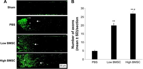
Our results indicated very few axons beyond the crush site in the PBS-treated group. However, a large number of GAP-43-positive axons passed through the crush site in the BMSC-treated group (low or high transplantation group) on day 15 after ONC (). Rats in the sham surgery group showed no significant staining (). The data analysis suggested that more axons were present in the two BMSC groups (low and high) than in the PBS group (P<0.001). Moreover, the high BMSC transplantation group was associated with higher numbers of regenerative axons in comparison with the low BMSC transplantation group.
Figure 1 Representative photomicrographs of regenerating axons in all groups after ONC.
Abbreviations: ONC, optic nerve crush; SD, standard deviation; BMSC, bone marrow mesenchymal stem cell; PBS, phosphate buffered saline.
BMSCs increase GAP-43 expression
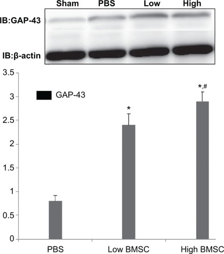
According to Western blot analysis, GAP-43 expression was weak in the sham surgery group, but GAP-43 expression was differentially increased in the PBS and BMSC (low and high) groups (P<0.001; ). Moreover, GAP-43 expression in the low and high BMSC transplantation groups was higher than that in the PBS group (). In addition, the group with a high number of transplanted cells demonstrated increased axonal regeneration compared with the group with a low number of transplanted cells (P>0.05; ).
Figure 2 Western blot analysis of GAP-43 expression.
Abbreviations: BMSC, bone marrow mesenchymal stem cell; IB, immunoblotting; PBS, phosphate buffered saline.
Discussion
The present study explored the effect of transplanted BMSCs on axonal-regeneration after the ONC. GAP-43 expression in rats in the PBS and BMSC groups was significantly increased compared with rats in the sham surgery group, but the GAP-43 protein levels were higher in both BMSC groups than in the PBS group. This result suggested that BMSCs promoted axonal regeneration of the injured optic nerve.
The mechanism of how BMSC therapy might be effective for axonal regeneration after ONC has not yet been elucidated. The function of transplanted BMSCs may involve the regulation of immune system-related inflammation in the injured tissues. In experimental autoimmune encephalomyelitis, Semon et al demonstrated a significant reduction in inflammatory infiltrates by BMSCs.Citation14 Furthermore, BMSC-conditioned medium significantly decreased cell proliferation and the secretion of pro-inflammatory factors through activated microglia.Citation15 Li et al also indicated that BMSCs enhanced axonal remodeling in a model of experimental stroke.Citation16 On the contrary, BMSCs promoted axonal regeneration via the increase in the production of trophic factors.Citation17 In addition, no significant adverse effects were observed in the two BMSC groups.
In summary, our study demonstrates that BMSCs may be a potential therapeutic option for disease of the central nervous system.
Disclosure
The authors report no conflicts of interest in this work.
References
- TangZZhangSLeeCAn optic nerve crush injury murine model to study retinal ganglion cell survivalJ Vis Exp201150268521540827
- YolesESchwartzMDegeneration of spared axons following partial white matter lesion: implications for optic nerve neuropathiesExp Neurol19981531179743562
- PlanchampVBermelCTöngesLBAG1 promotes axonal outgrowth and regeneration in vivo via Raf-1 and reduction of ROCK activityBrain2008131Pt 102606261918757464
- LevinLAAxonal loss and neuroprotection in optic neuropathiesCan J Ophthalmol200742340340817508035
- ZalishMLavieVDuvdevaniRYolesESchwartzMGangliosides attenuate axonal loss after optic nerve injuryRetina19931321451478337497
- FitzgeraldMPayneSCBartlettCAEvillLHarveyARDunlopSASecondary retinal ganglion cell death and the neuroprotective effects of the calcium channel blocker lomerizineInvest Ophthalmol Vis Sci200950115456546219474405
- ThalerSFiedorowiczMRejdakRNeuroprotective effects of tempol on retinal ganglion cells in a partial optic nerve crush rat model with and without iron loadExp Eye Res201090225426019883642
- TsaiRKChangCHSheuMMHuangZLAnti-apoptotic effects of human granulocyte colony-stimulating factor (G-CSF) on retinal ganglion cells after optic nerve crush are PI3K/AKT-dependentExp Eye Res201090553754520144610
- HouSNicholsonLvan NiekerkEMotschMBleschADependence of regenerated sensory axons on continuous neurotrophin-3 deliveryJ Neurosci20123238132061322022993437
- JohnsonTVMartinKRCell transplantation approaches to retinal ganglion cell neuroprotection in glaucomaCurr Opin Pharmacol2013131788222939899
- BouhenniRADunmireJSewellAEdwardDPAnimal models of glaucomaJ Biomed Biotechnol2012201269260922665989
- HuYTanHBWangXMRongHCuiHPCuiHBone marrow mesenchymal stem cells protect against retinal ganglion cell loss in aged rats with glaucomaClin Interv Aging201381467147024204132
- TanHZhongYShenXChengYJiaoQDengLErythropoietin promotes axonal regeneration after optic nerve crush in vivo by inhibition of RhoA/ROCK signaling pathwayNeuropharmacology20126361182119022750411
- SemonJAManessCZhangXComparison of human adult stem cells from adipose tissue and bone marrow in the treatment of experimental autoimmune encephalomyelitisStem Cell Res Ther201451224405805
- YanKZhangRSunCBone marrow-derived mesenchymal stem cells maintain the resting phenotype of microglia and inhibit microglial activationPLoS One2013812e8411624391898
- LiYMcIntoshKChenJAllogeneic bone marrow stromal cells promoteglial-axonal remodeling without immunologic sensitization after stroke in ratsExp Neurol2006198231332516455080
- NovikovaLNBrohlinMKinghamPJNovikovLNWibergMNeuroprotective and growth-promoting effects of bone marrow stromal cells after cervical spinal cord injury in adult ratsCytotherapy201113787388721521004