
Abstract
Purpose
Previous studies demonstrated changes in sensorimotor network activation over time after stroke that have been interpreted as partly compensatory. Locomotor and balance trainings may improve both mobility and cognition even in chronic stroke and thereby impact on cerebral activation patterns. We here aimed at testing these assumptions in an exploratory study to inform subsequent larger intervention studies.
Patients and methods
Eight patients (73.3±4.4 years) with a chronic lacunar stroke (mean interval 3.7 years after the acute event with a range from 2 to 4 years) and residual leg paresis leading to gait disturbance received a guided 5-week training focusing on mobility, endurance, and coordination. Before and afterward, they underwent clinical, neuropsychological, and gait assessments and brain MRI at 3 T including a functional ankle movement paradigm. Sixteen healthy controls (HCs; 68.8±5.4 years) followed the same protocol without intervention.
Results
After training, patients had improved in mobility, memory, and delayed recall of memory. While cerebral activations in HC remained completely unaltered, patients showed increased activations in the right precentral gyrus, the right and left superior frontal gyri, and the right frontal lobe, with bipedal ankle movements after training.
Conclusion
In this exploratory study of chronic stroke, we found not only significant effects of physical training on mobility but also distinct aspects of cognition already with a small number of highly selected patients. These improvements were paralleled by alterations in cerebral activity possibly reflecting neuronal plasticity. Larger studies including randomization are needed.
Introduction
There are reports that physical training may have positive effects even with more advanced age and in the chronic phase of a stroke.Citation1 Such effects have been shown not only concerning mobility but also for cognition, and it has been suggested that training may partially normalize brain activation in motor networks after stroke.Citation2 In contrast, especially increased contralesional and extramotor network activations have been correlated with decreased motor abilities.Citation3,Citation4 Previously termed lacunar infarcts (now preferably called recent small subcortical infarcts)Citation5 represent a morphologically well-characterized subtype of stroke that may cause such impairments in mobility and cognition.Citation1,Citation6,Citation7 More than 60% of patients with lacunar stroke still have motor dysfunction after 12 months.Citation8
In contrast to other studies, we thus examined training effects on mobility and cognition in healthy controls (HCs) and patients with lacunar stroke, because this patient group has a higher chance for intact compensatory networks and residual impairments of mobility.Citation9 We here explored 1) the behavioral effects of a dedicated mobility training on i) motor and ii) cognitive abilities in chronic stroke patients with residual gait impairment and 2) whether parallel changes in cerebral activation measured by functional magnetic resonance imaging (fMRI), using an apparatus for bipedal ankle movements, occur.
Patients and methods
Patients
We included eight patients from our stroke outpatient department, aged between 69 and 81 years, who were willing to undergo a 5-week training and who satisfied the following inclusion criteria: mobility impairment (impaired gait function – three with a right-sided paresis and five with a left-sided paresis) caused by an imaging-proven ischemic subcortical infarct, which had occurred at least 6 months (mean time interval =3.7 years, range =2–4 years) before inclusion. Mobility impairment was defined by m/s in the 10 meter walk test ([10 MWT] scores <0.93±0.64 m/s).Citation10 One patient used a cane for longer distances, but all reported difficulties in walking long distances, such as the need for pauses and decreased strength and security. Three patients had a left-hemispheric lacunar subcortical infarct (located in the corona radiata or basal ganglia), four patients had a right-hemispheric lacunar subcortical infarct (located in the capsula interna, thalamus, or basal ganglia), and a single patient had a right-sided pontine lacunar infarct. Patients were not considered for participation if there was a contraindication for MRI or mobility training (eg, orthopedic or rheumatic diseases) or if there were severe cognitive (Montreal Cognitive Assessment [MoCa] score of <27), affective (Geriatric Depression Scale [GDS] score of >5) or sensory impairments, which could have affected engagement in the study or other competing medical disorders.
Controls
As controls (HCs), we included 16 healthy people in the same age range (between 62 and 80 years). Exclusion criteria were analogous to those in patients.
The Ethics committee of the Medical University of Graz approved the study and all participants gave informed written consent.
Intervention
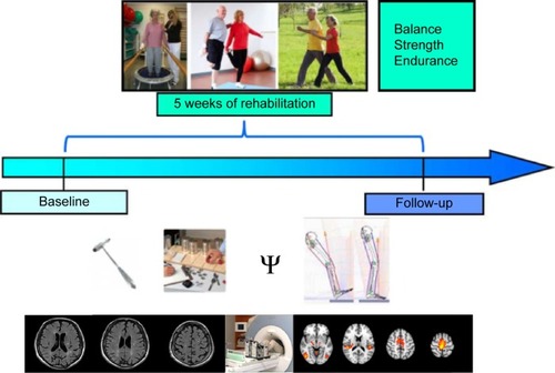
During the 5 consecutive weeks of training, patients received 15 training units, thrice per week, with each domain lasting for 20–30 minutes (including balance, strength, and endurance), rendering a total training time per participant of 1,350 minutes (22.5 hours). The training was guided by experienced physical therapists at a geriatric institution, the Albert Schweitzer Clinic, Graz, and supervised by physicians experienced in neurorehabilitation and geriatric medicine. The study design is shown in .
Figure 1 Design of the intervention study in chronic stroke patients undergoing 5 weeks of ambulatory training focusing on gait and coordination.
Abbreviation: MRI, magnetic resonance imaging.
Behavioral outcome assessment
At baseline and follow-up after 5 weeks, study participants underwent clinical examination, assessment using two scales (GDSCitation11 and WHO Quality of Life [WHOQOL]Citation12), and were neuropsychologically assessed using the following six tests: the Wechsler Memory Scale-logical memory (WMS-LM) and the delayed recall (WMS-LM DR),Citation13 the Symbol Digit Modalities Test (SDMT),Citation14 the MoCa,Citation15 the Trail Making Test A (TMT-A), and the Trail Making Test B (TMT-B).Citation16
The GDS consists of 15 items to assess depressive mood, with higher scores indicating a higher degree of depression. The WHOQOL (0–100) measures four subtypes of quality of life, with higher scores indicating a higher quality. The WMS-LM consists of two stories that have to be reproduced instantly and at the end (WMS-LM DR) of neuropsychological assessment to measure logical memory. More reproduced items indicate better logical memory. The SDMT measures processing speed, with higher scores of solved items indicating higher processing speed. The MoCa consists of eight domains to test global cognition, with higher scores indicating higher cognitive abilities. The TMT-A measures attention, with lower processing time indicating a higher degree of attention. The TMT-B measures executive functions, with lower processing time indicating better executive functions.
Participants also underwent a 3-minute walking test and mobility assessment using the Demmi (De Morton Mobility Index)Citation17 and the TinettiCitation18 scales. Higher scores in the mobility scales indicate better mobility.
Magnetic resonance imaging
Structural and functional MRI data were acquired on a 3 Tesla Tim Trio Scanner (Siemens, Erlangen, Germany) using a 32-channel head coil. A high-resolution T1-weighted structural image (TR =1,900 ms, TE =2.63 ms, 1 mm isotropic resolution, 176 slices) was acquired at baseline to allow for functional image registration, precise localization of activation, and location of old infarcts. Furthermore, a T2-weigthed fluid-attenuated inversion recovery (FLAIR) sequence (TR/TE/TI =10,000/69/2,500 ms, in plane resolution =0.9×0.9 mm2, slice thickness =3 mm, no gap, slices =44) served to assess deep white matter hyperintensities (WMH), which were classified using the Fazekas scaleCitation19 (grade 0= no lesions, 1= punctuate lesions, 2= early confluent lesions, and 3= confluent lesions). fMRI data were acquired on two occasions, before and after training, using identical scanning parameters (TR =3,000 ms, TE =30 ms, slices =143) and the same paradigm with the same scanner.
fMRI paradigm
A “block design”, comprising bipedal ankle movements and periods of rest, modified based on paradigms used successfully in earlier studies,Citation4,Citation20,Citation21 was employed. Bipedal movements were paced by arrows presented on a screen. Patients moved their feet alternately to simulate gait closely as possible. Vision was corrected with prism lenses if necessary. Blocks of bipedal ankle movement of 30 seconds alternated with periods of rest (21 seconds). Each session included four bipedal movement blocks separated by four blocks of rest. The paradigm was programmed in Presentation 16.3. The total scanning time for functional and structural imaging was 25 minutes. Prior to entering the scanner, subjects practiced the paradigm. Visual cues for ankle movements were presented on a screen and the experimenter explained the condition, conducting a training including the same purpose-built apparatus outside the scanner.
fMRI data analysis
Functional imaging analysis was carried out using FEAT (fMRI Expert Analysis Tool; Version 6.00, part of FMRIB’s Software Library, www.fmrib.ox.ac.uk/fsl). The following prestatistics were applied: high pass filtering using a cutoff of 100 seconds; motion correction using MCFLIRT; brain extraction was done using Brain Extraction Tool; and spatial smoothing was performed using a Gaussian kernel of 5 mm full width at half maximum. Time-series statistical analysis was carried out using FMRIB’s Improved Linear Model. Registration to high-resolution and/or standard images was carried out using FMRIB’s Linear Image Registration Tool. Functional imaging data of the three patients with left-hemispheric strokes were flipped left to right by the midline, so all patients virtually had a right-sided stroke, as suggested previously.Citation22 In first-level analysis, the effects of the bipedal movement blocks were determined for each subject. Head motion was corrected for using the displacement of the functional images in any direction, as derived from the FEAT motion correction report. Higher-level analysis was done using FLAME 1 (FMRIB’s Local Analysis of Mixed Effects). Z-statistic images were thresholded using clusters determined by Z>2.0 for mean activations of bipedal movement and for post- versus precomparisons, with a corrected cluster significance threshold of P<0.05.Citation23 To examine functional changes between and within groups, we calculated unpaired and paired t-tests in second-level analysis.
General statistical analysis
Comparisons between patients and controls regarding behavioral and demographic data (mobility, neuropsychological assessment) were computed with the Statistical Package of Social Science (IBM SPSS Statistics 20; IBM Corporation, Armonk, NY, USA). The level of significance was set at 0.05. Baseline differences between groups and pre–post comparisons for each group were calculated with analysis of variance.
Results
Clinical features of the cohort
All participants were right handed. Patients and controls did not differ significantly with regard to sex, but controls tended to be younger (χ2sex1,17=0.486, P=0.667; Fage1,22=4.097, P=0.055). HCs had a higher level of education (F1,22=4.403, P<0.05, η2=0.17). Patients had mildly to moderately impaired walking abilities (10-meter walk: MW =0.9 m/s, SD =0.21 m/s). Patients tended to have a higher lesion WMH grade (F1,22=4.15, P=0.054) but lower executive functions (F1,22=4.45, P<0.05, η2=0.17) and processing speed (F1,22=15.39, P<0.05, η2=0.41) at baseline (details listed in and shown in ).
Table 1 Baseline comparisons between chronic stroke patients and HCs
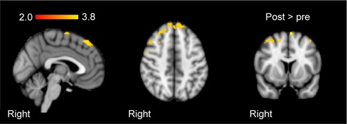
Figure 2 Group-mean activation changes from baseline to follow-up after training in the patient group.
Behavioral outcome assessment after training
After the intervention, patients showed increased mobility (Demmi F1,7=9.50, P<0.05, η2=0.58, Tinetti F1,7=4.59, P=0.07). They also showed improved logical memory (F1,7=5.67, P<0.05, η2=0.48) and logical memory recall (F1,7=21.06, P<0.05, η2=0.75). lists the remaining nonsignificant results.
Table 2 Changes from baseline to follow-up after 5 weeks of training in the patients
Changes in cerebral activation in patients after training
Subsequent to training, patients revealed increased activation in the right precentral gyrus, the right and left superior frontal gyri, and the right frontal lobe, with bipedal ankle movements (). Controls showed no significant changes in activation with the paradigm. At baseline, patients (compared to controls) showed higher activation in the contralesional hemisphere (postcentral gyrus, parietal operculum, superior temporal gyrus, and left cerebellum) and decreased activation in the ipsilesional precentral gyrus and supplemental motor area. After training, these differences disappeared.
Discussion
In this exploratory study, we found potential changes in mobility, cognition, and cerebral activation after 5 weeks of ambulatory training focusing on gait and coordination in chronic stroke patients with residual gait disturbance.
We noted mobility increases after training in our sample of chronic subcortical stroke patients, in line with an earlier study indicating that balance and basic motor abilities may be enhanced even in such “chronic” states.Citation22 Conforming previous reports, cognition can be augmented not only through cognitive but also through mobility training and that, in turn, cognitive resources are needed for adequate motor activity and mobility.Citation10,Citation24,Citation25 Supporting this notion, patients demonstrated improved memory after the training. Although causality with the training cannot be deferred and test–retest effects need to be considered, this observation deserves closer consideration in future studies. Moreover, we did not specifically select patients with impaired cognition in our study, although spontaneous improvements in cognitive abilities in the chronic stage of stroke appear unlikely.Citation25–Citation27 Indeed, in line with the reported profile of cognitive changes after symptomatic subcortical infarcts, our patients also fared worse than controls regarding executive functions and processing speed at baseline.
In this study, we could not safely conclude on the effects of WMH on rehabilitation success because of the small sample size, which should be taken into consideration in future studies. It appears to be of interest in this context that a recent review on small vessel disease highlighted the possibility that the functional effects of WMH, eg, on cognition, might be obscured due to an increased cognitive reserve measured by education years in some individuals.Citation28 This warrants further investigation with regard to training interventions.
On fMRI with bilateral ankle movements, patients demonstrated increased activation of the ipsi- and contralesional superior frontal gyri, ie, areas outside the motor network after the training. These brain regions have been implicated not only in self-consciousness, working memory, and executive functions but also in more complex motor functions.Citation29,Citation30 Increases were also noted in the ipsilesional frontal lobe and ipsilesional precentral gyrus, which are considered as important areas for initializing and preparing motor actions.Citation31 These results are partly in line with previous training studies,Citation32–Citation34 but mostly appear to indicate that the changes of brain function subsequent to a multimodal training focusing on such a complex behavior like gait but also affecting cognitive function may not be grasped by a simple motor paradigm. In this regard, using resting-state fMRI might be a superior approach as it obviates the need for restraining to a particular task and also leaves the possibility to assess changes in other networks.Citation35,Citation36 However, the fact that HCs who did not undergo training showed absolutely reproducible and unchanged cerebral activation at follow-up speaks for robustness of the used ankle movement fMRI paradigm.
Our study also has limitations. Importantly, the cognitive test battery should include validated parallel versions to avoid the learning effects that were also noted in our control group. The parallel versions should also be sensitive to educational differences between groups to measure only stroke or WMH-depending influences. These aspects need to be considered regarding the differences at baseline in cognition.
Furthermore, instead of testing a healthy cohort where ceiling effects are noted in the motor and gait tests, inclusion of a high-risk control group enriched for vascular risk factors might be more representative of the population in target. Extending the methodological limitations regarding fMRI (as already discussed), given the behavioral cognitive effects seen, additional inclusion of a cognitive paradigm appears favorable to more closely assess the effects of mobility training on cognitive brain function. In addition, more complex assessments like a dual task with walking while talking or providing cognitive challenges during gait assessment would be desirable to reveal more subtle deficits and effects.
It might also be possible that the overall results might have been biased, as our assessors were not blinded. However, patients were trained at the Albert Schweitzer Clinic, Graz, by experienced physiotherapists, whereas the MRI and behavioral outcome assessment took place at the Medical University of Graz by an unbiased researcher. In addition, social interaction might have increased motivation in our patients. Nevertheless, both groups were asked to daily record their physical activity for 5 weeks in a specific form. Thus, both groups focused on their physical function.
Because of our restricted inclusion criteria, we could only include a small sample size. Studying a larger cohort including randomization to intervention also comprising longer-term follow-up is mandatory to corroborate and extend our findings, with regard to the impact of preexisting brain damage as well. This should finally result in more refined insights regarding the effects of such a training and to predict responders.Citation37
Acknowledgments
Part of this study was funded by a project of the country of Styria (project ID 4321) called “Untersuchungen zu den Effekten eines Trainings von Gang und Gleichgewicht auf Gehirnfunktion im Alter – Einblicke anhand der funktionellen Magnetresonanztomographie (fMRT)”. The authors thank Karin Brodtrager for technical support and all involved persons from the Albert Schweitzer Clinic Graz, especially Antonella de Campo, MD; Martin Petio; and Katrin Glänzer. Deepest gratitude goes to all participants of this study.
Disclosure
The authors report no conflicts of interest in this work.
References
- LanghornePBernhardtJKwakkelGStroke rehabilitationLancet201137797781693170221571152
- EricksonKIHohmannTDie Effekte von Alter und Training auf die kognitive GesundheitZeitschrift für Sportpsychologie20132012532
- CalauttiCBaronJ-CFunctional neuroimaging studies of motor recovery after stroke in adults: a reviewStroke20033461553156612738893
- LinortnerPFazekasFSchmidtRWhite matter hyperintensities alter functional organization of the motor systemNeurobiol Aging201233119720724032
- WardlawJMSmithEEBiesselsGJNeuroimaging standards for research into small vessel disease and its contribution to ageing and neurodegenerationLancet Neurol201312882283823867200
- KimJSBaeYHPure or predominant sensory stroke due to brain stem lesionStroke1997289176117649303022
- LimanTGHeuschmannPUEndresMFlöelASchwabSKolominsky-RabasPLChanges in cognitive function over 3 years after first-ever stroke and predictors of cognitive impairment and long-term cognitive stability: the Erlangen Stroke ProjectDement Geriatr Cogn Disord201131429129921502760
- RosamondWFlegalKFurieKHeart disease and stroke statistics – 2008 update: a report from the American heart association statistics committee and stroke statistics subcommitteeCirculation20081177125146
- De LaatKFVan NordenAGGonsRAGait in elderly with cerebral small vessel diseaseStroke20104181652165820576951
- WardNSBrownMMThompsonAJFrackowiakRSThe influence of time after stroke on brain activations during a motor taskAnn Neurol200455682983415174016
- YesavageJASheikhJIGeriatric Depression Scale (GDS) recent evidence and development of a shorter versionClin Gerontol198651–2165173
- Development of the World Health Organization WHOQOL-BREF quality of life assessmentThe WHOQOL groupPsychol Med19982835515589626712
- HärtingCMarkowitschHNeufeldHCalabresePDeisingerKKesslerJWechsler Gedächtnistest – Revidierte Fassung. WMS-R. (Manual)Bern, SwitzerlandHans Huber2000
- RaoSA Manual for the Brief Repeatable Battery of Neuropsychological Tests in Multiple SclerosisMilwaukee, WIMedical College of Wisconsin1990
- NasreddineZSPhillipsNABédirianVThe Montreal Cognitive Assessment, MoCA: a brief screening tool for mild cognitive impairmentJ Am Geriatr Soc200553469569915817019
- TombaughTNTrail Making Test A and B: normative data stratified by age and educationArch Clin Neuropsychol200419220321415010086
- De MortonNDavidsonMKeatingJLThe de Morton Mobility Index (DEMMI): an essential health index for an ageing worldHealth Qual Life Outcomes200866318713451
- MarksDAssessment: Tinetti-Test Sturzrisiko erkennenPhysiopraxis200623233
- FazekasFChawlukJBAlaviAHurtigHZimmermanRMR signal abnormalities at 1.5T in Alzheimer’s dementia and normal aging defciencyAJR Am J Roentgenol198714923513563496763
- EnzingerCJohansen-BergHDawesHFunctional MRI correlates of lower limb function in stroke victims with gait impairmentStroke20083951507151318340092
- EnzingerCDawesHJohansen-BergHBrain activity changes associated with treadmill training after strokeStroke20094072460246719461018
- DawesHEnzingerCJohansen-BergHWalking performance and its recovery in chronic stroke in relation to extent of lesion overlap with the descending motor tractExp brain Res2008186232533318157670
- WorsleyKJStatistical analysis of activation imagesFunctional MRI: An Introduction to MethodsOxford, UKOxford University Press2001251270
- RazNRodrigueKMDifferential aging of the brain: patterns, cognitive correlates and modifiersNeurosci Biobehav Rev200630673074816919333
- Montero-OdassoMVergheseJBeauchetOHausdorffJMGait and cognition: a complementary approach to understanding brain function and the risk of fallingJ Am Geriatr Soc201260112127213623110433
- KludingPMTsengBYBillingerSAExercise and executive function in individuals with chronic stroke: a pilot studyJ Neurol Phys Ther2011351111721475079
- QuaneyBBoydLAerobic exercise improves cognition and motor function poststrokeNeurorehabil Neural Repair200923987988519541916
- PinterDEnzingerCFazekasFCerebral small vessel disease, cognitive reserve and cognitive dysfunctionJ Neurol2015262112411241925976029
- WenWZhuWHeYDiscrete neuroanatomical networks are associated with specific cognitive abilities in old ageJ Neurosci20113141204121221273405
- MartinoJGabarrósADeusJIntrasurgical mapping of complex motor function in the superior frontal gyrusNeuroscience201117913114221277357
- PorroCAFrancescatoMPCettoloVPrimary motor and sensory cortex activation during motor performance and motor imagery: a functional magnetic resonance imaging studyJ Neurosci19961623768876988922425
- ScholzJKleinMCBehrensTEJohansen-BergHTraining induces changes in white-matter architectureNat Neurosci200912111370137119820707
- BosnellRAKincsesTStaggCJMotor practice promotes increased activity in brain regions structurally disconnected after sub-cortical strokeNeurorehabil Neural Repair201325760761621646444
- CareyLMAbbottDFEganGFEvolution of brain activation with good and poor motor recovery after strokeNeurorehabil Neural Repair2006201244116467276
- Ovadia-CaroSVillringerKFiebachJLongitudinal effects of lesions on functional networks after strokeJ Cereb Blood Flow Metab20133381279128523715061
- WestlakeKPNagarajanSSFunctional connectivity in relation to motor performance and recovery after strokeFront Syst Neurosci2011581221472027
- NadkarniNKStudenskiSAPereraSWhite matter hyperintensities, exercise, and improvement in gait speed: does type of gait rehabilitation matter?J Am Geriatr Soc201361568669323590257